Pierro Luisa, Arrigo Alessandro, De Crescenzo Michele, Aragona Emanuela, Chiesa Roberto, Castellano Renata, Catenaccio Barbara, Bandello Francesco
Department of Ophthalmology, Scientific Institute San Raffaele Hospital, University Vita-Salute, Milan, Italy.
Department of Surgery, San Raffaele Hospital, Milan, Italy.
Front Neurosci. 2021 Apr 22;15:640666. doi: 10.3389/fnins.2021.640666. eCollection 2021.
Carotid artery stenosis (CAS) is a multifaceted disease characterized by possible ocular involvement. Treatment with carotid endarterectomy helps to restore cerebral perfusion, which may prevent ocular and cerebral complications. The main aim was to assess retinal and choroidal vascular perfusion changes before and after endarterectomy in patients affected by CAS.
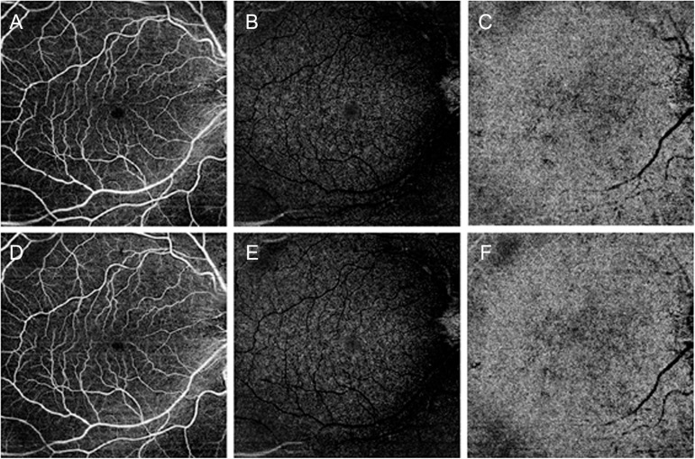
The design of the study was prospective and observational, including patients affected by CAS and healthy controls. The follow-up was 3 months. We performed quantitative optical coherence tomography (OCT) angiography (OCTA) analyses of retinal perfusion changes, before and after endarterectomy. The main outcome measures were the quantitative changes of choroidal thickness (CT), retinal nerve fiber layer (RNFL), and ganglion cell layer (GCL); vessel density (VD); and vessel tortuosity (VT) OCTA metrics were also measured.
Sixty eyes of 30 patients affected by CAS and 30 eyes of 30 controls were included. We separately considered the ipsilateral eyes to CAS, the contralateral eyes to CAS, and the healthy eyes. Visual symptoms were absent in all the patients. RNFL and GCL resulted similar between patients and controls ( > 0.05). CT was significantly thinner in ipsilateral eyes than controls ( < 0.01), and it resulted unchanged after surgery ( > 0.05). VD resulted significantly altered only in some plexa of the ipsilateral eyes ( < 0.01), whereas VT disclosed decreased values of the entire retinal vascular network, both in ipsilateral and contralateral eyes ( < 0.05). Endarterectomy was followed by statistically significant improvement of retinal perfusion ( < 0.05).
Optical coherence tomography angiography can noninvasively detect postendarterectomy retinal perfusion improvements in CAS patients with baseline diabetes and hypertension as a systemic risk factor.
颈动脉狭窄(CAS)是一种多方面的疾病,其特征可能包括眼部受累。颈动脉内膜切除术有助于恢复脑灌注,这可能预防眼部和脑部并发症。主要目的是评估CAS患者内膜切除术前和术后视网膜和脉络膜血管灌注的变化。
本研究设计为前瞻性观察性研究,纳入CAS患者和健康对照。随访时间为3个月。我们在内膜切除术前和术后对视网膜灌注变化进行了定量光学相干断层扫描(OCT)血管造影(OCTA)分析。主要观察指标包括脉络膜厚度(CT)、视网膜神经纤维层(RNFL)和神经节细胞层(GCL)的定量变化;血管密度(VD);还测量了血管迂曲度(VT)的OCTA指标。
纳入了30例CAS患者的60只眼和30例对照的30只眼。我们分别考虑了CAS同侧眼、CAS对侧眼和健康眼。所有患者均无视觉症状。患者和对照之间的RNFL和GCL相似(>0.05)。同侧眼的CT明显比对照薄(<0.01),术后无变化(>0.05)。仅同侧眼的某些层的VD有显著改变(<0.01),而VT显示同侧眼和对侧眼的整个视网膜血管网络的值均降低(<0.05)。内膜切除术后视网膜灌注有统计学意义的改善(<0.05)。
光学相干断层扫描血管造影可以无创地检测基线时有糖尿病和高血压作为全身危险因素的CAS患者内膜切除术后视网膜灌注的改善情况。