• 文献检索
  • 文档翻译
  • 深度研究
  • 学术资讯
  • Suppr Zotero 插件Zotero 插件
  • 邀请有礼
  • 套餐&价格
  • 历史记录
应用&插件
Suppr Zotero 插件Zotero 插件浏览器插件Mac 客户端Windows 客户端微信小程序
定价
高级版会员购买积分包购买API积分包
服务
文献检索文档翻译深度研究API 文档MCP 服务
关于我们
关于 Suppr公司介绍联系我们用户协议隐私条款
关注我们

Suppr 超能文献

核心技术专利:CN118964589B侵权必究
粤ICP备2023148730 号-1Suppr @ 2026

文献检索

告别复杂PubMed语法,用中文像聊天一样搜索,搜遍4000万医学文献。AI智能推荐,让科研检索更轻松。

立即免费搜索

文件翻译

保留排版,准确专业,支持PDF/Word/PPT等文件格式,支持 12+语言互译。

免费翻译文档

深度研究

AI帮你快速写综述,25分钟生成高质量综述,智能提取关键信息,辅助科研写作。

立即免费体验

视网膜厚度与颈动脉狭窄患者的脑血流动力学变化相关。

Retinal Thickness Correlates with Cerebral Hemodynamic Changes in Patients with Carotid Artery Stenosis.

作者信息

Kwapong William Robert, Liu Junfeng, Wan Jincheng, Tao Wendan, Ye Chen, Wu Bo

机构信息

Neurology Department, West China Hospital of Sichuan University, No. 37 Guo Xue Xiang, Chengdu 610041, China.

出版信息

Brain Sci. 2022 Jul 25;12(8):979. doi: 10.3390/brainsci12080979.

DOI:10.3390/brainsci12080979
PMID:35892420
原文链接:https://pmc.ncbi.nlm.nih.gov/articles/PMC9331379/
Abstract

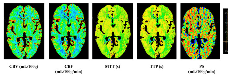
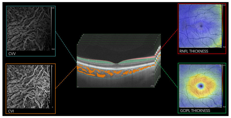
Background: We aimed to assess the retinal structural and choroidal changes in carotid artery stenosis (CAS) patients and their association with cerebral hemodynamic changes. Asymptomatic and symptomatic patients with unilateral CAS were enrolled in our study. Material and methods: Swept-source optical coherence tomography (SS-OCT) was used to image the retinal nerve fiber layer (RNFL), ganglion cell-inner plexiform layer (GCIPL), while SS-OCT angiography (SS-OCTA) was used to image and measure the choroidal vascular volume (CVV) and choroidal vascular index (CVI). Computed Tomography Perfusion (CTP) was used to assess the cerebral perfusion parameters; relative perfusion (r) was calculated as the ratio of the value on the contralateral side to that on the ipsilateral side. Results: Compared with contralateral eyes, ipsilateral eyes showed significantly thinner RNFL (p < 0.001), GCIPL (p = 0.013) and CVV (p = 0.001). Relative cerebral blood volume (rCBV) showed a significant correlation with RNFL (p < 0.001), GCIPL (p < 0.001) and CVI (p = 0.027), while the relative permeability surface (rPS) correlated with RNFL (p < 0.001) and GCIPL (p < 0.001). Conclusions: Our report suggests that retinal and choroidal changes have the potential to detect hemodynamic changes in CAS patients and could predict the risk of stroke.

摘要

背景

我们旨在评估颈动脉狭窄(CAS)患者的视网膜结构和脉络膜变化及其与脑血流动力学变化的关联。本研究纳入了单侧CAS的无症状和有症状患者。材料与方法:使用扫频光学相干断层扫描(SS-OCT)对视网膜神经纤维层(RNFL)、神经节细胞-内丛状层(GCIPL)进行成像,同时使用SS-OCT血管造影(SS-OCTA)对脉络膜血管容积(CVV)和脉络膜血管指数(CVI)进行成像和测量。计算机断层扫描灌注(CTP)用于评估脑灌注参数;相对灌注(r)计算为对侧值与同侧值的比值。结果:与对侧眼相比,同侧眼的RNFL(p < 0.001)、GCIPL(p = 0.013)和CVV(p = 0.001)明显更薄。相对脑血容量(rCBV)与RNFL(p < 0.001)、GCIPL(p < 0.001)和CVI(p = 0.027)显著相关,而相对通透表面(rPS)与RNFL(p < 0.001)和GCIPL(p < 0.001)相关。结论:我们 的报告表明,视网膜和脉络膜变化有可能检测出CAS患者的血流动力学变化,并可预测中风风险。

https://cdn.ncbi.nlm.nih.gov/pmc/blobs/87ed/9331379/c5236e62774f/brainsci-12-00979-g002.jpg
https://cdn.ncbi.nlm.nih.gov/pmc/blobs/87ed/9331379/a95e0e92ab69/brainsci-12-00979-g001.jpg
https://cdn.ncbi.nlm.nih.gov/pmc/blobs/87ed/9331379/c5236e62774f/brainsci-12-00979-g002.jpg
https://cdn.ncbi.nlm.nih.gov/pmc/blobs/87ed/9331379/a95e0e92ab69/brainsci-12-00979-g001.jpg
https://cdn.ncbi.nlm.nih.gov/pmc/blobs/87ed/9331379/c5236e62774f/brainsci-12-00979-g002.jpg

相似文献

1
Retinal Thickness Correlates with Cerebral Hemodynamic Changes in Patients with Carotid Artery Stenosis.视网膜厚度与颈动脉狭窄患者的脑血流动力学变化相关。
Brain Sci. 2022 Jul 25;12(8):979. doi: 10.3390/brainsci12080979.
2
Choroidal Changes in Carotid Stenosis Patients After Stenting Detected by Swept-source Optical Coherence Tomography Angiography.颈动脉狭窄患者支架置入术后脉络膜变化的扫频源光学相干断层血管造影研究。
Curr Neurovasc Res. 2022;19(1):100-107. doi: 10.2174/1567202619666220406092532.
3
Retinal microvasculature and cerebral hemodynamics in patients with internal carotid artery stenosis.颈内动脉狭窄患者的视网膜微血管和脑血流动力学。
BMC Neurol. 2022 Oct 13;22(1):386. doi: 10.1186/s12883-022-02908-7.
4
Influence of Carotid Artery Stenting on the Retina and Choroid.颈动脉支架置入术对视网膜和脉络膜的影响。
Transl Vis Sci Technol. 2024 Aug 1;13(8):5. doi: 10.1167/tvst.13.8.5.
5
Quantitative Optical Coherence Tomography Angiography Detects Retinal Perfusion Changes in Carotid Artery Stenosis.定量光学相干断层扫描血管造影术检测颈动脉狭窄中的视网膜灌注变化。
Front Neurosci. 2021 Apr 22;15:640666. doi: 10.3389/fnins.2021.640666. eCollection 2021.
6
Choroid and choriocapillaris changes in early-stage Parkinson's disease: a swept-source optical coherence tomography angiography-based cross-sectional study.基于扫频源光相干断层血管造影的早期帕金森病脉络膜和脉络膜毛细血管变化的横断面研究。
Alzheimers Res Ther. 2022 Aug 25;14(1):116. doi: 10.1186/s13195-022-01054-z.
7
Ganglion cell-inner plexiform layer and retinal nerve fiber layer thickness according to myopia and optic disc area: a quantitative and three-dimensional analysis.根据近视和视盘面积分析神经节细胞-内网状层及视网膜神经纤维层厚度:定量与三维分析
BMC Ophthalmol. 2017 Mar 11;17(1):22. doi: 10.1186/s12886-017-0419-1.
8
Ganglion cell-inner plexiform layer and retinal nerve fibre layer changes within the macula in retinitis pigmentosa: a spectral domain optical coherence tomography study.色素性视网膜炎黄斑部神经节细胞-内丛状层和视网膜神经纤维层的改变:频域光学相干断层扫描研究。
Acta Ophthalmol. 2018 Mar;96(2):e180-e188. doi: 10.1111/aos.13577. Epub 2017 Nov 2.
9
Quantitative assessment of retinochoroidal microvasculature in patients with carotid artery stenosis using OCT angiography.使用光学相干断层扫描血管造影术对颈动脉狭窄患者视网膜脉络膜微血管进行定量评估。
Photodiagnosis Photodyn Ther. 2024 Apr;46:104082. doi: 10.1016/j.pdpdt.2024.104082. Epub 2024 Apr 6.
10
Influence of Carotid Endarterectomy on Choroidal Perfusion: The INFLATE Study.颈动脉内膜切除术对脉络膜灌注的影响:INFLATE 研究。
Ophthalmol Retina. 2024 Jan;8(1):62-71. doi: 10.1016/j.oret.2023.07.026. Epub 2023 Jul 31.

引用本文的文献

1
Interocular retinal asymmetry and cognitive frailty: A prospective cohort study.双眼视网膜不对称与认知衰弱:一项前瞻性队列研究。
Alzheimers Dement. 2025 Feb;21(2):e14437. doi: 10.1002/alz.14437. Epub 2024 Dec 23.
2
Choroidal vascularity index in health and systemic diseases: a systematic review.健康与全身性疾病中的脉络膜血管指数:一项系统评价
Int J Retina Vitreous. 2024 Nov 18;10(1):87. doi: 10.1186/s40942-024-00607-8.
3
Advances in retinal imaging biomarkers for the diagnosis of cerebrovascular disease.用于诊断脑血管疾病的视网膜成像生物标志物的进展。

本文引用的文献

1
Changes in the choroid plexuses and brain barriers associated with high blood pressure and ageing.高血压和衰老相关脉络丛和脑屏障的变化。
Neurologia (Engl Ed). 2022 Jun;37(5):371-382. doi: 10.1016/j.nrleng.2020.05.007. Epub 2020 Jun 3.
2
Choroidal Changes in Carotid Stenosis Patients After Stenting Detected by Swept-source Optical Coherence Tomography Angiography.颈动脉狭窄患者支架置入术后脉络膜变化的扫频源光学相干断层血管造影研究。
Curr Neurovasc Res. 2022;19(1):100-107. doi: 10.2174/1567202619666220406092532.
3
Macular Microvasculature Is Associated With Total Cerebral Small Vessel Disease Burden in Recent Single Subcortical Infarction.
Front Neurol. 2024 Sep 19;15:1393899. doi: 10.3389/fneur.2024.1393899. eCollection 2024.
4
Insight into the Brain: Application of the Retinal Microvasculature as a Biomarker for Cerebrovascular Diseases through Optical Coherence Tomography Angiography.洞察大脑:通过光学相干断层扫描血管造影术将视网膜微血管系统用作脑血管疾病生物标志物的应用
Curr Ophthalmol Rep. 2024 Mar;12(1):1-11. doi: 10.1007/s40135-023-00320-z. Epub 2023 Nov 28.
5
Influence of Carotid Artery Stenting on the Retina and Choroid.颈动脉支架置入术对视网膜和脉络膜的影响。
Transl Vis Sci Technol. 2024 Aug 1;13(8):5. doi: 10.1167/tvst.13.8.5.
6
Changes in the retina and choroid in patients with internal carotid artery stenosis: a systematic review and meta-analysis.颈内动脉狭窄患者视网膜和脉络膜的变化:系统评价与荟萃分析
Front Neurosci. 2024 Apr 15;18:1368957. doi: 10.3389/fnins.2024.1368957. eCollection 2024.
7
Optical Coherence Tomography Angiography: Revolutionizing Clinical Diagnostics and Treatment in Central Nervous System Disease.光学相干断层扫描血管造影术:革新中枢神经系统疾病的临床诊断与治疗
Aging Dis. 2024 Jan 20;16(1):77-114. doi: 10.14336/AD.2024.0112.
8
Retinal microvascular and structural changes in intracranial hypertension patients correlate with intracranial pressure.颅内高压患者的视网膜微血管和结构变化与颅内压相关。
CNS Neurosci Ther. 2023 Dec;29(12):4093-4101. doi: 10.1111/cns.14298. Epub 2023 Jun 8.
9
Association of retinal thickness and microvasculature with cognitive performance and brain volumes in elderly adults.老年人视网膜厚度和微血管与认知能力及脑容量的关联
Front Aging Neurosci. 2022 Nov 16;14:1010548. doi: 10.3389/fnagi.2022.1010548. eCollection 2022.
黄斑部微血管与近期单发性皮质下梗死中的全脑小血管疾病负担相关。
Front Aging Neurosci. 2022 Jan 20;13:787775. doi: 10.3389/fnagi.2021.787775. eCollection 2021.
4
The effect of systemic factors on retinal blood flow in patients with carotid stenosis: an optical coherence tomography angiography study.系统性因素对颈动脉狭窄患者视网膜血流的影响:一项光学相干断层扫描血管造影研究。
Geroscience. 2022 Feb;44(1):389-401. doi: 10.1007/s11357-021-00492-1. Epub 2021 Nov 27.
5
Optical Coherence Tomography Angiography as a Noninvasive Assessment of Cerebral Microcirculatory Disorders Caused by Carotid Artery Stenosis.光学相干断层扫描血管造影术作为一种非侵入性评估颈动脉狭窄引起的脑微循环障碍的方法。
Dis Markers. 2021 Jul 5;2021:2662031. doi: 10.1155/2021/2662031. eCollection 2021.
6
A Case of Anterior Choroidal Artery Occlusion and Stroke Secondary to External Compression.一例继发于外部压迫的脉络膜前动脉闭塞和中风病例。
Case Rep Neurol. 2021 Jun 14;13(2):369-374. doi: 10.1159/000515574. eCollection 2021 May-Aug.
7
Carotid Stenosis and Recurrent Ischemic Stroke: A Post-Hoc Analysis of the POINT Trial.颈动脉狭窄与复发性缺血性卒中:POINT 试验的事后分析。
Stroke. 2021 Jul;52(7):2414-2417. doi: 10.1161/STROKEAHA.121.034089. Epub 2021 May 4.
8
Asymptomatic carotid artery stenosis is associated with cerebral hypoperfusion.无症状颈动脉狭窄与脑灌注不足有关。
J Vasc Surg. 2021 May;73(5):1611-1621.e2. doi: 10.1016/j.jvs.2020.10.063. Epub 2020 Nov 7.
9
Three-dimensional choroidal vascularity index in acute central serous chorioretinopathy using swept-source optical coherence tomography.应用扫频源光学相干断层扫描测量急性中心性浆液性脉络膜视网膜病变的脉络膜血流指数
Graefes Arch Clin Exp Ophthalmol. 2020 Feb;258(2):241-247. doi: 10.1007/s00417-019-04524-7. Epub 2019 Nov 14.
10
Optical Coherence Tomography Angiography Evaluation of Retinal Microvasculature Before and After Carotid Angioplasty and Stenting.颈动脉血管成形术及支架置入术前、后视网膜微血管的光相干断层扫描血管造影评估。
Sci Rep. 2019 Oct 14;9(1):14755. doi: 10.1038/s41598-019-51382-8.