Hansen Niels, Singh Aditya, Bartels Claudia, Brosseron Frederic, Buerger Katharina, Cetindag Arda C, Dobisch Laura, Dechent Peter, Ertl-Wagner Birgit B, Fliessbach Klaus, Haynes John D, Heneka Michael T, Janowitz Daniel, Kilimann Ingo, Laske Christoph, Metzger Coraline D, Munk Matthias H, Peters Oliver, Priller Josef, Roy Nina, Scheffler Klaus, Schneider Anja, Spottke Annika, Spruth Eike J, Teipel Stefan, Tscheuschler Maike, Vukovich Ruth, Wiltfang Jens, Duezel Emrah, Jessen Frank, Goya-Maldonado Roberto
Department of Psychiatry and Psychotherapy, Göttingen, Germany.
Laboratory of Systems Neuroscience and Imaging in Psychiatry, University Medical Center Göttingen, Göttingen, Germany.
Front Aging Neurosci. 2021 Apr 21;13:626974. doi: 10.3389/fnagi.2021.626974. eCollection 2021.
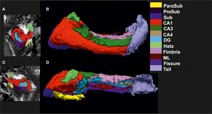
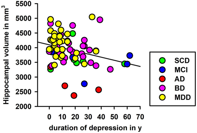
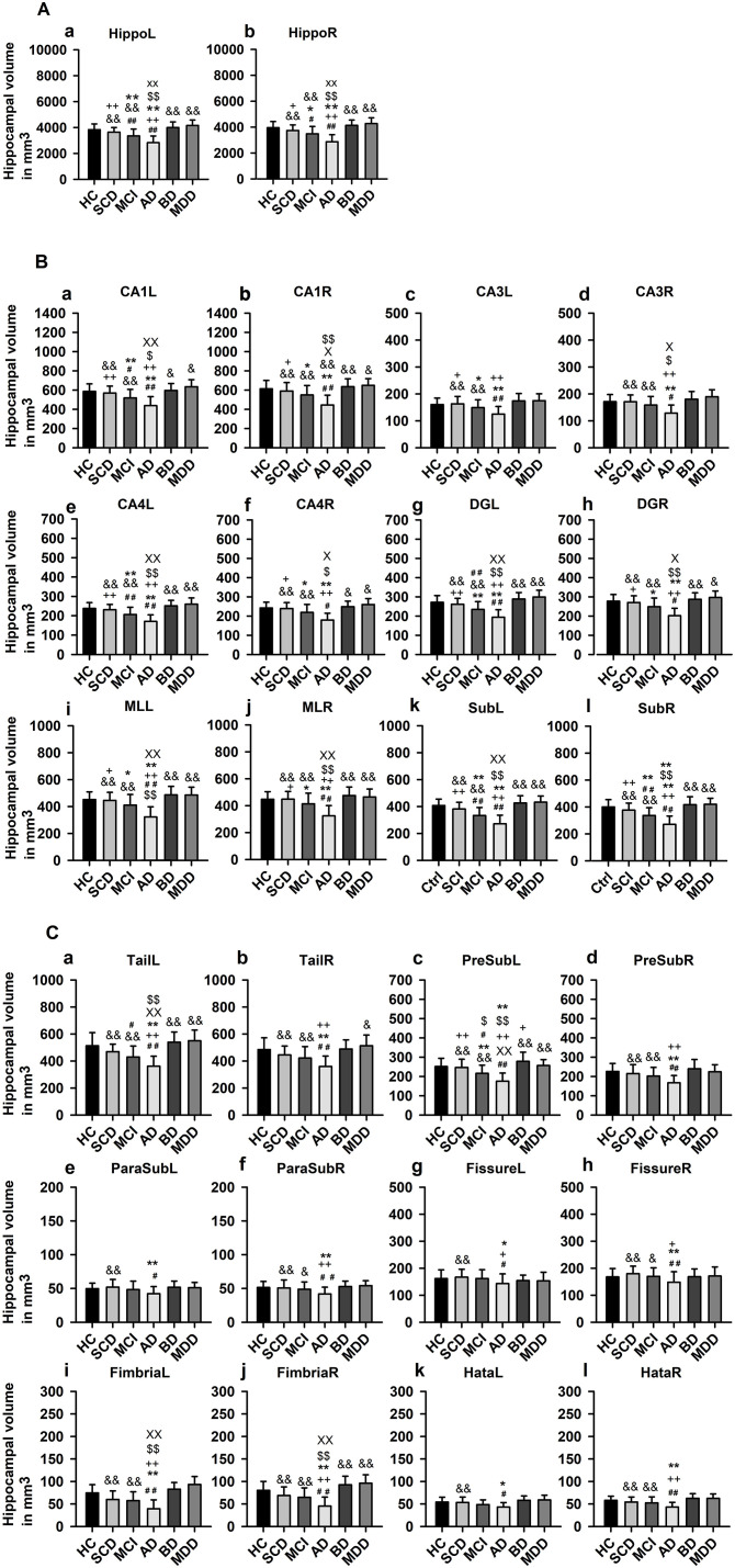
The hippocampus and its subfields (HippSub) are reported to be diminished in patients with Alzheimer's disease (AD), bipolar disorder (BD), and major depressive disorder (MDD). We examined these groups vs healthy controls (HC) to reveal HippSub alterations between diseases. We segmented 3T-MRI T2-weighted hippocampal images of 67 HC, 58 BD, and MDD patients from the AFFDIS study and 137 patients from the DELCODE study assessing cognitive decline, including subjective cognitive decline (SCD), amnestic mild cognitive impairment (aMCI), and AD, via Free Surfer 6.0 to compare volumes across groups. Groups differed significantly in several HippSub volumes, particularly between patients with AD and mood disorders. In comparison to HC, significant lower volumes appear in aMCI and AD groups in specific subfields. Smaller volumes in the left presubiculum are detected in aMCI and AD patients, differing from the BD group. A significant linear regression is seen between left hippocampus volume and duration since the first depressive episode. HippSub volume alterations were observed in AD, but not in early-onset MDD and BD, reinforcing the notion of different neural mechanisms in hippocampal degeneration. Moreover, duration since the first depressive episode was a relevant factor explaining the lower left hippocampal volumes present in groups.
据报道,阿尔茨海默病(AD)、双相情感障碍(BD)和重度抑郁症(MDD)患者的海马体及其亚区(HippSub)会减小。我们对这些组与健康对照(HC)进行了检查,以揭示不同疾病之间HippSub的变化。我们通过Free Surfer 6.0对来自AFFDIS研究的67名HC、58名BD和MDD患者以及来自DELCODE研究的137名评估认知衰退(包括主观认知衰退(SCD)、遗忘型轻度认知障碍(aMCI)和AD)的患者的3T-MRI T2加权海马体图像进行分割,以比较各组之间的体积。几组在几个HippSub体积上存在显著差异,尤其是AD患者与情绪障碍患者之间。与HC相比,aMCI组和AD组的特定亚区体积明显更低。在aMCI和AD患者中检测到左侧前扣带回体积较小,这与BD组不同。左侧海马体体积与首次抑郁发作后的持续时间之间存在显著的线性回归。在AD中观察到HippSub体积变化,但在早发性MDD和BD中未观察到,这强化了海马体退化中不同神经机制的观点。此外,首次抑郁发作后的持续时间是解释各组中左侧海马体体积较低的一个相关因素。