Michels Lars, Riese Florian, Meyer Rafael, Kälin Andrea M, Leh Sandra E, Unschuld Paul G, Luechinger Roger, Hock Christoph, O'Gorman Ruth, Kollias Spyros, Gietl Anton
Department of Neuroradiology, Clinical Neuroscience Center, University Hospital Zurich, Zurich, Switzerland.
Department of Geriatric Psychiatry, Psychiatric University Hospital Zurich (PUK), Zurich, Switzerland.
Front Aging Neurosci. 2021 Apr 23;13:631172. doi: 10.3389/fnagi.2021.631172. eCollection 2021.
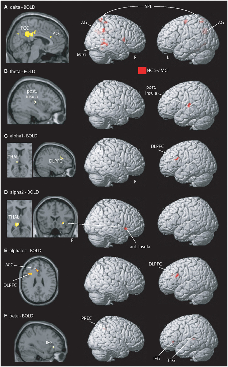
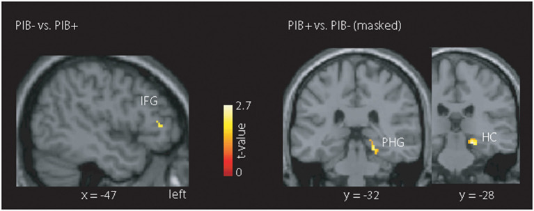
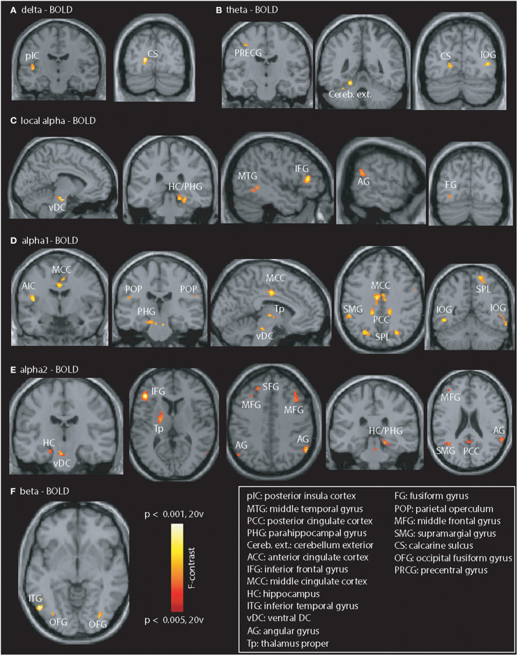
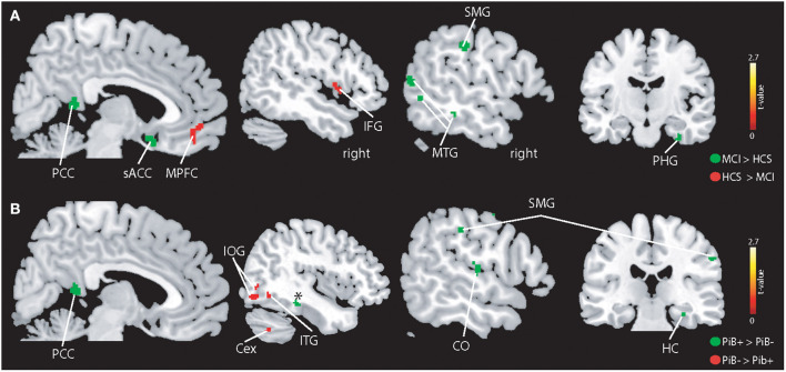
Cognitive impairment indicates disturbed brain physiology which can be due to various mechanisms including Alzheimer's pathology. Combined functional magnetic resonance imaging (fMRI) and electroencephalography (EEG) recordings (EEG-fMRI) can assess the interplay between complementary measures of brain activity and EEG changes to be localized to specific brain regions. We used a two-step approach, where we first examined changes related to a syndrome of mild cognitive impairment irrespective of pathology and then studied the specific impact of amyloid pathology. After detailed clinical and neuropsychological characterization as well as a positron emission tomography (PET) scans with the tracer 11-[C]-Pittsburgh Compound B to estimate cerebral amyloid deposition, 14 subjects with mild cognitive impairment (MCI) (mean age 75.6 SD: 8.9) according to standard criteria and 21 cognitively healthy controls (HCS) (mean age 71.8 SD: 4.2) were assessed with EEG-fMRI. Thalamo-cortical alpha-fMRI signal coupling was only observed in HCS. Additional EEG-fMRI signal coupling differences between HCS and MCI were observed in parts of the default mode network, salience network, fronto-parietal network, and thalamus. Individuals with significant cerebral amyloid deposition (amyloid-positive MCI and HCS combined compared to amyloid-negative HCS) displayed abnormal EEG-fMRI signal coupling in visual, fronto-parietal regions but also in the parahippocampus, brain stem, and cerebellum. This finding was paralleled by stronger absolute fMRI signal in the parahippocampus and weaker absolute fMRI signal in the inferior frontal gyrus in amyloid-positive subjects. We conclude that the thalamocortical coupling in the alpha band in HCS more closely reflects previous findings observed in younger adults, while in MCI there is a clearly aberrant coupling in several networks dominated by an anticorrelation in the posterior cingulate cortex. While these findings may broadly indicate physiological changes in MCI, amyloid pathology was specifically associated with abnormal fMRI signal responses and disrupted coupling between brain oscillations and fMRI signal responses, which especially involve core regions of memory: the hippocampus, para-hippocampus, and lateral prefrontal cortex.
认知障碍表明大脑生理功能紊乱,这可能由多种机制引起,包括阿尔茨海默病病理。功能磁共振成像(fMRI)和脑电图(EEG)记录相结合(EEG-fMRI)可以评估大脑活动的互补测量与EEG变化之间的相互作用,以便将EEG变化定位到特定脑区。我们采用了两步法,首先检查与轻度认知障碍综合征相关的变化,而不考虑病理情况,然后研究淀粉样蛋白病理的具体影响。在进行详细的临床和神经心理学特征描述以及使用示踪剂11-[C]-匹兹堡化合物B进行正电子发射断层扫描(PET)以估计脑淀粉样蛋白沉积后,根据标准标准,对14名轻度认知障碍(MCI)患者(平均年龄75.6,标准差:8.9)和21名认知健康对照者(HCS)(平均年龄71.8,标准差:4.2)进行了EEG-fMRI评估。丘脑-皮质α-fMRI信号耦合仅在HCS中观察到。在默认模式网络、突显网络、额顶网络和丘脑的部分区域观察到HCS和MCI之间额外的EEG-fMRI信号耦合差异。有显著脑淀粉样蛋白沉积的个体(淀粉样蛋白阳性的MCI和HCS合并与淀粉样蛋白阴性的HCS相比)在视觉、额顶区域以及海马旁回、脑干和小脑中显示出异常的EEG-fMRI信号耦合。这一发现与淀粉样蛋白阳性受试者海马旁回中更强的绝对fMRI信号和额下回中较弱的绝对fMRI信号相平行。我们得出结论,HCS中α波段的丘脑皮质耦合更紧密地反映了在年轻人中观察到的先前发现,而在MCI中,在几个以后扣带回皮质的反相关为主导的网络中存在明显异常的耦合。虽然这些发现可能广泛表明MCI中的生理变化,但淀粉样蛋白病理与异常的fMRI信号反应以及脑振荡与fMRI信号反应之间的耦合破坏特别相关,这尤其涉及记忆的核心区域:海马体、海马旁回和外侧前额叶皮质。