• 文献检索
  • 文档翻译
  • 深度研究
  • 学术资讯
  • Suppr Zotero 插件Zotero 插件
  • 邀请有礼
  • 套餐&价格
  • 历史记录
应用&插件
Suppr Zotero 插件Zotero 插件浏览器插件Mac 客户端Windows 客户端微信小程序
定价
高级版会员购买积分包购买API积分包
服务
文献检索文档翻译深度研究API 文档MCP 服务
关于我们
关于 Suppr公司介绍联系我们用户协议隐私条款
关注我们

Suppr 超能文献

核心技术专利:CN118964589B侵权必究
粤ICP备2023148730 号-1Suppr @ 2026

文献检索

告别复杂PubMed语法,用中文像聊天一样搜索,搜遍4000万医学文献。AI智能推荐,让科研检索更轻松。

立即免费搜索

文件翻译

保留排版,准确专业,支持PDF/Word/PPT等文件格式,支持 12+语言互译。

免费翻译文档

深度研究

AI帮你快速写综述,25分钟生成高质量综述,智能提取关键信息,辅助科研写作。

立即免费体验

相似文献

1
Multiple Particle Tracking Detects Changes in Brain Extracellular Matrix and Predicts Neurodevelopmental Age.多粒子追踪检测大脑细胞外基质变化,预测神经发育年龄。
ACS Nano. 2021 May 25;15(5):8559-8573. doi: 10.1021/acsnano.1c00394. Epub 2021 May 10.
2
High-fidelity predictions of diffusion in the brain microenvironment.高保真预测大脑微环境中的扩散。
Biophys J. 2024 Nov 19;123(22):3935-3950. doi: 10.1016/j.bpj.2024.10.005. Epub 2024 Oct 10.
3
Organotypic whole hemisphere brain slice models to study the effects of donor age and oxygen-glucose-deprivation on the extracellular properties of cortical and striatal tissue.用于研究供体年龄和氧糖剥夺对皮质和纹状体组织细胞外特性影响的器官型全脑半球脑片模型。
J Biol Eng. 2022 Jun 13;16(1):14. doi: 10.1186/s13036-022-00293-w.
4
Formation and remodeling of the brain extracellular matrix in neural plasticity: Roles of chondroitin sulfate and hyaluronan.脑外细胞基质在神经可塑性中的形成和重塑:硫酸软骨素和透明质酸的作用。
Biochim Biophys Acta Gen Subj. 2017 Oct;1861(10):2420-2434. doi: 10.1016/j.bbagen.2017.06.010. Epub 2017 Jun 15.
5
Dilation and degradation of the brain extracellular matrix enhances penetration of infused polymer nanoparticles.脑细胞外基质的扩张和降解增强了注入的聚合物纳米颗粒的渗透。
Brain Res. 2007 Nov 14;1180:121-32. doi: 10.1016/j.brainres.2007.08.050. Epub 2007 Aug 29.
6
Erasure of striatal chondroitin sulfate proteoglycan-associated extracellular matrix rescues aging-dependent decline of motor learning.消除纹状体软骨素硫酸蛋白聚糖相关细胞外基质可挽救运动学习随衰老的下降。
Neurobiol Aging. 2018 Nov;71:61-71. doi: 10.1016/j.neurobiolaging.2018.07.008. Epub 2018 Jul 23.
7
Extracellular Matrix Limits Nanoparticle Diffusion and Cellular Uptake in a Tissue-Specific Manner.细胞外基质以组织特异性的方式限制纳米颗粒的扩散和细胞摄取。
ACS Nano. 2024 Nov 19;18(46):32045-32055. doi: 10.1021/acsnano.4c10381. Epub 2024 Nov 5.
8
Activity-dependent switches between dynamic regimes of extracellular matrix expression.活动依赖性地切换细胞外基质表达的动态状态。
PLoS One. 2020 Jan 24;15(1):e0227917. doi: 10.1371/journal.pone.0227917. eCollection 2020.
9
Fractal dimension and directional analysis of elastic and collagen fiber arrangement in unsectioned arterial tissues affected by atherosclerosis and aging.分形维数和弹性纤维及胶原纤维在动脉粥样硬化和老化未切片组织中的排列方向分析。
J Appl Physiol (1985). 2019 Mar 1;126(3):638-646. doi: 10.1152/japplphysiol.00497.2018. Epub 2019 Jan 10.
10
Extracellular matrix protein expression is brain region dependent.细胞外基质蛋白表达具有脑区依赖性。
J Comp Neurol. 2016 May 1;524(7):1309-36. doi: 10.1002/cne.23965. Epub 2016 Feb 1.

引用本文的文献

1
Design Principles of Nanosensors for Multiplex Detection of Contaminants in Food.用于食品中污染物多重检测的纳米传感器设计原理
Small. 2025 Jul;21(26):e2412271. doi: 10.1002/smll.202412271. Epub 2025 May 7.
2
A rotenone organotypic whole hemisphere slice model of mitochondrial abnormalities in the neonatal brain.新生大脑线粒体异常的鱼藤酮器官型全脑半球切片模型。
J Biol Eng. 2024 Nov 14;18(1):67. doi: 10.1186/s13036-024-00465-w.
3
High-fidelity predictions of diffusion in the brain microenvironment.高保真预测大脑微环境中的扩散。
Biophys J. 2024 Nov 19;123(22):3935-3950. doi: 10.1016/j.bpj.2024.10.005. Epub 2024 Oct 10.
4
Mind the gap: Exploring extracellular spaces in the brain with particle tracking and AI.注意差距:利用粒子追踪和人工智能探索大脑中的细胞外空间。
Biophys J. 2024 Nov 19;123(22):3857-3858. doi: 10.1016/j.bpj.2024.09.023. Epub 2024 Sep 26.
5
Hydroxylated polychlorinated biphenyls may affect the thyroid hormone-induced brain development during metamorphosis of Xenopus laevis by disturbing the expression of matrix metalloproteinases.羟基化多氯联苯可能通过干扰基质金属蛋白酶的表达,影响甲状腺激素诱导的非洲爪蟾变态期大脑发育。
Mol Biol Rep. 2024 May 7;51(1):624. doi: 10.1007/s11033-024-09555-w.
6
Novel extracellular matrix architecture on excitatory neurons revealed by HaloTag-HAPLN1.通过HaloTag-HAPLN1揭示的兴奋性神经元上的新型细胞外基质结构
bioRxiv. 2024 Mar 30:2024.03.29.587384. doi: 10.1101/2024.03.29.587384.
7
Glycation-mediated tissue-level remodeling of brain meningeal membrane by aging.衰老通过糖基化介导的脑膜组织水平重塑。
Aging Cell. 2023 May;22(5):e13805. doi: 10.1111/acel.13805. Epub 2023 Feb 28.
8
Transport in the Brain Extracellular Space: Diffusion, but Which Kind?脑外细胞外空间的转运:弥散,但哪种弥散?
Int J Mol Sci. 2022 Oct 17;23(20):12401. doi: 10.3390/ijms232012401.
9
Organotypic whole hemisphere brain slice models to study the effects of donor age and oxygen-glucose-deprivation on the extracellular properties of cortical and striatal tissue.用于研究供体年龄和氧糖剥夺对皮质和纹状体组织细胞外特性影响的器官型全脑半球脑片模型。
J Biol Eng. 2022 Jun 13;16(1):14. doi: 10.1186/s13036-022-00293-w.
10
Toward dynamic, anisotropic, high-resolution, and functional measurement in the brain extracellular space.迈向对脑细胞外空间进行动态、各向异性、高分辨率和功能性测量。
Neurophotonics. 2022 Jul;9(3):032210. doi: 10.1117/1.NPh.9.3.032210. Epub 2022 May 11.

本文引用的文献

1
Nanoparticle-microglial interaction in the ischemic brain is modulated by injury duration and treatment.缺血性脑中的纳米颗粒与小胶质细胞的相互作用受损伤持续时间和治疗的调节。
Bioeng Transl Med. 2020 Aug 15;5(3):e10175. doi: 10.1002/btm2.10175. eCollection 2020 Sep.
2
Fibrotic Scar in Neurodegenerative Diseases.神经退行性疾病中的纤维瘢痕
Front Immunol. 2020 Aug 14;11:1394. doi: 10.3389/fimmu.2020.01394. eCollection 2020.
3
Synucleinopathy alters nanoscale organization and diffusion in the brain extracellular space through hyaluronan remodeling.突触核蛋白病通过透明质酸重塑改变脑细胞外空间的纳米级组织和扩散。
Nat Commun. 2020 Jul 10;11(1):3440. doi: 10.1038/s41467-020-17328-9.
4
Quantum Dot Cellular Uptake and Toxicity in the Developing Brain: Implications for Use as Imaging Probes.量子点在发育中大脑的细胞摄取与毒性:作为成像探针的应用意义
Nanoscale Adv. 2019 Sep 1;1(9):3424-3442. doi: 10.1039/C9NA00334G. Epub 2019 Jul 30.
5
Functional maturation of human neural stem cells in a 3D bioengineered brain model enriched with fetal brain-derived matrix.三维生物工程脑模型中富含胎脑来源基质对人神经干细胞功能成熟的影响。
Sci Rep. 2019 Nov 29;9(1):17874. doi: 10.1038/s41598-019-54248-1.
6
Predicting in situ nanoparticle behavior using multiple particle tracking and artificial neural networks.使用多粒子跟踪和人工神经网络预测原位纳米颗粒行为。
Nanoscale. 2019 Nov 28;11(46):22515-22530. doi: 10.1039/c9nr06327g.
7
diff_classifier: Parallelization of multi-particle tracking video analyses.差异分类器:多粒子跟踪视频分析的并行化
J Open Source Softw. 2019;4(36). doi: 10.21105/joss.00989. Epub 2019 Apr 10.
8
Basement membrane and blood-brain barrier.基底膜和血脑屏障。
Stroke Vasc Neurol. 2018 Dec 5;4(2):78-82. doi: 10.1136/svn-2018-000198. eCollection 2019 Jul.
9
Imaging striatal dopamine release using a nongenetically encoded near infrared fluorescent catecholamine nanosensor.使用非基因编码的近红外荧光儿茶酚胺纳米传感器成像纹状体多巴胺释放。
Sci Adv. 2019 Jul 10;5(7):eaaw3108. doi: 10.1126/sciadv.aaw3108. eCollection 2019 Jul.
10
Single-Particle Diffusion Characterization by Deep Learning.基于深度学习的单颗粒扩散特征分析。
Biophys J. 2019 Jul 23;117(2):185-192. doi: 10.1016/j.bpj.2019.06.015. Epub 2019 Jun 22.

多粒子追踪检测大脑细胞外基质变化,预测神经发育年龄。

Multiple Particle Tracking Detects Changes in Brain Extracellular Matrix and Predicts Neurodevelopmental Age.

机构信息

Department of Chemical Engineering, University of Washington, Seattle, Washington 98195, United States.

Department of Radiology, University of Washington, Seattle, Washington 98195, United States.

出版信息

ACS Nano. 2021 May 25;15(5):8559-8573. doi: 10.1021/acsnano.1c00394. Epub 2021 May 10.

DOI:10.1021/acsnano.1c00394
PMID:33969999
原文链接:https://pmc.ncbi.nlm.nih.gov/articles/PMC8281364/
Abstract

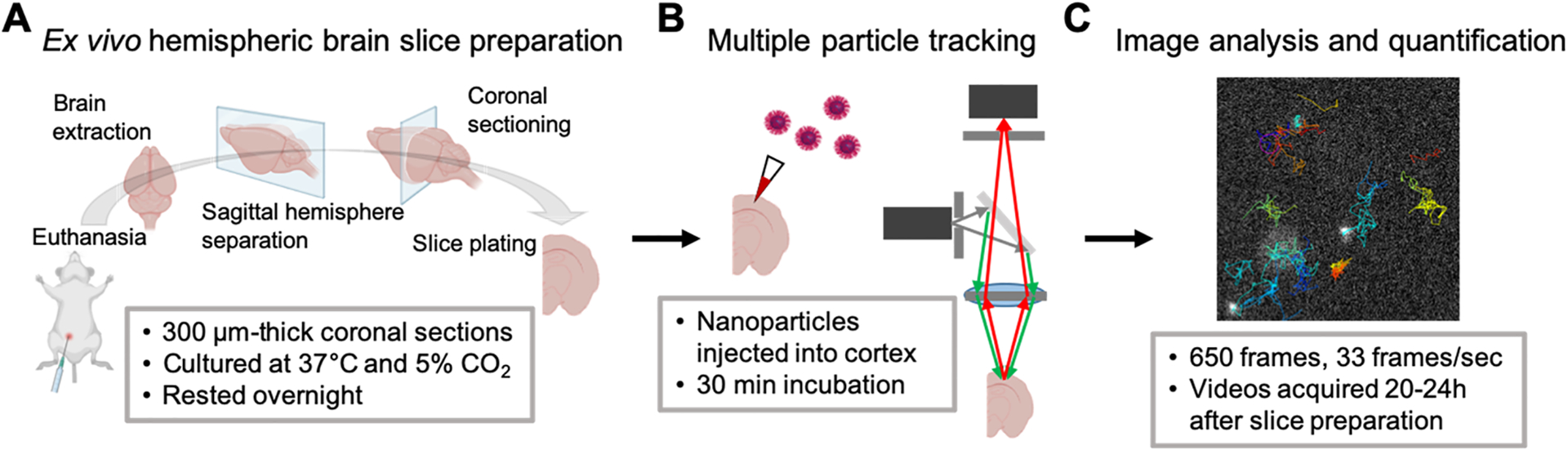
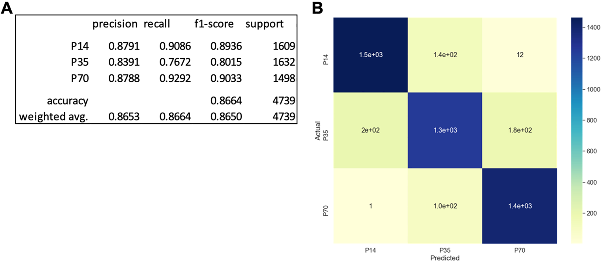
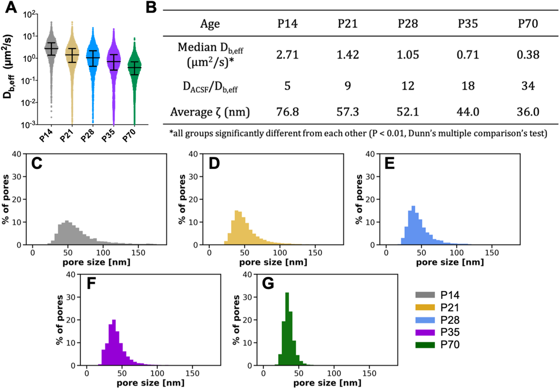
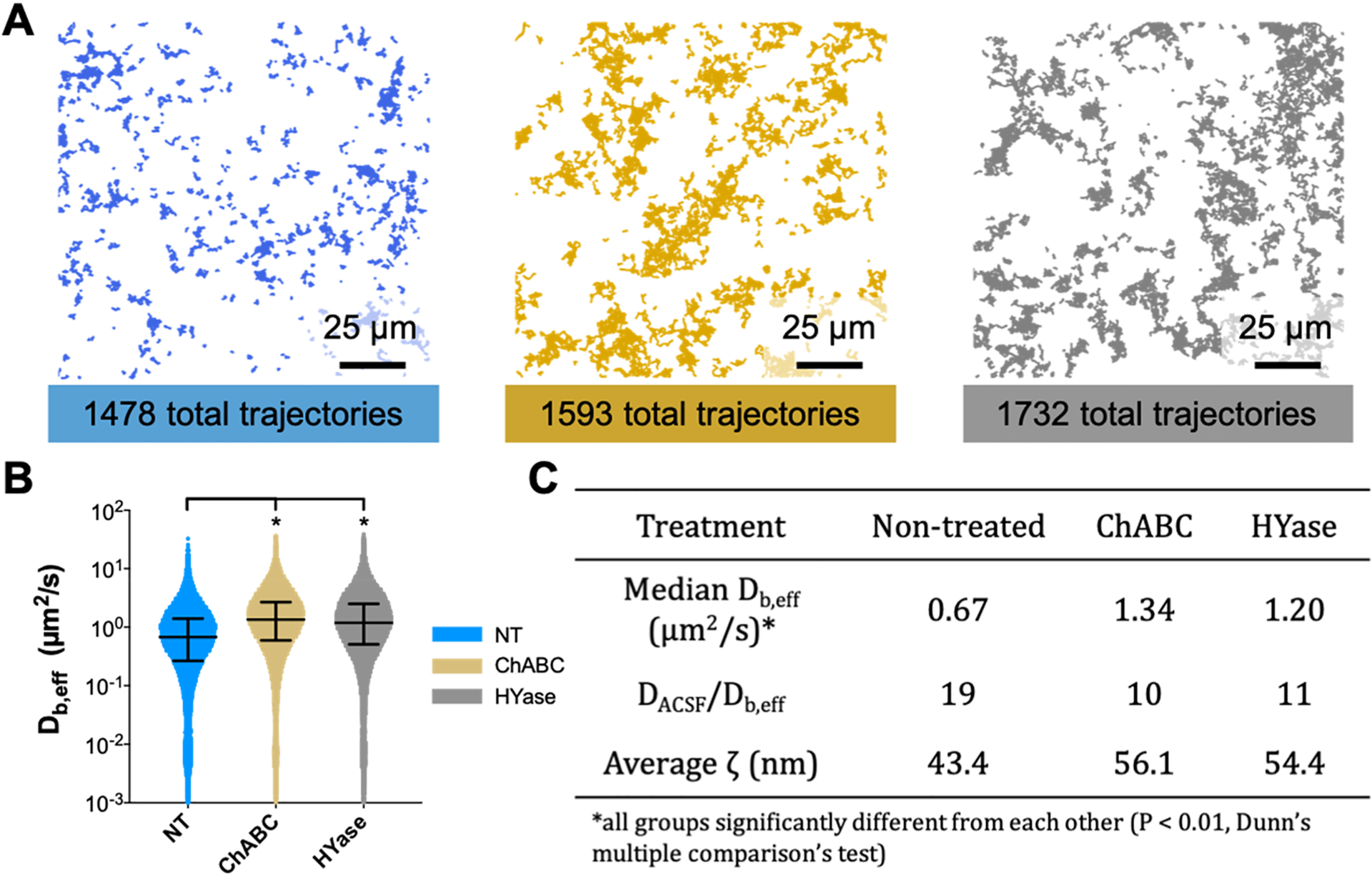
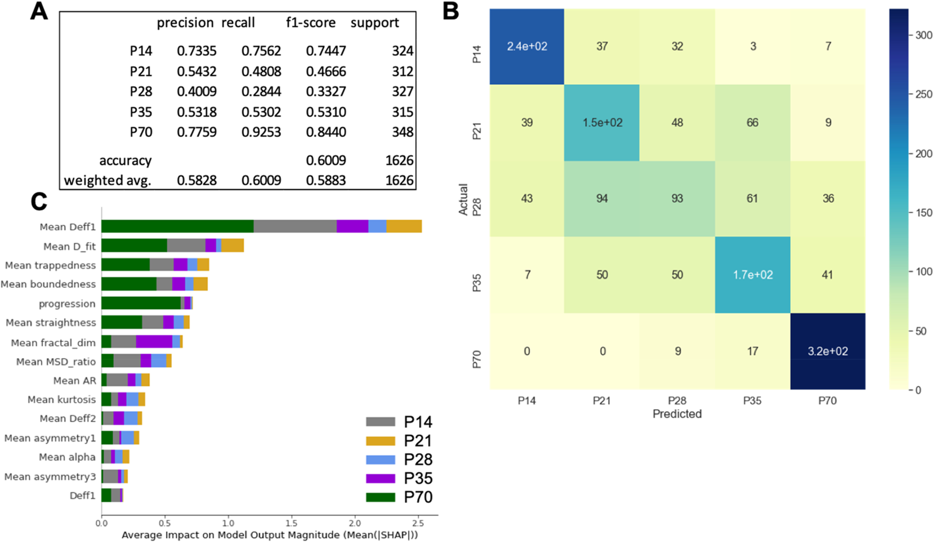
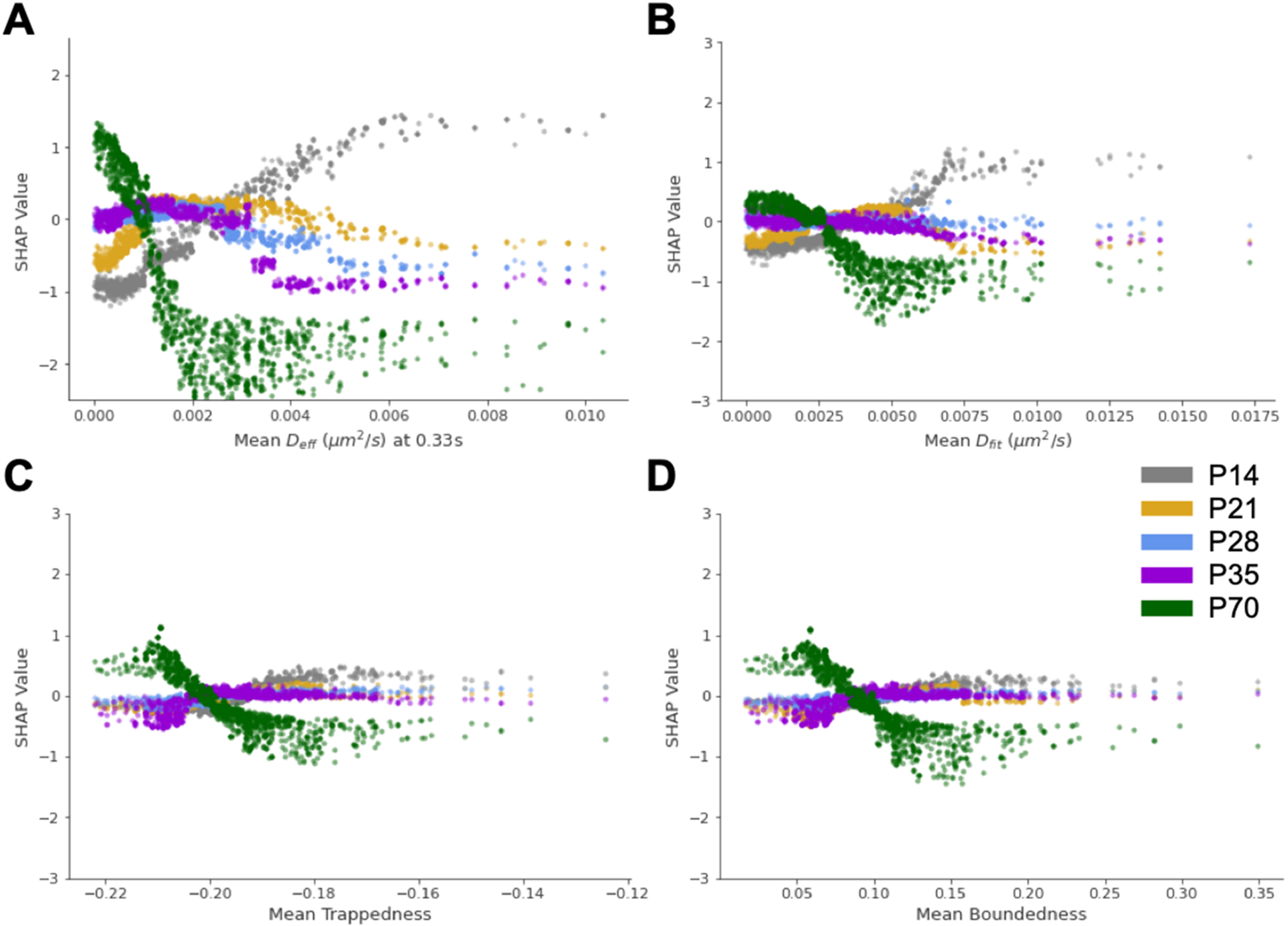
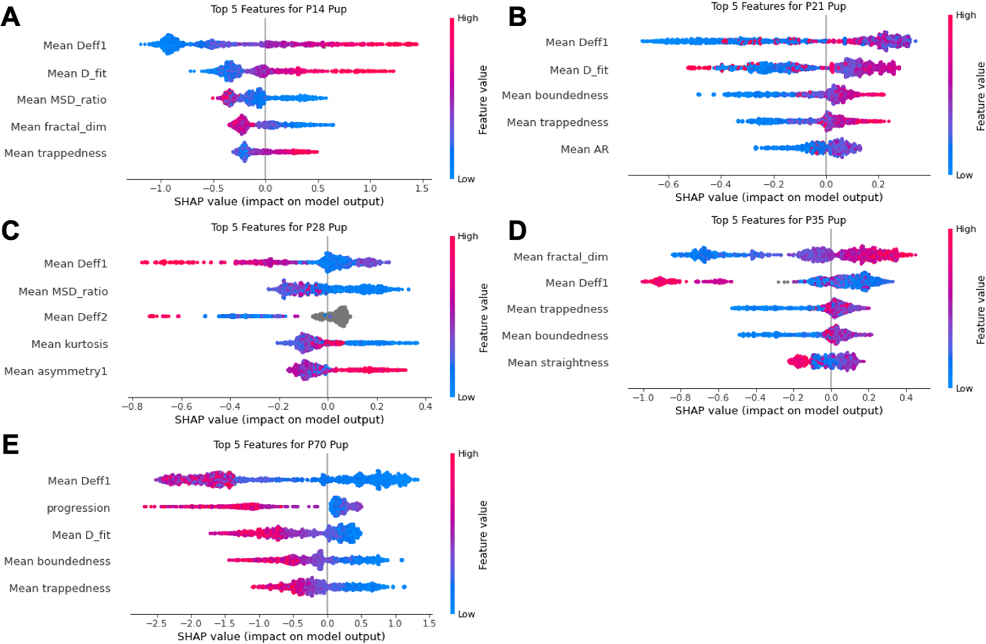
Brain extracellular matrix (ECM) structure mediates many aspects of neural development and function. Probing structural changes in brain ECM could thus provide insights into mechanisms of neurodevelopment, the loss of neural function in response to injury, and the detrimental effects of pathological aging and neurological disease. We demonstrate the ability to probe changes in brain ECM microstructure using multiple particle tracking (MPT). We performed MPT of colloidally stable polystyrene nanoparticles in organotypic rat brain slices collected from rats aged 14-70 days old. Our analysis revealed an inverse relationship between nanoparticle diffusive ability in the brain extracellular space and age. Additionally, the distribution of effective ECM pore sizes in the cortex shifted to smaller pores throughout development. We used the raw data and features extracted from nanoparticle trajectories to train a boosted decision tree capable of predicting chronological age with high accuracy. Collectively, this work demonstrates the utility of combining MPT with machine learning for measuring changes in brain ECM structure and predicting associated complex features such as chronological age. This will enable further understanding of the roles brain ECM play in development and aging and the specific mechanisms through which injuries cause aberrant neuronal function. Additionally, this approach has the potential to develop machine learning models capable of detecting the presence of injury or indicating the extent of injury based on changes in the brain microenvironment microstructure.

摘要

脑细胞外基质(ECM)结构介导了神经发育和功能的许多方面。因此,探测脑 ECM 的结构变化可以深入了解神经发育的机制、损伤导致的神经功能丧失以及病理性衰老和神经疾病的有害影响。我们展示了使用多粒子跟踪(MPT)探测脑 ECM 微结构变化的能力。我们在从 14-70 天大的大鼠中收集的器官型大鼠脑切片中进行了胶体稳定聚苯乙烯纳米颗粒的 MPT。我们的分析表明,脑细胞外空间中纳米颗粒扩散能力与年龄呈反比关系。此外,皮层中有效 ECM 孔径的分布在整个发育过程中向较小的孔径转移。我们使用原始数据和从纳米颗粒轨迹中提取的特征来训练一个能够高精度预测年龄的提升决策树。总的来说,这项工作证明了将 MPT 与机器学习相结合来测量脑 ECM 结构变化并预测相关复杂特征(如年龄)的有效性。这将进一步了解脑 ECM 在发育和衰老中的作用,以及损伤导致异常神经元功能的具体机制。此外,这种方法有可能开发出基于脑微环境微结构变化来检测损伤存在或指示损伤程度的机器学习模型。

https://cdn.ncbi.nlm.nih.gov/pmc/blobs/7fbb/8281364/ad8302b4f604/nihms-1717710-f0009.jpg
https://cdn.ncbi.nlm.nih.gov/pmc/blobs/7fbb/8281364/e4f4aa93b091/nihms-1717710-f0002.jpg
https://cdn.ncbi.nlm.nih.gov/pmc/blobs/7fbb/8281364/752b1f7c34db/nihms-1717710-f0003.jpg
https://cdn.ncbi.nlm.nih.gov/pmc/blobs/7fbb/8281364/1432f833f408/nihms-1717710-f0004.jpg
https://cdn.ncbi.nlm.nih.gov/pmc/blobs/7fbb/8281364/6de11f7bd333/nihms-1717710-f0005.jpg
https://cdn.ncbi.nlm.nih.gov/pmc/blobs/7fbb/8281364/7e981fd6ac30/nihms-1717710-f0006.jpg
https://cdn.ncbi.nlm.nih.gov/pmc/blobs/7fbb/8281364/694e44c482a1/nihms-1717710-f0007.jpg
https://cdn.ncbi.nlm.nih.gov/pmc/blobs/7fbb/8281364/e4b639bb347d/nihms-1717710-f0008.jpg
https://cdn.ncbi.nlm.nih.gov/pmc/blobs/7fbb/8281364/ad8302b4f604/nihms-1717710-f0009.jpg