Department of Radiology, University of Pittsburgh, Pittsburgh, PA, USA.
Helen Wills Neuroscience Institute, University of California Berkeley, Berkeley, CA, USA.
Alzheimers Res Ther. 2021 May 10;13(1):99. doi: 10.1186/s13195-021-00836-1.
Inconsistent positivity thresholds, image analysis pipelines, and quantitative outcomes are key challenges of multisite studies using more than one β-amyloid (Aβ) radiotracer in positron emission tomography (PET). Variability related to these factors contributes to disagreement and lack of replicability in research and clinical trials. To address these problems and promote Aβ PET harmonization, we used [F]florbetaben (FBB) and [F]florbetapir (FBP) data from the Alzheimer's Disease Neuroimaging Initiative (ADNI) to derive (1) standardized Centiloid (CL) transformations and (2) internally consistent positivity thresholds based on separate young control samples.
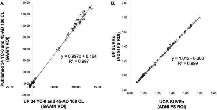
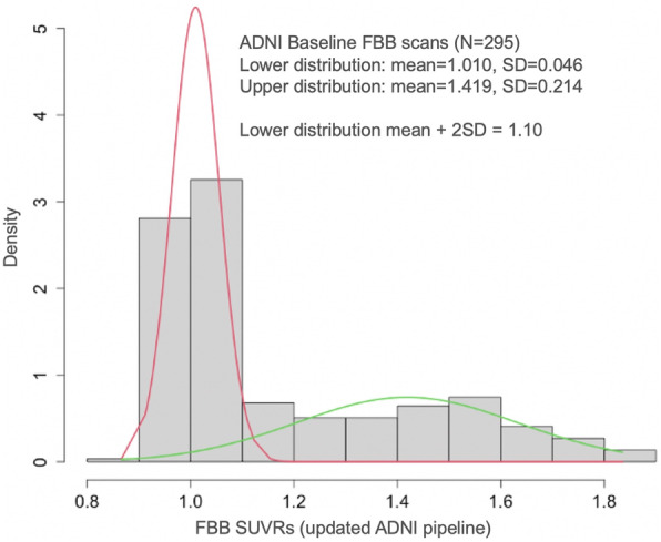
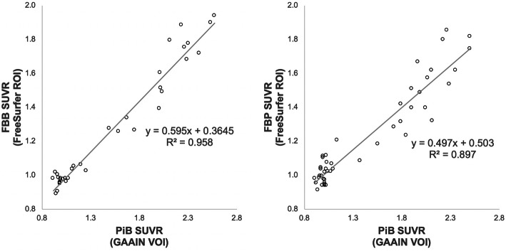
We analyzed Aβ PET data using a native-space, automated image processing pipeline that is used for PET quantification in many large, multisite AD studies and trials and made available to the research community. With this pipeline, we derived SUVR-to-CL transformations using the Global Alzheimer's Association Interactive Network data; we used reference regions for cross-sectional (whole cerebellum) and longitudinal (subcortical white matter, brain stem, whole cerebellum) analyses. Finally, we developed a FBB positivity threshold using an independent young control sample (N=62) with methods parallel to our existing FBP positivity threshold and validated the FBB threshold using a data-driven approach in ADNI participants (N=295).
The FBB threshold based on the young sample (1.08; 18 CL) was consistent with that of the data-driven approach (1.10; 21 CL), and the existing FBP threshold converted to CL with the derived transformation (1.11; 20 CL). The following equations can be used to convert whole cerebellum- (cross-sectional) and composite- (longitudinal) normalized FBB and FBP data quantified with the native-space pipeline to CL units: [F]FBB: CL = 157.15 × SUVR - 151.87; threshold=1.08, 18 CL [F]FBP: CL = 188.22 × SUVR - 189.16; threshold=1.11, 20 CL [F]FBB: CL = 244.20 × SUVR - 170.80 [F]FBP: CL = 300.66 × SUVR - 208.84 CONCLUSIONS: FBB and FBP positivity thresholds derived from independent young control samples and quantified using an automated, native-space approach result in similar CL values. These findings are applicable to thousands of available and anticipated outcomes analyzed using this pipeline and shared with the scientific community. This work demonstrates the feasibility of harmonized PET acquisition and analysis in multisite PET studies and internal consistency of positivity thresholds in standardized units.
在使用正电子发射断层扫描 (PET) 中的不止一种 β-淀粉样蛋白 (Aβ) 示踪剂进行多中心研究时,阳性阈值、图像分析流程和定量结果不一致是关键挑战。这些因素相关的变异性导致研究和临床试验中的分歧和缺乏可重复性。为了解决这些问题并促进 Aβ PET 协调,我们使用了来自阿尔茨海默病神经影像学倡议 (ADNI) 的 [F]florbetaben (FBB) 和 [F]florbetapir (FBP) 数据,得出了 (1) 标准化 Centiloid (CL) 转换,以及 (2) 基于单独的年轻对照组样本的内部一致的阳性阈值。
我们使用基于本地空间的自动化图像处理流水线分析 Aβ PET 数据,该流水线用于许多大型多中心 AD 研究和试验中的 PET 定量,并且可供研究界使用。使用此流水线,我们使用全球阿尔茨海默病协会交互网络数据得出了 SUVR 到 CL 的转换;我们使用参考区域进行了横断面(整个小脑)和纵向(皮质下白质、脑干、整个小脑)分析。最后,我们使用独立的年轻对照组样本(N=62)开发了 FBB 阳性阈值,并使用与我们现有的 FBP 阳性阈值平行的方法进行验证,并使用 ADNI 参与者(N=295)的基于数据驱动的方法验证了 FBB 阈值。
基于年轻样本的 FBB 阈值(1.08;18 CL)与数据驱动方法的阈值(1.10;21 CL)一致,并且通过推导的转换转换为 CL 的现有 FBP 阈值(1.11;20 CL)。以下方程可用于将使用本地空间流水线定量的整个小脑(横断面)和复合(纵向)归一化 FBB 和 FBP 数据转换为 CL 单位:[F]FBB:CL = 157.15 × SUVR - 151.87;阈值=1.08,18 CL [F]FBP:CL = 188.22 × SUVR - 189.16;阈值=1.11,20 CL [F]FBB:CL = 244.20 × SUVR - 170.80 [F]FBP:CL = 300.66 × SUVR - 208.84
从独立的年轻对照组样本中得出的 FBB 和 FBP 阳性阈值,并使用自动化的本地空间方法进行定量,得到了相似的 CL 值。这些发现适用于使用此流水线分析的数千个现有和预期的结果,并与科学界共享。这项工作证明了在多中心 PET 研究中进行协调的 PET 采集和分析以及在标准化单位中保持阳性阈值的一致性是可行的。