• 文献检索
  • 文档翻译
  • 深度研究
  • 学术资讯
  • Suppr Zotero 插件Zotero 插件
  • 邀请有礼
  • 套餐&价格
  • 历史记录
应用&插件
Suppr Zotero 插件Zotero 插件浏览器插件Mac 客户端Windows 客户端微信小程序
定价
高级版会员购买积分包购买API积分包
服务
文献检索文档翻译深度研究API 文档MCP 服务
关于我们
关于 Suppr公司介绍联系我们用户协议隐私条款
关注我们

Suppr 超能文献

核心技术专利:CN118964589B侵权必究
粤ICP备2023148730 号-1Suppr @ 2026

文献检索

告别复杂PubMed语法,用中文像聊天一样搜索,搜遍4000万医学文献。AI智能推荐,让科研检索更轻松。

立即免费搜索

文件翻译

保留排版,准确专业,支持PDF/Word/PPT等文件格式,支持 12+语言互译。

免费翻译文档

深度研究

AI帮你快速写综述,25分钟生成高质量综述,智能提取关键信息,辅助科研写作。

立即免费体验

多发性硬化症患者视网膜的体内分子变化。

In Vivo Molecular Changes in the Retina of Patients With Multiple Sclerosis.

机构信息

Center of Neuroimmunology, Institut d'Investigacions Biomediques August Pi Sunyer, Barcelona, Spain.

Department of Ophthalmology, Hospital Clinic, University of Barcelona, Barcelona, Spain.

出版信息

Invest Ophthalmol Vis Sci. 2021 May 3;62(6):11. doi: 10.1167/iovs.62.6.11.

DOI:10.1167/iovs.62.6.11
PMID:33974046
原文链接:https://pmc.ncbi.nlm.nih.gov/articles/PMC8114005/
Abstract

PURPOSE

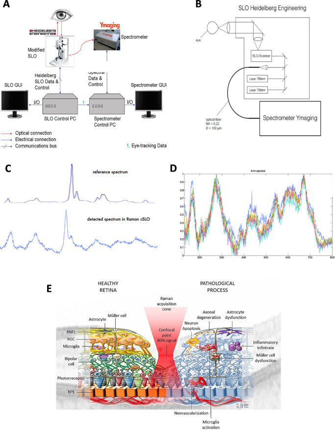
Raman spectroscopy allows molecular changes to be quantified in vivo from the tissues like the retina. Here we aimed to assess the metabolic changes in the retina of patients with multiple sclerosis (MS).

METHODS

We built a Raman spectroscopy prototype by connecting a scanning laser ophthalmoscope to a spectrophotometer. We defined the spectra of 10 molecules participating on energy supply, axon biology, or synaptic damage, which have been shown to be altered in the brain of patients with MS: cytochrome C, flavin adenine dinucleotide (FAD), nicotinamide adenine dinucleotide (NADH), N-acetyl-aspartate (NAA), excitotoxicity, glutamate, amyloid β (Aβ), τ and α-synuclein (SNCA), phosphatidyl-ethanolamine, and phosphatidyl-choline. We studied these molecules in a prospective cohort of patients with MS, either in the chronic phase or during relapses of acute optic neuritis (AON).

RESULTS

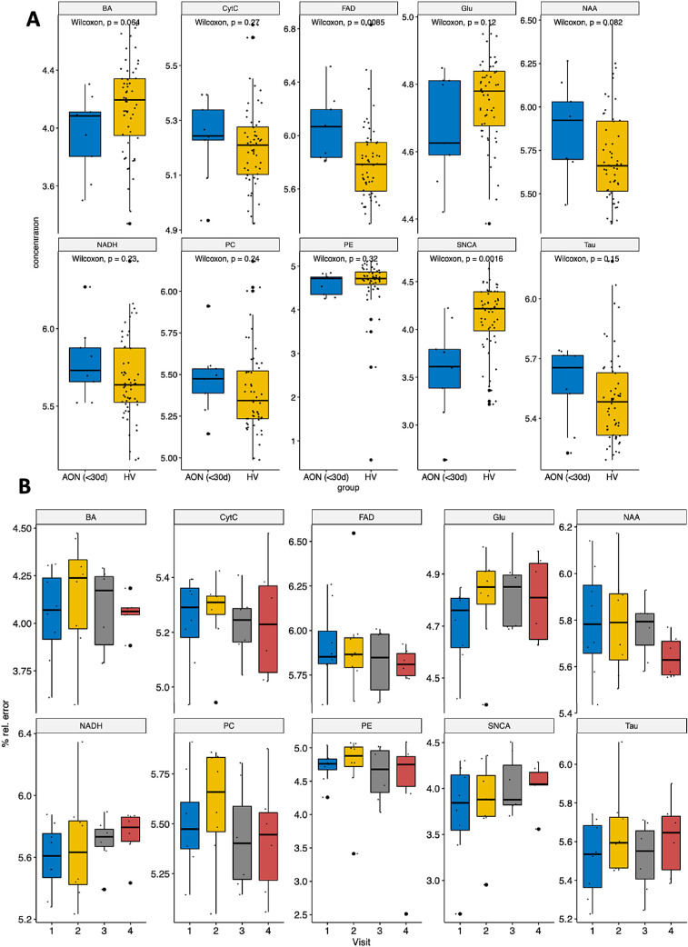
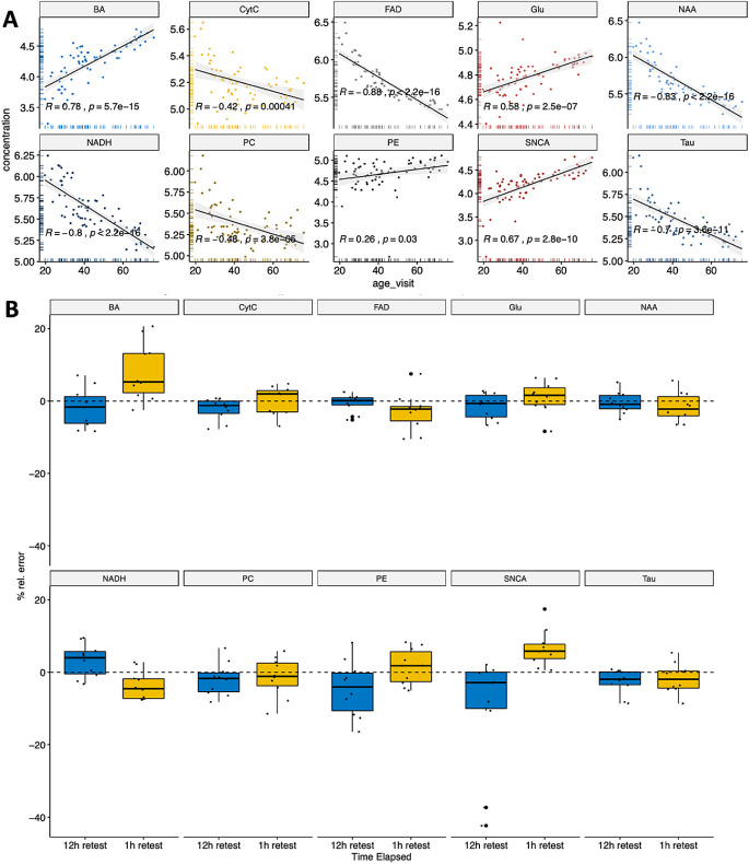
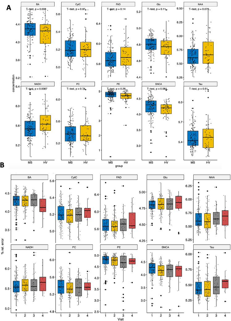
Significant changes to all these molecules were associated with age in healthy individuals. There was a significant decrease in NADH and a trend toward a decrease in NAA in patients with MS, as well as an increase in Aβ compared with healthy controls. Moreover, NADH and FAD increased over time in a longitudinal analysis of patients with MS, whereas Aβ diminished. In patients with acute retinal inflammation due to AON, there was a significant increase in FAD and a decrease in SNCA in the affected retina. Moreover, glutamate levels increased in the affected eyes after a 6-month follow-up.

CONCLUSIONS

Alterations of molecules related to axonal degeneration are observed during neuroinflammation and show dynamic changes over time, suggesting progressive neurodegeneration.

摘要

目的

拉曼光谱允许从视网膜等组织中定量检测分子变化。在这里,我们旨在评估多发性硬化症(MS)患者视网膜的代谢变化。

方法

我们通过将扫描激光检眼镜连接到分光光度计来构建拉曼光谱原型。我们定义了参与能量供应、轴突生物学或突触损伤的 10 种分子的光谱,这些分子在 MS 患者的大脑中已经显示出改变:细胞色素 C、黄素腺嘌呤二核苷酸(FAD)、烟酰胺腺嘌呤二核苷酸(NADH)、N-乙酰天冬氨酸(NAA)、兴奋性毒性、谷氨酸、β-淀粉样蛋白(Aβ)、τ 和α-突触核蛋白(SNCA)、磷脂乙醇胺和磷脂酰胆碱。我们在 MS 患者的前瞻性队列中研究了这些分子,无论是在慢性期还是在急性视神经炎(AON)复发期间。

结果

在健康个体中,所有这些分子的显著变化都与年龄相关。与健康对照组相比,MS 患者的 NADH 显著降低,NAA 呈下降趋势,Aβ 增加。此外,在 MS 患者的纵向分析中,NADH 和 FAD 随时间增加,而 Aβ 减少。在由于 AON 引起的急性视网膜炎症患者中,受影响视网膜中的 FAD 显著增加,SNCA 减少。此外,在 6 个月的随访后,受影响眼睛中的谷氨酸水平增加。

结论

在神经炎症期间观察到与轴突变性相关的分子的改变,并随着时间的推移显示出动态变化,提示进行性神经退行性变。

https://cdn.ncbi.nlm.nih.gov/pmc/blobs/d909/8114005/eaff3e8d1957/iovs-62-6-11-f004.jpg
https://cdn.ncbi.nlm.nih.gov/pmc/blobs/d909/8114005/5204516b460f/iovs-62-6-11-f001.jpg
https://cdn.ncbi.nlm.nih.gov/pmc/blobs/d909/8114005/ec442d4e6eab/iovs-62-6-11-f002.jpg
https://cdn.ncbi.nlm.nih.gov/pmc/blobs/d909/8114005/9132a4f45102/iovs-62-6-11-f003.jpg
https://cdn.ncbi.nlm.nih.gov/pmc/blobs/d909/8114005/eaff3e8d1957/iovs-62-6-11-f004.jpg

相似文献

1
In Vivo Molecular Changes in the Retina of Patients With Multiple Sclerosis.多发性硬化症患者视网膜的体内分子变化。
Invest Ophthalmol Vis Sci. 2021 May 3;62(6):11. doi: 10.1167/iovs.62.6.11.
2
The association between retinal nerve fibre layer thickness and N-acetyl aspartate levels in multiple sclerosis brain normal-appearing white matter: a longitudinal study using magnetic resonance spectroscopy and optical coherence tomography.多发性硬化症脑正常外观白质中视网膜神经纤维层厚度与 N-乙酰天门冬氨酸水平的相关性:使用磁共振波谱和光学相干断层扫描的纵向研究。
Eur J Neurol. 2016 Dec;23(12):1769-1774. doi: 10.1111/ene.13116. Epub 2016 Sep 3.
3
Predictors of retinal atrophy in multiple sclerosis: A longitudinal study using spectral domain optical coherence tomography with segmentation analysis.多发性硬化症中视网膜萎缩的预测因素:应用谱域光学相干断层扫描结合分割分析的纵向研究。
Mult Scler Relat Disord. 2018 Apr;21:56-62. doi: 10.1016/j.msard.2018.02.010. Epub 2018 Feb 11.
4
Peripapillary retinal nerve fiber layer thickness measured by optical coherence tomography in different clinical subtypes of multiple sclerosis.光学相干断层扫描测量多发性硬化不同临床亚型的视盘周围视网膜神经纤维层厚度。
Mult Scler Relat Disord. 2019 Jan;27:260-268. doi: 10.1016/j.msard.2018.11.003. Epub 2018 Nov 5.
5
Exploring experimental autoimmune optic neuritis using multimodal imaging.探索实验性自身免疫性视神经炎的多模态成像。
Neuroimage. 2018 Jul 15;175:327-339. doi: 10.1016/j.neuroimage.2018.04.004. Epub 2018 Apr 6.
6
Axonal damage in the optic radiation assessed by white matter tract integrity metrics is associated with retinal thinning in multiple sclerosis.轴突损伤在评估视辐射中的白质束完整性指标与多发性硬化症中的视网膜变薄有关。
Neuroimage Clin. 2020;27:102293. doi: 10.1016/j.nicl.2020.102293. Epub 2020 May 26.
7
Optical Coherence Tomography for the Detection of Remote Optic Neuritis in Multiple Sclerosis.光学相干断层扫描用于检测多发性硬化症中的远隔性视神经炎
J Neuroimaging. 2016 May;26(3):283-8. doi: 10.1111/jon.12326. Epub 2016 Jan 15.
8
[Retinal atrophy using optical coherence tomography (OCT) in 15 patients with multiple sclerosis and comparison with healthy subjects].[利用光学相干断层扫描(OCT)对15例多发性硬化症患者视网膜萎缩情况的研究及与健康受试者的比较]
Rev Neurol (Paris). 2008 Nov;164(11):927-34. doi: 10.1016/j.neurol.2008.03.008. Epub 2008 Jun 6.
9
Retinal imaging by laser polarimetry and optical coherence tomography evidence of axonal degeneration in multiple sclerosis.通过激光偏振imetry和光学相干断层扫描进行视网膜成像:多发性硬化症中轴突退变的证据
Arch Neurol. 2008 Jul;65(7):924-8. doi: 10.1001/archneur.65.7.924.
10
Using Acute Optic Neuritis Trials to Assess Neuroprotective and Remyelinating Therapies in Multiple Sclerosis.利用急性视神经炎试验评估多发性硬化症的神经保护和髓鞘修复疗法。
JAMA Neurol. 2020 Feb 1;77(2):234-244. doi: 10.1001/jamaneurol.2019.3283.

引用本文的文献

1
Raman liquid biopsy: a new approach to the multiple sclerosis diagnostics.拉曼液体活检:多发性硬化症诊断的新方法。
Front Neurol. 2025 Apr 16;16:1516712. doi: 10.3389/fneur.2025.1516712. eCollection 2025.
2
Development of a label-free, functional, molecular and structural imaging system combining optical coherence tomography and Raman spectroscopy for in vivo measurement of rat retina.开发一种结合光学相干断层扫描和拉曼光谱的无标记、功能性、分子和结构成像系统,用于大鼠视网膜的体内测量。
Biomed Opt Express. 2025 Jan 16;16(2):566-577. doi: 10.1364/BOE.541315. eCollection 2025 Feb 1.
3
Imaging the eye as a window to brain health: frontier approaches and future directions.

本文引用的文献

1
Comparability of Raman Spectroscopic Configurations: A Large Scale Cross-Laboratory Study.拉曼光谱配置的可比性:大规模的跨实验室研究。
Anal Chem. 2020 Dec 15;92(24):15745-15756. doi: 10.1021/acs.analchem.0c02696. Epub 2020 Nov 21.
2
Retinal and brain damage during multiple sclerosis course: inflammatory activity is a key factor in the first 5 years.多发性硬化症病程中的视网膜和大脑损伤:炎症活动是前 5 年的关键因素。
Sci Rep. 2020 Aug 7;10(1):13333. doi: 10.1038/s41598-020-70255-z.
3
alpha-Synuclein: a Modulator During Inflammatory CNS Demyelination.
将眼睛视为大脑健康之窗:前沿方法和未来方向。
J Neuroinflammation. 2024 Nov 29;21(1):309. doi: 10.1186/s12974-024-03304-3.
4
Retinal Damage and Visual Network Reconfiguration Defines Visual Function Recovery in Optic Neuritis.视网膜损伤和视觉网络重构定义了视神经炎中的视觉功能恢复。
Neurol Neuroimmunol Neuroinflamm. 2024 Nov;11(6):e200288. doi: 10.1212/NXI.0000000000200288. Epub 2024 Aug 30.
5
Retinal metabolism displays evidence for uncoupling of glycolysis and oxidative phosphorylation via Cori-, Cahill-, and mini-Krebs-cycle.视网膜代谢通过科里、卡希尔和微型克雷布斯循环表现出糖酵解和氧化磷酸化解偶联的证据。
Elife. 2024 May 13;12:RP91141. doi: 10.7554/eLife.91141.
6
Neuroimaging to monitor worsening of multiple sclerosis: advances supported by the grant for multiple sclerosis innovation.用于监测多发性硬化症病情恶化的神经影像学:多发性硬化症创新基金支持下的进展
Front Neurol. 2023 Dec 1;14:1319869. doi: 10.3389/fneur.2023.1319869. eCollection 2023.
7
Combining electromyography and Raman spectroscopy: optical EMG.结合肌电图和拉曼光谱:光学 EMG。
Muscle Nerve. 2023 Oct;68(4):464-470. doi: 10.1002/mus.27937. Epub 2023 Jul 21.
8
From Research to Diagnostic Application of Raman Spectroscopy in Neurosciences: Past and Perspectives.从拉曼光谱在神经科学中的研究到诊断应用:过去与展望
Free Neuropathol. 2022 Aug 5;3:19. doi: 10.17879/freeneuropathology-2022-4210. eCollection 2022 Jan.
9
Thinking outside the box: non-canonical targets in multiple sclerosis.跳出固有思维:多发性硬化症中的非经典靶点。
Nat Rev Drug Discov. 2022 Aug;21(8):578-600. doi: 10.1038/s41573-022-00477-5. Epub 2022 Jun 6.
10
Retinal Glutamate Neurotransmission: From Physiology to Pathophysiological Mechanisms of Retinal Ganglion Cell Degeneration.视网膜谷氨酸神经传递:从生理学到视网膜神经节细胞变性的病理生理机制
Life (Basel). 2022 Apr 25;12(5):638. doi: 10.3390/life12050638.
α-突触核蛋白:中枢神经系统炎症性脱髓鞘中的调节因子。
J Mol Neurosci. 2020 Jul;70(7):1038-1049. doi: 10.1007/s12031-020-01498-8. Epub 2020 Mar 23.
4
Anterior visual system imaging to investigate energy failure in multiple sclerosis.利用前视系统成像研究多发性硬化症中的能量衰竭。
Brain. 2020 Jul 1;143(7):1999-2008. doi: 10.1093/brain/awaa049.
5
Imaging Biomarkers of Alzheimer Disease in Multiple Sclerosis.阿尔茨海默病的多发性硬化症的成像生物标志物。
Ann Neurol. 2020 Apr;87(4):556-567. doi: 10.1002/ana.25684. Epub 2020 Feb 8.
6
Using Acute Optic Neuritis Trials to Assess Neuroprotective and Remyelinating Therapies in Multiple Sclerosis.利用急性视神经炎试验评估多发性硬化症的神经保护和髓鞘修复疗法。
JAMA Neurol. 2020 Feb 1;77(2):234-244. doi: 10.1001/jamaneurol.2019.3283.
7
Nonresonant Raman spectroscopy of isolated human retina samples complying with laser safety regulations for measurements.符合激光安全测量规定的分离人视网膜样本的非共振拉曼光谱。
Neurophotonics. 2019 Oct;6(4):041106. doi: 10.1117/1.NPh.6.4.041106. Epub 2019 Sep 3.
8
Mechanisms of injury-induced axon degeneration.损伤诱导的轴突变性的机制。
Curr Opin Neurobiol. 2019 Aug;57:171-178. doi: 10.1016/j.conb.2019.03.006. Epub 2019 May 6.
9
β-Synuclein-reactive T cells induce autoimmune CNS grey matter degeneration.β-突触核蛋白反应性 T 细胞诱导自身免疫性中枢神经系统灰质变性。
Nature. 2019 Feb;566(7745):503-508. doi: 10.1038/s41586-019-0964-2. Epub 2019 Feb 20.
10
CSF β-amyloid predicts prognosis in patients with multiple sclerosis.脑脊液β-淀粉样蛋白可预测多发性硬化症患者的预后。
Mult Scler. 2019 Aug;25(9):1223-1231. doi: 10.1177/1352458518791709. Epub 2018 Aug 7.