• 文献检索
  • 文档翻译
  • 深度研究
  • 学术资讯
  • Suppr Zotero 插件Zotero 插件
  • 邀请有礼
  • 套餐&价格
  • 历史记录
应用&插件
Suppr Zotero 插件Zotero 插件浏览器插件Mac 客户端Windows 客户端微信小程序
定价
高级版会员购买积分包购买API积分包
服务
文献检索文档翻译深度研究API 文档MCP 服务
关于我们
关于 Suppr公司介绍联系我们用户协议隐私条款
关注我们

Suppr 超能文献

核心技术专利:CN118964589B侵权必究
粤ICP备2023148730 号-1Suppr @ 2026

文献检索

告别复杂PubMed语法,用中文像聊天一样搜索,搜遍4000万医学文献。AI智能推荐,让科研检索更轻松。

立即免费搜索

文件翻译

保留排版,准确专业,支持PDF/Word/PPT等文件格式,支持 12+语言互译。

免费翻译文档

深度研究

AI帮你快速写综述,25分钟生成高质量综述,智能提取关键信息,辅助科研写作。

立即免费体验

计算机断层扫描在评估急诊就诊的 COVID-19 患者死亡风险中的价值。

The value of computed tomography in assessing the risk of death in COVID-19 patients presenting to the emergency room.

机构信息

Clinical and Experimental Medicine PhD Program, University of Modena and Reggio Emilia, Modena, Italy.

Radiology Unit, Department of Diagnostic Imaging and Laboratory Medicine, AUSL-IRCCS di Reggio Emilia, Via Risorgimento 80, 42123, Reggio Emilia, Italy.

出版信息

Eur Radiol. 2021 Dec;31(12):9164-9175. doi: 10.1007/s00330-021-07993-9. Epub 2021 May 12.

DOI:10.1007/s00330-021-07993-9
PMID:33978822
原文链接:https://pmc.ncbi.nlm.nih.gov/articles/PMC8113019/
Abstract

OBJECTIVE

The aims of this study were to develop a multiparametric prognostic model for death in COVID-19 patients and to assess the incremental value of CT disease extension over clinical parameters.

METHODS

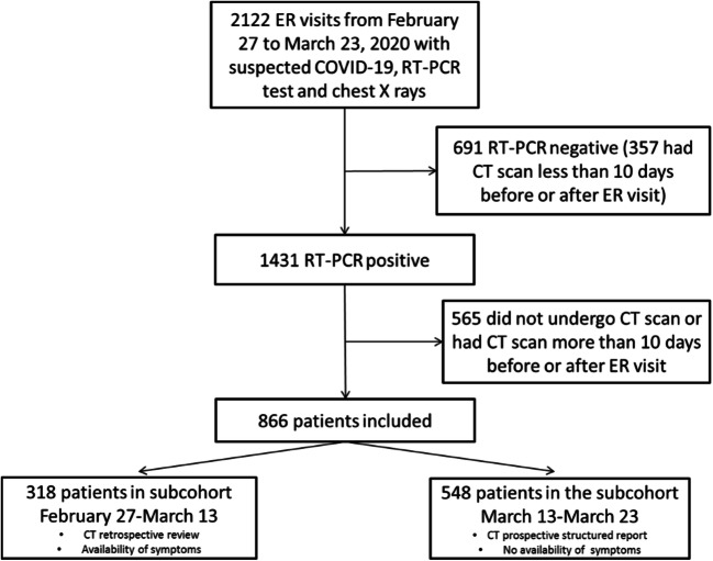
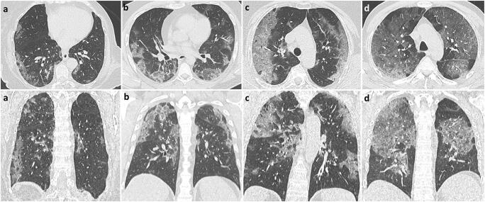
Consecutive patients who presented to all five of the emergency rooms of the Reggio Emilia province between February 27 and March 23, 2020, for suspected COVID-19, underwent chest CT, and had a positive swab within 10 days were included in this retrospective study. Age, sex, comorbidities, days from symptom onset, and laboratory data were retrieved from institutional information systems. CT disease extension was visually graded as < 20%, 20-39%, 40-59%, or ≥ 60%. The association between clinical and CT variables with death was estimated with univariable and multivariable Cox proportional hazards models; model performance was assessed using k-fold cross-validation for the area under the ROC curve (cvAUC).

RESULTS

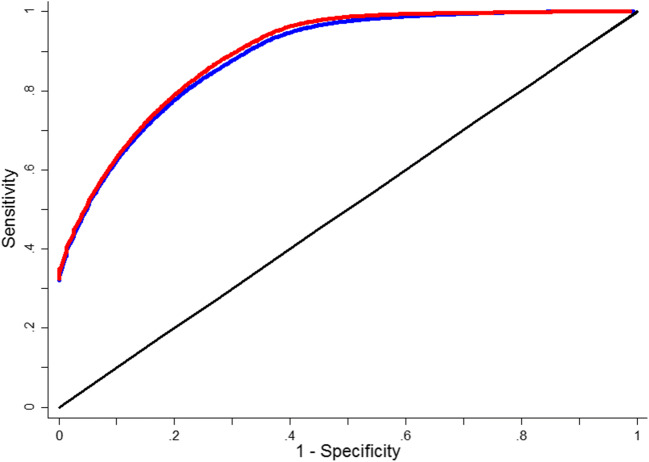
Of the 866 included patients (median age 59.8, women 39.2%), 93 (10.74%) died. Clinical variables significantly associated with death in multivariable model were age, male sex, HDL cholesterol, dementia, heart failure, vascular diseases, time from symptom onset, neutrophils, LDH, and oxygen saturation level. CT disease extension was also independently associated with death (HR = 7.56, 95% CI = 3.49; 16.38 for ≥ 60% extension). cvAUCs were 0.927 (bootstrap bias-corrected 95% CI = 0.899-0.947) for the clinical model and 0.936 (bootstrap bias-corrected 95% CI = 0.912-0.953) when adding CT extension.

CONCLUSIONS

A prognostic model based on clinical variables is highly accurate in predicting death in COVID-19 patients. Adding CT disease extension to the model scarcely improves its accuracy.

KEY POINTS

• Early identification of COVID-19 patients at higher risk of disease progression and death is crucial; the role of CT scan in defining prognosis is unclear. • A clinical model based on age, sex, comorbidities, days from symptom onset, and laboratory results was highly accurate in predicting death in COVID-19 patients presenting to the emergency room. • Disease extension assessed with CT was independently associated with death when added to the model but did not produce a valuable increase in accuracy.

摘要

目的

本研究旨在建立一个针对 COVID-19 患者死亡的多参数预后模型,并评估 CT 疾病扩展相对于临床参数的增量价值。

方法

本回顾性研究纳入了 2020 年 2 月 27 日至 3 月 23 日期间因疑似 COVID-19 而前往雷焦艾米利亚省五个急诊室的连续患者,这些患者均接受了胸部 CT 检查,并且在 10 天内进行了阳性拭子检测。从机构信息系统中检索年龄、性别、合并症、症状出现后天数和实验室数据。通过视觉评分将 CT 疾病扩展分为<20%、20-39%、40-59%或≥60%。使用单变量和多变量 Cox 比例风险模型估计临床和 CT 变量与死亡之间的关联;使用 K 折交叉验证评估 ROC 曲线下面积(cvAUC)的模型性能。

结果

在 866 名纳入患者中(中位年龄 59.8 岁,女性占 39.2%),有 93 人(10.74%)死亡。多变量模型中与死亡显著相关的临床变量包括年龄、男性、高密度脂蛋白胆固醇、痴呆、心力衰竭、血管疾病、症状出现后时间、中性粒细胞、乳酸脱氢酶和氧饱和度水平。CT 疾病扩展也与死亡独立相关(HR=7.56,95%CI=3.49;16.38 为≥60%扩展)。临床模型的 cvAUC 为 0.927(自举偏置校正 95%CI=0.899-0.947),添加 CT 扩展后为 0.936(自举偏置校正 95%CI=0.912-0.953)。

结论

基于临床变量的预后模型在预测 COVID-19 患者死亡方面具有很高的准确性。在模型中添加 CT 疾病扩展几乎不会提高其准确性。

关键点

  1. 早期识别 COVID-19 患者中疾病进展和死亡风险较高的患者至关重要;CT 扫描在确定预后方面的作用尚不清楚。

  2. 基于年龄、性别、合并症、症状出现后时间和实验室结果的临床模型在预测 COVID-19 患者就诊急诊室时的死亡方面具有很高的准确性。

  3. 通过 CT 评估的疾病扩展与死亡独立相关,但添加到模型中并未产生有价值的准确性提高。

https://cdn.ncbi.nlm.nih.gov/pmc/blobs/4f4d/8113019/121a2203370a/330_2021_7993_Fig3_HTML.jpg
https://cdn.ncbi.nlm.nih.gov/pmc/blobs/4f4d/8113019/57295064989c/330_2021_7993_Fig1_HTML.jpg
https://cdn.ncbi.nlm.nih.gov/pmc/blobs/4f4d/8113019/1221608ea38c/330_2021_7993_Fig2_HTML.jpg
https://cdn.ncbi.nlm.nih.gov/pmc/blobs/4f4d/8113019/121a2203370a/330_2021_7993_Fig3_HTML.jpg
https://cdn.ncbi.nlm.nih.gov/pmc/blobs/4f4d/8113019/57295064989c/330_2021_7993_Fig1_HTML.jpg
https://cdn.ncbi.nlm.nih.gov/pmc/blobs/4f4d/8113019/1221608ea38c/330_2021_7993_Fig2_HTML.jpg
https://cdn.ncbi.nlm.nih.gov/pmc/blobs/4f4d/8113019/121a2203370a/330_2021_7993_Fig3_HTML.jpg

相似文献

1
The value of computed tomography in assessing the risk of death in COVID-19 patients presenting to the emergency room.计算机断层扫描在评估急诊就诊的 COVID-19 患者死亡风险中的价值。
Eur Radiol. 2021 Dec;31(12):9164-9175. doi: 10.1007/s00330-021-07993-9. Epub 2021 May 12.
2
The association between clinical laboratory data and chest CT findings explains disease severity in a large Italian cohort of COVID-19 patients.临床实验室数据与胸部 CT 表现之间的关联可解释意大利大型 COVID-19 患者队列中疾病的严重程度。
BMC Infect Dis. 2021 Feb 8;21(1):157. doi: 10.1186/s12879-021-05855-9.
3
Multicenter Study of Temporal Changes and Prognostic Value of a CT Visual Severity Score in Hospitalized Patients With Coronavirus Disease (COVID-19).多中心研究:住院的新型冠状病毒肺炎(COVID-19)患者 CT 视觉严重程度评分的时间变化及其预后价值。
AJR Am J Roentgenol. 2021 Jul;217(1):83-92. doi: 10.2214/AJR.20.24044. Epub 2020 Sep 9.
4
Thoracic imaging tests for the diagnosis of COVID-19.用于诊断新型冠状病毒肺炎的胸部影像学检查
Cochrane Database Syst Rev. 2020 Sep 30;9:CD013639. doi: 10.1002/14651858.CD013639.pub2.
5
Clinical and imaging characteristics of patients with COVID-19 predicting hospital readmission after emergency department discharge: a single-centre cohort study in Italy.预测急诊科出院后COVID-19患者再次入院的临床和影像学特征:意大利一项单中心队列研究
BMJ Open. 2022 Apr 6;12(4):e052665. doi: 10.1136/bmjopen-2021-052665.
6
Chest CT opportunistic biomarkers for phenotyping high-risk COVID-19 patients: a retrospective multicentre study.胸部 CT 机会性生物标志物用于表型分析 COVID-19 高危患者:一项回顾性多中心研究。
Eur Radiol. 2023 Nov;33(11):7756-7768. doi: 10.1007/s00330-023-09702-0. Epub 2023 May 11.
7
CT-derived Chest Muscle Metrics for Outcome Prediction in Patients with COVID-19.基于 CT 的胸部肌肉指标预测 COVID-19 患者的预后。
Radiology. 2021 Aug;300(2):E328-E336. doi: 10.1148/radiol.2021204141. Epub 2021 Mar 16.
8
Chest CT in the Emergency Department for Diagnosis of COVID-19 Pneumonia: Dutch Experience.急诊科胸部 CT 诊断 COVID-19 肺炎:荷兰经验。
Radiology. 2021 Feb;298(2):E98-E106. doi: 10.1148/radiol.2020203465. Epub 2020 Nov 17.
9
Prognostic value of CT integrated with clinical and laboratory data during the first peak of the COVID-19 pandemic in Northern Italy: A nomogram to predict unfavorable outcome.在意大利北部 COVID-19 大流行的第一个高峰期,CT 与临床和实验室数据的综合预后价值:预测不良结局的列线图。
Eur J Radiol. 2021 Apr;137:109612. doi: 10.1016/j.ejrad.2021.109612. Epub 2021 Feb 26.
10
Chest CT-derived pulmonary artery enlargement at the admission predicts overall survival in COVID-19 patients: insight from 1461 consecutive patients in Italy.入院时胸部CT显示的肺动脉增宽可预测COVID-19患者的总生存期:来自意大利1461例连续患者的见解
Eur Radiol. 2021 Jun;31(6):4031-4041. doi: 10.1007/s00330-020-07622-x. Epub 2020 Dec 23.

引用本文的文献

1
Elevated NET, Calprotectin, and Neopterin Levels Discriminate between Disease Activity in COVID-19, as Evidenced by Need for Hospitalization among Patients in Northern Italy.较高的中性粒细胞胞外诱捕网、钙卫蛋白和新蝶呤水平可区分新冠病毒疾病的活动情况,意大利北部患者的住院需求即为明证。
Biomedicines. 2024 Mar 30;12(4):766. doi: 10.3390/biomedicines12040766.
2
Epicardial Adipose Tissue as a Prognostic Marker in COVID-19.心外膜脂肪组织作为 COVID-19 的预后标志物。
In Vivo. 2024 Jan-Feb;38(1):281-285. doi: 10.21873/invivo.13436.
3
CT coronary artery calcification score as a prognostic marker in COVID-19.

本文引用的文献

1
Accuracy of CT in a cohort of symptomatic patients with suspected COVID-19 pneumonia during the outbreak peak in Italy.在意大利疫情高峰期,对疑似 COVID-19 肺炎的有症状患者队列进行 CT 检查的准确性。
Eur Radiol. 2020 Dec;30(12):6818-6827. doi: 10.1007/s00330-020-07050-x. Epub 2020 Jul 14.
2
CT features of COVID-19 patients with two consecutive negative RT-PCR tests after treatment.治疗后连续两次 RT-PCR 检测阴性的 COVID-19 患者的 CT 特征。
Sci Rep. 2020 Jul 14;10(1):11548. doi: 10.1038/s41598-020-68509-x.
3
Development of a clinical decision support system for severity risk prediction and triage of COVID-19 patients at hospital admission: an international multicentre study.
CT冠状动脉钙化评分作为COVID-19的预后标志物
J Thorac Dis. 2023 Oct 31;15(10):5559-5565. doi: 10.21037/jtd-23-728. Epub 2023 Oct 20.
4
Prognostic models in COVID-19 infection that predict severity: a systematic review.COVID-19 感染中预测严重程度的预后模型:系统评价。
Eur J Epidemiol. 2023 Apr;38(4):355-372. doi: 10.1007/s10654-023-00973-x. Epub 2023 Feb 25.
5
Complement activation predicts negative outcomes in COVID-19: The experience from Northen Italian patients.补体激活可预测 COVID-19 的不良结局:来自意大利北部患者的经验。
Autoimmun Rev. 2023 Jan;22(1):103232. doi: 10.1016/j.autrev.2022.103232. Epub 2022 Nov 19.
6
Point-of-care Lung Ultrasound, Lung CT and NEWS to Predict Adverse Outcomes and Mortality in COVID-19 Associated Pneumonia.床旁肺部超声、肺部 CT 和 NEWS 预测 COVID-19 相关肺炎的不良结局和死亡率。
J Intensive Care Med. 2022 Dec;37(12):1614-1624. doi: 10.1177/08850666221111731. Epub 2022 Nov 1.
7
Predicting In-Hospital Mortality in Severe COVID-19: A Systematic Review and External Validation of Clinical Prediction Rules.预测重症 COVID-19 的院内死亡率:临床预测规则的系统评价与外部验证
Biomedicines. 2022 Sep 27;10(10):2414. doi: 10.3390/biomedicines10102414.
8
Heart failure during the COVID-19 pandemic: clinical, diagnostic, management, and organizational dilemmas.COVID-19 大流行期间的心力衰竭:临床、诊断、管理和组织方面的困境。
ESC Heart Fail. 2022 Dec;9(6):3713-3736. doi: 10.1002/ehf2.14118. Epub 2022 Sep 16.
9
Imaging-based indices combining disease severity and time from disease onset to predict COVID-19 mortality: A cohort study.基于影像学的疾病严重程度和从发病到预测 COVID-19 死亡率的时间的综合指数:一项队列研究。
PLoS One. 2022 Jun 16;17(6):e0270111. doi: 10.1371/journal.pone.0270111. eCollection 2022.
10
Computed tomography-defined body composition as prognostic markers for unfavourable outcomes and in-hospital mortality in coronavirus disease 2019.计算机断层扫描定义的身体成分作为 2019 年冠状病毒病不良结局和住院死亡率的预后标志物。
J Cachexia Sarcopenia Muscle. 2022 Feb;13(1):159-168. doi: 10.1002/jcsm.12868. Epub 2022 Jan 12.
开发一种临床决策支持系统,用于预测 COVID-19 患者入院时的严重程度风险和分诊:一项国际多中心研究。
Eur Respir J. 2020 Aug 20;56(2). doi: 10.1183/13993003.01104-2020. Print 2020 Aug.
4
A fully automatic deep learning system for COVID-19 diagnostic and prognostic analysis.一种用于 COVID-19 诊断和预后分析的全自动深度学习系统。
Eur Respir J. 2020 Aug 6;56(2). doi: 10.1183/13993003.00775-2020. Print 2020 Aug.
5
Association Between Clinical Manifestations and Prognosis in Patients with COVID-19.新型冠状病毒肺炎患者临床表现与预后的相关性研究。
Clin Ther. 2020 Jun;42(6):964-972. doi: 10.1016/j.clinthera.2020.04.009. Epub 2020 Apr 27.
6
Risk factors of critical & mortal COVID-19 cases: A systematic literature review and meta-analysis.危重症和死亡 COVID-19 病例的风险因素:系统文献回顾和荟萃分析。
J Infect. 2020 Aug;81(2):e16-e25. doi: 10.1016/j.jinf.2020.04.021. Epub 2020 Apr 23.
7
D-dimer levels on admission to predict in-hospital mortality in patients with Covid-19.入院时 D-二聚体水平预测 COVID-19 患者住院死亡率。
J Thromb Haemost. 2020 Jun;18(6):1324-1329. doi: 10.1111/jth.14859.
8
The Performance of Chest CT in Evaluating the Clinical Severity of COVID-19 Pneumonia: Identifying Critical Cases Based on CT Characteristics.胸部 CT 对评估 COVID-19 肺炎临床严重程度的性能:基于 CT 特征识别重症病例。
Invest Radiol. 2020 Jul;55(7):412-421. doi: 10.1097/RLI.0000000000000689.
9
Well-aerated Lung on Admitting Chest CT to Predict Adverse Outcome in COVID-19 Pneumonia.胸部 CT 显示充气良好的肺可预测 COVID-19 肺炎的不良结局。
Radiology. 2020 Aug;296(2):E86-E96. doi: 10.1148/radiol.2020201433. Epub 2020 Apr 17.
10
A Tool for Early Prediction of Severe Coronavirus Disease 2019 (COVID-19): A Multicenter Study Using the Risk Nomogram in Wuhan and Guangdong, China.一种用于早期预测严重 2019 冠状病毒病(COVID-19)的工具:来自中国武汉和广东的多中心研究使用风险列线图。
Clin Infect Dis. 2020 Jul 28;71(15):833-840. doi: 10.1093/cid/ciaa443.