• 文献检索
  • 文档翻译
  • 深度研究
  • 学术资讯
  • Suppr Zotero 插件Zotero 插件
  • 邀请有礼
  • 套餐&价格
  • 历史记录
应用&插件
Suppr Zotero 插件Zotero 插件浏览器插件Mac 客户端Windows 客户端微信小程序
定价
高级版会员购买积分包购买API积分包
服务
文献检索文档翻译深度研究API 文档MCP 服务
关于我们
关于 Suppr公司介绍联系我们用户协议隐私条款
关注我们

Suppr 超能文献

核心技术专利:CN118964589B侵权必究
粤ICP备2023148730 号-1Suppr @ 2026

文献检索

告别复杂PubMed语法,用中文像聊天一样搜索,搜遍4000万医学文献。AI智能推荐,让科研检索更轻松。

立即免费搜索

文件翻译

保留排版,准确专业,支持PDF/Word/PPT等文件格式,支持 12+语言互译。

免费翻译文档

深度研究

AI帮你快速写综述,25分钟生成高质量综述,智能提取关键信息,辅助科研写作。

立即免费体验

成骨细胞作为低强度脉冲超声波治疗骨折愈合过程中的主要反应细胞。

Osteocytes as main responders to low-intensity pulsed ultrasound treatment during fracture healing.

机构信息

Oral Biochemistry and Molecular Biology, Graduate School of Dental Medicine, Hokkaido University, Kita 13, Nishi 7, Kita-ku, Sapporo, 060-8586, Japan.

Oral Diagnosis and Medicine, Graduate School of Dental Medicine, Hokkaido University, Sapporo, Japan.

出版信息

Sci Rep. 2021 May 13;11(1):10298. doi: 10.1038/s41598-021-89672-9.

DOI:10.1038/s41598-021-89672-9
PMID:33986415
原文链接:https://pmc.ncbi.nlm.nih.gov/articles/PMC8119462/
Abstract

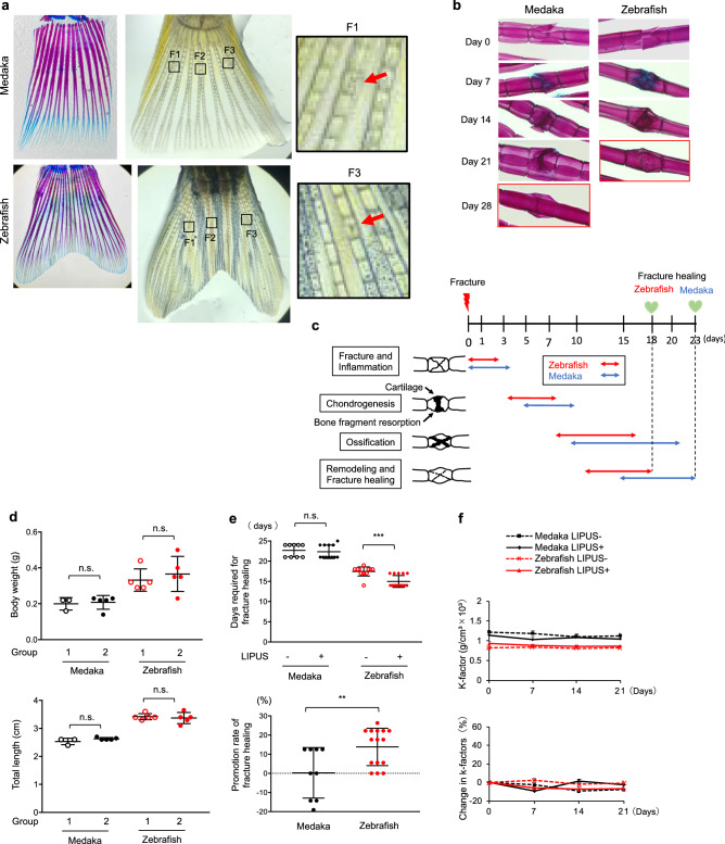
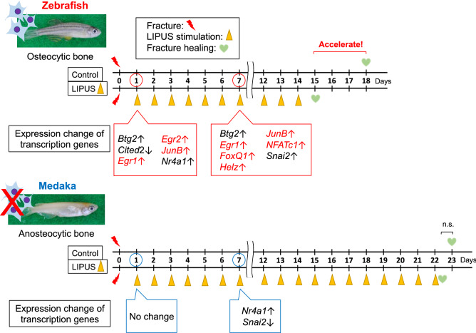
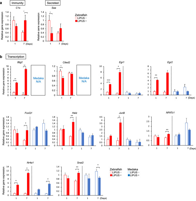
Ultrasound stimulation is a type of mechanical stress, and low-intensity pulsed ultrasound (LIPUS) devices have been used clinically to promote fracture healing. However, it remains unclear which skeletal cells, in particular osteocytes or osteoblasts, primarily respond to LIPUS stimulation and how they contribute to fracture healing. To examine this, we utilized medaka, whose bone lacks osteocytes, and zebrafish, whose bone has osteocytes, as in vivo models. Fracture healing was accelerated by ultrasound stimulation in zebrafish, but not in medaka. To examine the molecular events induced by LIPUS stimulation in osteocytes, we performed RNA sequencing of a murine osteocytic cell line exposed to LIPUS. 179 genes reacted to LIPUS stimulation, and functional cluster analysis identified among them several molecular signatures related to immunity, secretion, and transcription. Notably, most of the isolated transcription-related genes were also modulated by LIPUS in vivo in zebrafish. However, expression levels of early growth response protein 1 and 2 (Egr1, 2), JunB, forkhead box Q1 (FoxQ1), and nuclear factor of activated T cells c1 (NFATc1) were not altered by LIPUS in medaka, suggesting that these genes are key transcriptional regulators of LIPUS-dependent fracture healing via osteocytes. We therefore show that bone-embedded osteocytes are necessary for LIPUS-induced promotion of fracture healing via transcriptional control of target genes, which presumably activates neighboring cells involved in fracture healing processes.

摘要

超声刺激是一种机械应力,低强度脉冲超声(LIPUS)设备已在临床上用于促进骨折愈合。然而,尚不清楚哪种骨骼细胞(特别是成骨细胞或骨细胞)对 LIPUS 刺激做出主要反应,以及它们如何促进骨折愈合。为了研究这个问题,我们利用了骨骼中缺乏骨细胞的斑马鱼和骨骼中有骨细胞的牙鲆作为体内模型。超声刺激可加速斑马鱼的骨折愈合,但不能加速牙鲆的骨折愈合。为了研究 LIPUS 刺激在成骨细胞中诱导的分子事件,我们对暴露于 LIPUS 的小鼠成骨细胞系进行了 RNA 测序。179 个基因对 LIPUS 刺激有反应,功能聚类分析从中鉴定出了几个与免疫、分泌和转录相关的分子特征。值得注意的是,在体内的斑马鱼中,LIPUS 也调节了分离出的大多数与转录相关的基因。然而,早期生长反应蛋白 1 和 2(Egr1、2)、JunB、叉头框 Q1(FoxQ1)和激活 T 细胞核因子 c1(NFATc1)的表达水平在牙鲆中不受 LIPUS 影响,这表明这些基因是通过成骨细胞介导的 LIPUS 依赖性骨折愈合的关键转录调控因子。因此,我们表明,嵌入骨骼中的成骨细胞是 LIPUS 诱导促进骨折愈合所必需的,通过对靶基因的转录控制,可能激活参与骨折愈合过程的邻近细胞。

https://cdn.ncbi.nlm.nih.gov/pmc/blobs/33be/8119462/0d88bba7d338/41598_2021_89672_Fig4_HTML.jpg
https://cdn.ncbi.nlm.nih.gov/pmc/blobs/33be/8119462/42ac2e17ae73/41598_2021_89672_Fig1_HTML.jpg
https://cdn.ncbi.nlm.nih.gov/pmc/blobs/33be/8119462/11942fdffdd3/41598_2021_89672_Fig2_HTML.jpg
https://cdn.ncbi.nlm.nih.gov/pmc/blobs/33be/8119462/18eb43693833/41598_2021_89672_Fig3_HTML.jpg
https://cdn.ncbi.nlm.nih.gov/pmc/blobs/33be/8119462/0d88bba7d338/41598_2021_89672_Fig4_HTML.jpg

相似文献

1
Osteocytes as main responders to low-intensity pulsed ultrasound treatment during fracture healing.成骨细胞作为低强度脉冲超声波治疗骨折愈合过程中的主要反应细胞。
Sci Rep. 2021 May 13;11(1):10298. doi: 10.1038/s41598-021-89672-9.
2
Osteocytes exposed to far field of therapeutic ultrasound promotes osteogenic cellular activities in pre-osteoblasts through soluble factors.远场治疗超声作用于成骨细胞可通过可溶性因子促进前成骨细胞的成骨细胞活性。
Ultrasonics. 2014 Jul;54(5):1358-65. doi: 10.1016/j.ultras.2014.02.003. Epub 2014 Feb 11.
3
Low-intensity pulsed ultrasound regulates proliferation and differentiation of osteoblasts through osteocytes.低强度脉冲超声通过成骨细胞调节成骨细胞的增殖和分化。
Biochem Biophys Res Commun. 2012 Feb 10;418(2):296-300. doi: 10.1016/j.bbrc.2012.01.014. Epub 2012 Jan 13.
4
Higher-intensity ultrasound accelerates fracture healing via mechanosensitive ion channel Piezo1.高强度超声通过机械敏感离子通道 Piezo1 加速骨折愈合。
Bone. 2023 Dec;177:116916. doi: 10.1016/j.bone.2023.116916. Epub 2023 Sep 28.
5
Low-intensity pulsed ultrasound promotes endothelial cell-mediated osteogenesis in a conditioned medium coculture system with osteoblasts.低强度脉冲超声在与成骨细胞的条件培养基共培养系统中促进内皮细胞介导的骨生成。
Medicine (Baltimore). 2017 Oct;96(43):e8397. doi: 10.1097/MD.0000000000008397.
6
Latest Progress of LIPUS in Fracture Healing: A Mini-Review.超声刺激在骨折愈合中的最新进展:一篇小型综述。
J Ultrasound Med. 2024 Apr;43(4):643-655. doi: 10.1002/jum.16403. Epub 2024 Jan 15.
7
A Numerical Study on Mechanical Effects of Low-Intensity Pulsed Ultrasound on Trabecular Bone and Osteoblasts.低频脉冲超声对小梁骨和成骨细胞力学效应的数值研究。
J Biomech Eng. 2023 May 1;145(5). doi: 10.1115/1.4056658.
8
Inhibition of myostatin signal pathway may be involved in low-intensity pulsed ultrasound promoting bone healing.抑制肌生成抑制素信号通路可能参与低强度脉冲超声促进骨愈合的过程。
J Med Ultrason (2001). 2019 Oct;46(4):377-388. doi: 10.1007/s10396-019-00962-2. Epub 2019 Aug 3.
9
The Effectiveness of Human Parathyroid Hormone and Low-Intensity Pulsed Ultrasound on the Fracture Healing in Osteoporotic Bones.人甲状旁腺激素与低强度脉冲超声对骨质疏松性骨折愈合的有效性
Ann Biomed Eng. 2016 Aug;44(8):2480-2488. doi: 10.1007/s10439-015-1533-y. Epub 2016 Jan 21.
10
Comparison Between Clinically Available Low-Intensity Pulsed Ultrasound (LIPUS) and a Novel Bimodal Acoustic Signal System for Accelerating Fracture Healing.临床可用的低强度脉冲超声(LIPUS)与新型双模态声信号系统加速骨折愈合的比较。
IEEE Trans Ultrason Ferroelectr Freq Control. 2022 Feb;69(2):629-636. doi: 10.1109/TUFFC.2021.3130554. Epub 2022 Jan 27.

引用本文的文献

1
Low Intensity Pulsed Ultrasounds Modulate Adipose Stem Cells Differentiation.低强度脉冲超声调节脂肪干细胞分化。
Stem Cell Rev Rep. 2025 May 23. doi: 10.1007/s12015-025-10896-7.
2
[Advances of low-intensity pulsed ultrasound for treatment of musculoskeletal disorders in the past decade].[低强度脉冲超声治疗肌肉骨骼疾病在过去十年的进展]
Nan Fang Yi Ke Da Xue Xue Bao. 2025 Mar 20;45(3):661-668. doi: 10.12122/j.issn.1673-4254.2025.03.24.
3
The pivotal role of the Hes1/Piezo1 pathway in the pathophysiology of glucocorticoid-induced osteoporosis.

本文引用的文献

1
FOXQ1 promotes the osteogenic differentiation of bone mesenchymal stem cells via Wnt/β-catenin signaling by binding with ANXA2.FOXQ1 通过与 ANXA2 结合,通过 Wnt/β-catenin 信号促进骨髓间充质干细胞的成骨分化。
Stem Cell Res Ther. 2020 Sep 17;11(1):403. doi: 10.1186/s13287-020-01928-9.
2
Methionine Enkephalin Suppresses Osteocyte Apoptosis Induced by Compressive Force through Regulation of Nuclear Translocation of NFATc1.甲硫氨酸脑啡肽通过调节NFATc1的核转位抑制压缩力诱导的骨细胞凋亡。
JBMR Plus. 2020 May 26;4(7):e10369. doi: 10.1002/jbm4.10369. eCollection 2020 Jul.
3
A comparison of the structure, composition and mechanical properties of anosteocytic vertebrae of medaka (O. latipes) and osteocytic vertebrae of zebrafish (D. rerio).
Hes1/Piezo1信号通路在糖皮质激素性骨质疏松症病理生理学中的关键作用。
JCI Insight. 2024 Dec 6;9(23):e179963. doi: 10.1172/jci.insight.179963.
4
Focused Low-Intensity Pulsed Ultrasound (FLIPUS) Mitigates Apoptosis of MLO-Y4 Osteocyte-like Cells.聚焦低强度脉冲超声(FLIPUS)减轻MLO-Y4骨细胞样细胞的凋亡。
Bioengineering (Basel). 2023 Mar 21;10(3):387. doi: 10.3390/bioengineering10030387.
5
LIPUS as a potential strategy for periodontitis treatment: A review of the mechanisms.低强度脉冲超声作为牙周炎治疗的潜在策略:作用机制综述
Front Bioeng Biotechnol. 2023 Feb 22;11:1018012. doi: 10.3389/fbioe.2023.1018012. eCollection 2023.
6
Contributions of Resin Cast Etching to Visualising the Osteocyte Lacuno-Canalicular Network Architecture in Bone Biology and Tissue Engineering.树脂铸型蚀刻技术在骨生物学和组织工程中可视化骨细胞腔隙-管道网络结构中的作用。
Calcif Tissue Int. 2023 May;112(5):525-542. doi: 10.1007/s00223-022-01058-9. Epub 2023 Jan 7.
7
Zebrafish models for glucocorticoid-induced osteoporosis.用于糖皮质激素诱导性骨质疏松症的斑马鱼模型
Tzu Chi Med J. 2022 Aug 23;34(4):373-380. doi: 10.4103/tcmj.tcmj_80_22. eCollection 2022 Oct-Dec.
8
Emerging roles of growth factors in osteonecrosis of the femoral head.生长因子在股骨头坏死中的新作用
Front Genet. 2022 Nov 14;13:1037190. doi: 10.3389/fgene.2022.1037190. eCollection 2022.
9
Genetically engineered zebrafish as models of skeletal development and regeneration.基因工程斑马鱼作为骨骼发育和再生的模型。
Bone. 2023 Feb;167:116611. doi: 10.1016/j.bone.2022.116611. Epub 2022 Nov 14.
10
Low Intensity Pulsed Ultrasound for Bone Tissue Engineering.用于骨组织工程的低强度脉冲超声
Micromachines (Basel). 2021 Nov 30;12(12):1488. doi: 10.3390/mi12121488.
比较斑马鱼(D. rerio)和牙鲆(O. latipes)的骨细胞椎体的结构、组成和力学性能。
J Fish Biol. 2021 Apr;98(4):995-1006. doi: 10.1111/jfb.14334. Epub 2020 Apr 19.
4
The phylogenetic origin and evolution of acellular bone in teleost fishes: insights into osteocyte function in bone metabolism.硬骨鱼类无细胞骨的系统发生起源和进化:对骨代谢中骨细胞功能的认识。
Biol Rev Camb Philos Soc. 2019 Aug;94(4):1338-1363. doi: 10.1111/brv.12505. Epub 2019 Mar 28.
5
A novel nonosteocytic regulatory mechanism of bone modeling.一种新型的非成骨细胞调控骨形成的机制。
PLoS Biol. 2019 Feb 1;17(2):e3000140. doi: 10.1371/journal.pbio.3000140. eCollection 2019 Feb.
6
ChIP-Atlas: a data-mining suite powered by full integration of public ChIP-seq data.ChIP-Atlas:一款数据挖掘套件,其功能由公共 ChIP-seq 数据的全面整合提供支持。
EMBO Rep. 2018 Dec;19(12). doi: 10.15252/embr.201846255. Epub 2018 Nov 9.
7
Cellular biology of fracture healing.骨折愈合的细胞生物学。
J Orthop Res. 2019 Jan;37(1):35-50. doi: 10.1002/jor.24170. Epub 2018 Nov 30.
8
Tumor suppressors BTG1 and BTG2: Beyond growth control.肿瘤抑制因子 BTG1 和 BTG2:超越生长控制。
J Cell Physiol. 2019 May;234(5):5379-5389. doi: 10.1002/jcp.27407. Epub 2018 Oct 23.
9
Osteocyte-Intrinsic TGF-β Signaling Regulates Bone Quality through Perilacunar/Canalicular Remodeling.骨细胞内源性 TGF-β 信号通过骨陷窝/骨小管改建调节骨质量。
Cell Rep. 2017 Nov 28;21(9):2585-2596. doi: 10.1016/j.celrep.2017.10.115.
10
Bone Regulates Browning and Energy Metabolism Through Mature Osteoblast/Osteocyte PPARγ Expression.骨骼通过成熟成骨细胞/骨细胞中过氧化物酶体增殖物激活受体γ(PPARγ)的表达来调节褐色化和能量代谢。
Diabetes. 2017 Oct;66(10):2541-2554. doi: 10.2337/db17-0116. Epub 2017 Jul 7.