• 文献检索
  • 文档翻译
  • 深度研究
  • 学术资讯
  • Suppr Zotero 插件Zotero 插件
  • 邀请有礼
  • 套餐&价格
  • 历史记录
应用&插件
Suppr Zotero 插件Zotero 插件浏览器插件Mac 客户端Windows 客户端微信小程序
定价
高级版会员购买积分包购买API积分包
服务
文献检索文档翻译深度研究API 文档MCP 服务
关于我们
关于 Suppr公司介绍联系我们用户协议隐私条款
关注我们

Suppr 超能文献

核心技术专利:CN118964589B侵权必究
粤ICP备2023148730 号-1Suppr @ 2026

文献检索

告别复杂PubMed语法,用中文像聊天一样搜索,搜遍4000万医学文献。AI智能推荐,让科研检索更轻松。

立即免费搜索

文件翻译

保留排版,准确专业,支持PDF/Word/PPT等文件格式,支持 12+语言互译。

免费翻译文档

深度研究

AI帮你快速写综述,25分钟生成高质量综述,智能提取关键信息,辅助科研写作。

立即免费体验

用于石蜡包埋肿瘤组织免疫细胞分析的多重免疫荧光酪胺信号放大技术

Multiplex Immunofluorescence Tyramide Signal Amplification for Immune Cell Profiling of Paraffin-Embedded Tumor Tissues.

作者信息

Hernandez Sharia, Rojas Frank, Laberiano Caddie, Lazcano Rossana, Wistuba Ignacio, Parra Edwin Roger

机构信息

Department of Translational Molecular Pathology, The University of Texas MD Anderson Cancer Center, Houston, TX, United States.

出版信息

Front Mol Biosci. 2021 Apr 29;8:667067. doi: 10.3389/fmolb.2021.667067. eCollection 2021.

DOI:10.3389/fmolb.2021.667067
PMID:33996912
原文链接:https://pmc.ncbi.nlm.nih.gov/articles/PMC8118604/
Abstract

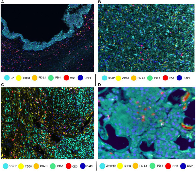
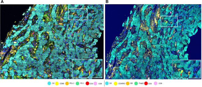
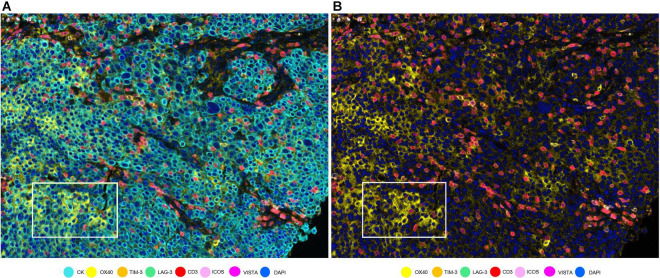
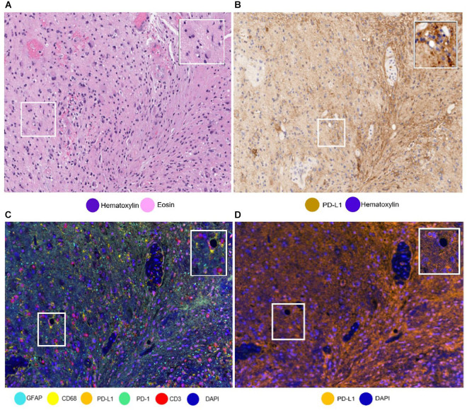
Every day, more evidence is revealed regarding the importance of the relationship between the response to cancer immunotherapy and the cancer immune microenvironment. It is well established that a profound characterization of the immune microenvironment is needed to identify prognostic and predictive immune biomarkers. To this end, we find phenotyping cells by multiplex immunofluorescence (mIF) a powerful and useful tool to identify cell types in biopsy specimens. Here, we describe the use of mIF tyramide signal amplification for labeling up to eight markers on a single slide of formalin-fixed, paraffin-embedded tumor tissue to phenotype immune cells in tumor tissues. Different panels show different markers, and the different panels can be used to characterize immune cells and relevant checkpoint proteins. The panel design depends on the research hypothesis, the cell population of interest, or the treatment under investigation. To phenotype the cells, image analysis software is used to identify individual marker expression or specific co-expression markers, which can differentiate already selected phenotypes. The individual-markers approach identifies a broad number of cell phenotypes, including rare cells, which may be helpful in a tumor microenvironment study. To accurately interpret results, it is important to recognize which receptors are expressed on different cell types and their typical location (i.e., nuclear, membrane, and/or cytoplasm). Furthermore, the amplification system of mIF may allow us to see weak marker signals, such as programmed cell death ligand 1, more easily than they are seen with single-marker immunohistochemistry (IHC) labeling. Finally, mIF technologies are promising resources for discovery of novel cancer immunotherapies and related biomarkers. In contrast with conventional IHC, which permits only the labeling of one single marker per tissue sample, mIF can detect multiple markers from a single tissue sample, and at the same time, deliver extensive information about the cell phenotypes composition and their spatial localization. In this matter, the phenotyping process is critical and must be done accurately by a highly trained personal with knowledge of immune cell protein expression and tumor pathology.

摘要

每天,越来越多的证据表明癌症免疫治疗反应与癌症免疫微环境之间关系的重要性。众所周知,为了识别预后和预测性免疫生物标志物,需要对免疫微环境进行深入表征。为此,我们发现通过多重免疫荧光(mIF)对细胞进行表型分析是一种在活检标本中识别细胞类型的强大且有用的工具。在此,我们描述了使用mIF酪胺信号放大技术在福尔马林固定、石蜡包埋的肿瘤组织的单个载玻片上标记多达八个标志物,以对肿瘤组织中的免疫细胞进行表型分析。不同的 panel 显示不同的标志物,不同的 panel 可用于表征免疫细胞和相关的检查点蛋白。panel 的设计取决于研究假设、感兴趣的细胞群体或所研究的治疗方法。为了对细胞进行表型分析,使用图像分析软件来识别单个标志物表达或特定的共表达标志物,这些标志物可以区分已经选定的表型。单标志物方法可识别大量细胞表型,包括罕见细胞,这在肿瘤微环境研究中可能会有所帮助。为了准确解释结果,重要的是要识别不同细胞类型上表达的哪些受体及其典型位置(即细胞核、细胞膜和/或细胞质)。此外,mIF 的放大系统可能使我们比单标志物免疫组织化学(IHC)标记更容易看到弱标志物信号,如程序性细胞死亡配体 1。最后,mIF 技术是发现新型癌症免疫疗法和相关生物标志物的有前景的资源。与传统 IHC 不同,传统 IHC 每个组织样本仅允许标记一个单一标志物,mIF 可以从单个组织样本中检测多个标志物,同时提供有关细胞表型组成及其空间定位的广泛信息。在这方面,表型分析过程至关重要,必须由具有免疫细胞蛋白表达和肿瘤病理学知识的训练有素的人员准确完成。

https://cdn.ncbi.nlm.nih.gov/pmc/blobs/1ac9/8118604/e4b31966ecfa/fmolb-08-667067-g007.jpg
https://cdn.ncbi.nlm.nih.gov/pmc/blobs/1ac9/8118604/312146e8f7b4/fmolb-08-667067-g001.jpg
https://cdn.ncbi.nlm.nih.gov/pmc/blobs/1ac9/8118604/e87e2f7452a9/fmolb-08-667067-g002.jpg
https://cdn.ncbi.nlm.nih.gov/pmc/blobs/1ac9/8118604/7ae84f8ef023/fmolb-08-667067-g003.jpg
https://cdn.ncbi.nlm.nih.gov/pmc/blobs/1ac9/8118604/c157c184091f/fmolb-08-667067-g004.jpg
https://cdn.ncbi.nlm.nih.gov/pmc/blobs/1ac9/8118604/13bc80dab410/fmolb-08-667067-g005.jpg
https://cdn.ncbi.nlm.nih.gov/pmc/blobs/1ac9/8118604/506a50733268/fmolb-08-667067-g006.jpg
https://cdn.ncbi.nlm.nih.gov/pmc/blobs/1ac9/8118604/e4b31966ecfa/fmolb-08-667067-g007.jpg

相似文献

1
Multiplex Immunofluorescence Tyramide Signal Amplification for Immune Cell Profiling of Paraffin-Embedded Tumor Tissues.用于石蜡包埋肿瘤组织免疫细胞分析的多重免疫荧光酪胺信号放大技术
Front Mol Biosci. 2021 Apr 29;8:667067. doi: 10.3389/fmolb.2021.667067. eCollection 2021.
2
Immuno-profiling and cellular spatial analysis using five immune oncology multiplex immunofluorescence panels for paraffin tumor tissue.使用五个免疫肿瘤多重免疫荧光面板对石蜡肿瘤组织进行免疫分析和细胞空间分析。
Sci Rep. 2021 Apr 19;11(1):8511. doi: 10.1038/s41598-021-88156-0.
3
Characterizing the Tumor Immune Microenvironment with Tyramide-Based Multiplex Immunofluorescence.基于酪胺的多重免疫荧光法描绘肿瘤免疫微环境。
J Mammary Gland Biol Neoplasia. 2020 Dec;25(4):417-432. doi: 10.1007/s10911-021-09479-2. Epub 2021 Feb 15.
4
Validation of multiplex immunofluorescence panels using multispectral microscopy for immune-profiling of formalin-fixed and paraffin-embedded human tumor tissues.利用多光谱显微镜对福尔马林固定石蜡包埋的人类肿瘤组织进行免疫分析的多重免疫荧光试剂盒验证。
Sci Rep. 2017 Oct 17;7(1):13380. doi: 10.1038/s41598-017-13942-8.
5
Impact of Region-of-Interest Size on Immune Profiling Using Multiplex Immunofluorescence Tyramide Signal Amplification for Paraffin-Embedded Tumor Tissues.使用多重免疫荧光酪胺信号放大技术对石蜡包埋肿瘤组织进行免疫分析时,感兴趣区域大小的影响。
Pathobiology. 2023;90(1):1-12. doi: 10.1159/000523751. Epub 2022 May 24.
6
Immune profiling of mouse lung adenocarcinoma paraffin tissues using multiplex immunofluorescence panel: a pilot study.使用多重免疫荧光组对小鼠肺腺癌石蜡组织进行免疫谱分析:一项初步研究。
Lab Anim Res. 2024 Jun 14;40(1):24. doi: 10.1186/s42826-024-00210-w.
7
Procedural Requirements and Recommendations for Multiplex Immunofluorescence Tyramide Signal Amplification Assays to Support Translational Oncology Studies.支持转化肿瘤学研究的多重免疫荧光酪胺信号放大检测的程序要求和建议。
Cancers (Basel). 2020 Jan 21;12(2):255. doi: 10.3390/cancers12020255.
8
Automated Multiplex Immunofluorescence Panel for Immuno-oncology Studies on Formalin-fixed Carcinoma Tissue Specimens.用于福尔马林固定癌组织标本免疫肿瘤学研究的自动化多重免疫荧光检测板
J Vis Exp. 2019 Jan 21(143). doi: 10.3791/58390.
9
Validation of an Accurate Automated Multiplex Immunofluorescence Method for Immuno-Profiling Melanoma.一种用于黑色素瘤免疫分析的准确自动化多重免疫荧光方法的验证
Front Mol Biosci. 2022 May 19;9:810858. doi: 10.3389/fmolb.2022.810858. eCollection 2022.
10
Identification of distinct immune landscapes using an automated nine-color multiplex immunofluorescence staining panel and image analysis in paraffin tumor tissues.使用自动化九色多重免疫荧光染色面板和石蜡肿瘤组织的图像分析鉴定不同的免疫图谱。
Sci Rep. 2021 Feb 25;11(1):4530. doi: 10.1038/s41598-021-83858-x.

引用本文的文献

1
Yes-associated protein plays oncogenic roles in human sporadic colorectal adenomas.Yes相关蛋白在人类散发性结肠直肠腺瘤中发挥致癌作用。
Carcinogenesis. 2025 Jan 20;46(1). doi: 10.1093/carcin/bgaf007.
2
Research progress and perspectives on the application of tyramide signal amplification-based multiplex immunohistochemistry/immunofluorescence: a bibliometrics analysis.基于酪胺信号放大的多重免疫组织化学/免疫荧光应用的研究进展与展望:一项文献计量学分析
Front Oncol. 2025 Jan 24;14:1473414. doi: 10.3389/fonc.2024.1473414. eCollection 2024.
3
Wanted: Dead or Alive Cells with Propidium Iodide Staining in Liver Tissue.

本文引用的文献

1
Neoadjuvant chemotherapy and nivolumab in resectable non-small-cell lung cancer (NADIM): an open-label, multicentre, single-arm, phase 2 trial.可切除非小细胞肺癌新辅助化疗联合纳武利尤单抗(NADIM):一项开放标签、多中心、单臂、Ⅱ期临床试验。
Lancet Oncol. 2020 Nov;21(11):1413-1422. doi: 10.1016/S1470-2045(20)30453-8. Epub 2020 Sep 24.
2
Myeloid Cell-Derived Arginase in Cancer Immune Response.髓系细胞衍生的精氨酸酶在癌症免疫反应中的作用
Front Immunol. 2020 May 15;11:938. doi: 10.3389/fimmu.2020.00938. eCollection 2020.
3
The Society for Immunotherapy of Cancer statement on best practices for multiplex immunohistochemistry (IHC) and immunofluorescence (IF) staining and validation.
征寻:肝组织中经碘化丙啶染色的死活细胞。
Int J Mol Sci. 2024 Dec 17;25(24):13521. doi: 10.3390/ijms252413521.
4
Pembrolizumab plus chemotherapy in frontline treatment of advanced ovarian cancer: Clinical and translational results from a phase 2 trial.帕博利珠单抗联合化疗用于晚期卵巢癌一线治疗:一项2期试验的临床和转化结果
Med. 2025 Jan 10;6(1):100494. doi: 10.1016/j.medj.2024.07.022. Epub 2024 Aug 15.
5
Increased Multiplexity in Optical Tissue Clearing-Based Three-Dimensional Immunofluorescence Microscopy of the Tumor Microenvironment by Light-Emitting Diode Photobleaching.通过发光二极管光漂白提高基于光学组织通透的三维免疫荧光显微镜检测肿瘤微环境的多重性。
Lab Invest. 2024 Jun;104(6):102072. doi: 10.1016/j.labinv.2024.102072. Epub 2024 Apr 26.
6
Advancements in technology for characterizing the tumor immune microenvironment.肿瘤免疫微环境特征分析技术的进展
Int J Biol Sci. 2024 Mar 25;20(6):2151-2167. doi: 10.7150/ijbs.92525. eCollection 2024.
7
Exploratory pilot study to characterize the immune landscapes of malignant pleural effusions and their corresponding primary tumors from patients with breast carcinoma and lung adenocarcinoma.探索性初步研究,以描绘乳腺癌和肺腺癌患者的恶性胸腔积液及其相应原发性肿瘤的免疫图谱。
J Am Soc Cytopathol. 2024 May-Jun;13(3):161-173. doi: 10.1016/j.jasc.2024.02.005. Epub 2024 Feb 29.
8
Intracerebral hemorrhage-induced brain injury in mice: The role of peroxiredoxin 2-Toll-like receptor 4 inflammatory axis.脑内出血诱导的小鼠脑损伤:过氧化物酶 2-Toll 样受体 4 炎症轴的作用。
CNS Neurosci Ther. 2024 Mar;30(3):e14681. doi: 10.1111/cns.14681.
9
Digital analysis of the prostate tumor microenvironment with high-order chromogenic multiplexing.基于高阶显色多重技术的前列腺肿瘤微环境数字分析
J Pathol Inform. 2023 Nov 21;15:100352. doi: 10.1016/j.jpi.2023.100352. eCollection 2024 Dec.
10
Immune cellular patterns of distribution affect outcomes of patients with non-small cell lung cancer.免疫细胞的分布模式会影响非小细胞肺癌患者的预后。
Nat Commun. 2023 Apr 25;14(1):2364. doi: 10.1038/s41467-023-37905-y.
癌症免疫治疗学会关于多重免疫组织化学(IHC)和免疫荧光(IF)染色及验证最佳实践的声明。
J Immunother Cancer. 2020 May;8(1). doi: 10.1136/jitc-2019-000155.
4
Overview of multiplex immunohistochemistry/immunofluorescence techniques in the era of cancer immunotherapy.癌症免疫治疗时代的多重免疫组化/免疫荧光技术概述。
Cancer Commun (Lond). 2020 Apr;40(4):135-153. doi: 10.1002/cac2.12023. Epub 2020 Apr 17.
5
A Phase I/II Study of Neoadjuvant Cisplatin, Docetaxel, and Nintedanib for Resectable Non-Small Cell Lung Cancer.一项新辅助顺铂、多西他赛和尼达尼布治疗可切除非小细胞肺癌的 I/II 期研究。
Clin Cancer Res. 2020 Jul 15;26(14):3525-3536. doi: 10.1158/1078-0432.CCR-19-4180. Epub 2020 Mar 19.
6
Variants in Epithelial-Mesenchymal Transition and Immune Checkpoint Genes Are Associated With Immune Cell Profiles and Predict Survival in Non-Small Cell Lung Cancer.上皮-间质转化和免疫检查点基因变异与非小细胞肺癌的免疫细胞特征相关,并可预测生存。
Arch Pathol Lab Med. 2020 Oct 1;144(10):1234-1244. doi: 10.5858/arpa.2019-0419-OA.
7
High-dimensional immune-profiling in cancer: implications for immunotherapy.癌症的高维免疫分析:对免疫治疗的启示。
J Immunother Cancer. 2020 Feb;8(1). doi: 10.1136/jitc-2019-000363.
8
Procedural Requirements and Recommendations for Multiplex Immunofluorescence Tyramide Signal Amplification Assays to Support Translational Oncology Studies.支持转化肿瘤学研究的多重免疫荧光酪胺信号放大检测的程序要求和建议。
Cancers (Basel). 2020 Jan 21;12(2):255. doi: 10.3390/cancers12020255.
9
Computational Staining of Pathology Images to Study the Tumor Microenvironment in Lung Cancer.计算染色病理图像以研究肺癌中的肿瘤微环境。
Cancer Res. 2020 May 15;80(10):2056-2066. doi: 10.1158/0008-5472.CAN-19-1629. Epub 2020 Jan 8.
10
Highly multiplexed immunofluorescence images and single-cell data of immune markers in tonsil and lung cancer.高多重免疫荧光图像和扁桃体及肺癌免疫标志物的单细胞数据。
Sci Data. 2019 Dec 17;6(1):323. doi: 10.1038/s41597-019-0332-y.