Department of Medicine and.
Department of Medical and Molecular Genetics, Indiana University School of Medicine, Indianapolis, Indiana, USA.
JCI Insight. 2021 Jun 22;6(12):147703. doi: 10.1172/jci.insight.147703.
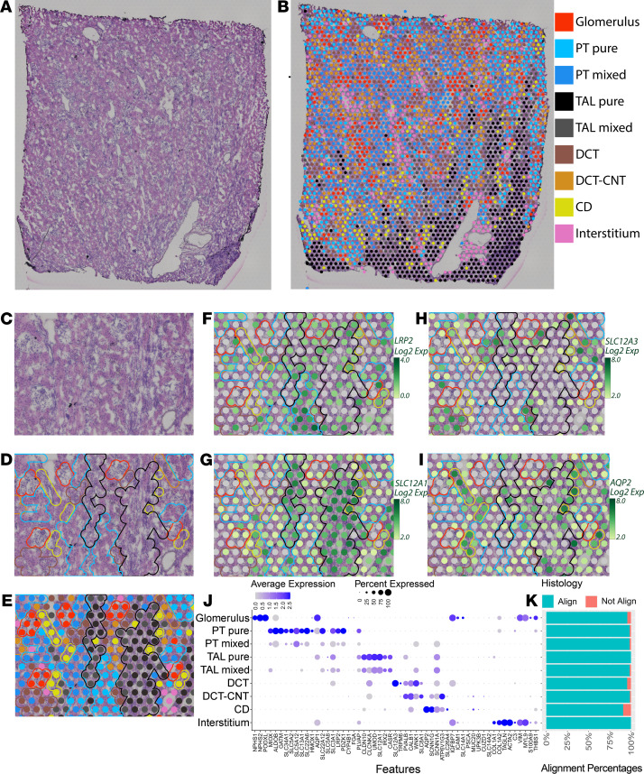
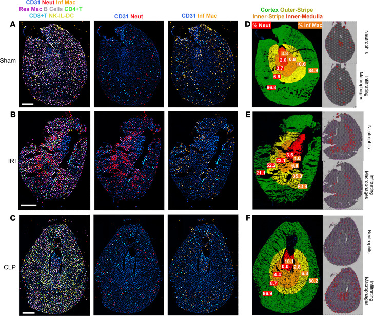
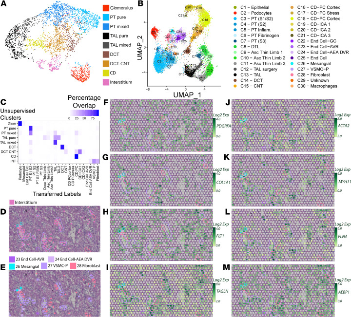
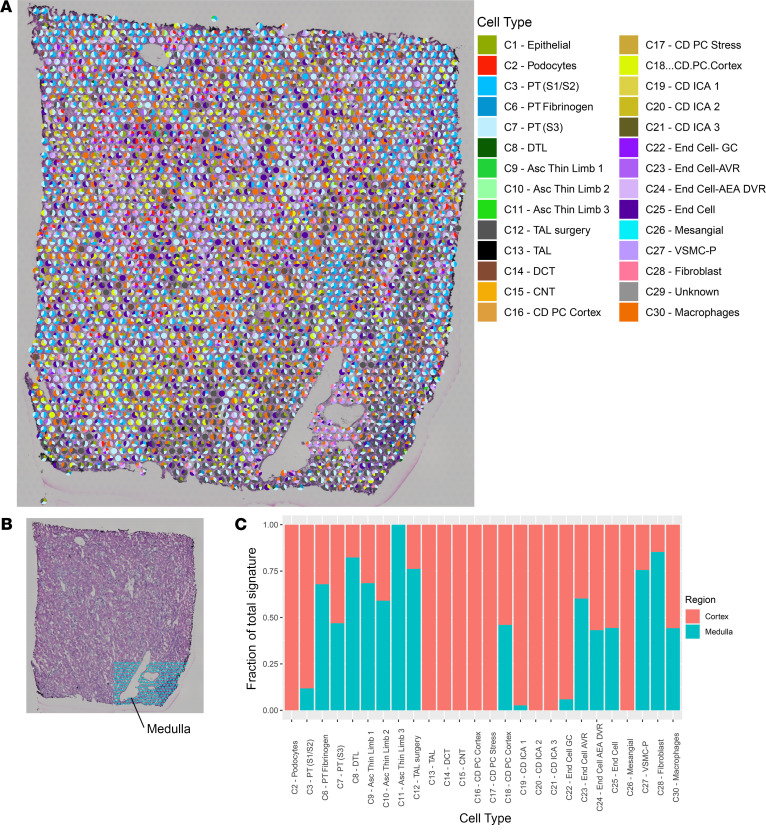
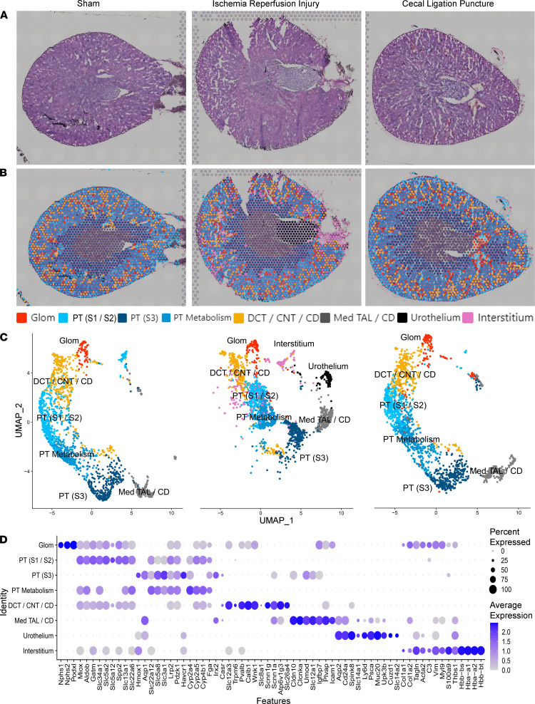
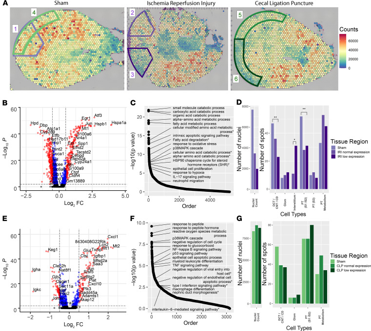
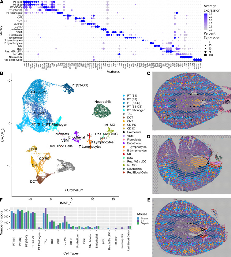
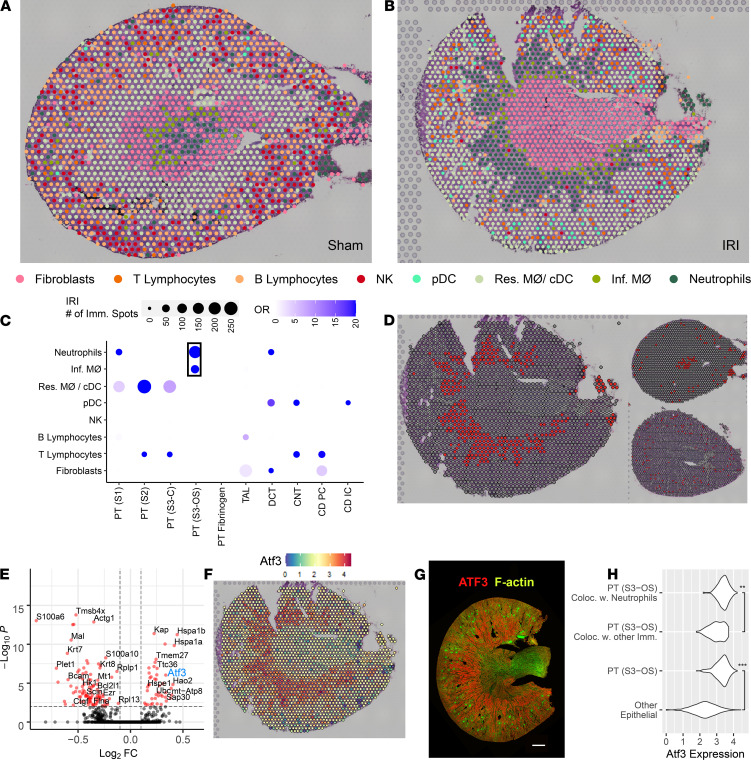
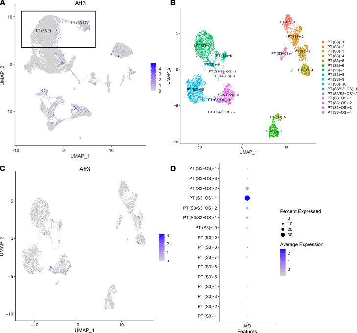
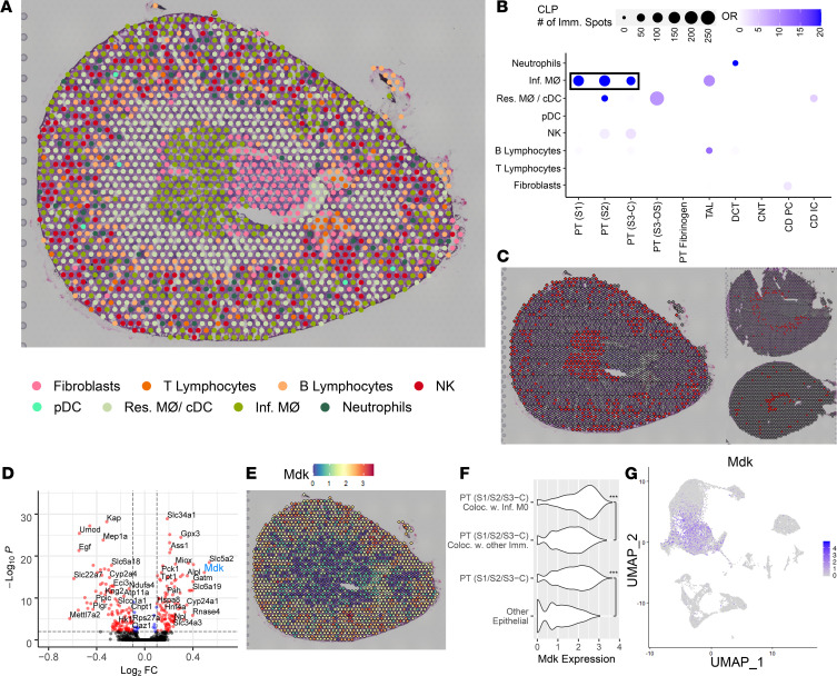
Single-cell sequencing studies have characterized the transcriptomic signature of cell types within the kidney. However, the spatial distribution of acute kidney injury (AKI) is regional and affects cells heterogeneously. We first optimized coordination of spatial transcriptomics and single-nuclear sequencing data sets, mapping 30 dominant cell types to a human nephrectomy. The predicted cell-type spots corresponded with the underlying histopathology. To study the implications of AKI on transcript expression, we then characterized the spatial transcriptomic signature of 2 murine AKI models: ischemia/reperfusion injury (IRI) and cecal ligation puncture (CLP). Localized regions of reduced overall expression were associated with injury pathways. Using single-cell sequencing, we deconvoluted the signature of each spatial transcriptomic spot, identifying patterns of colocalization between immune and epithelial cells. Neutrophils infiltrated the renal medulla in the ischemia model. Atf3 was identified as a chemotactic factor in S3 proximal tubules. In the CLP model, infiltrating macrophages dominated the outer cortical signature, and Mdk was identified as a corresponding chemotactic factor. The regional distribution of these immune cells was validated with multiplexed CO-Detection by indEXing (CODEX) immunofluorescence. Spatial transcriptomic sequencing complemented single-cell sequencing by uncovering mechanisms driving immune cell infiltration and detection of relevant cell subpopulations.
单细胞测序研究已经描述了肾脏内细胞类型的转录组特征。然而,急性肾损伤 (AKI) 的空间分布是区域性的,并且会不均匀地影响细胞。我们首先优化了空间转录组学和单核测序数据集的协调,将 30 种主要细胞类型映射到人类肾切除标本上。预测的细胞类型点与潜在的组织病理学相对应。为了研究 AKI 对转录表达的影响,我们随后描述了 2 种 AKI 模型(缺血再灌注损伤 [IRI] 和盲肠结扎穿刺 [CLP])的空间转录组特征。与损伤途径相关的是整体表达降低的局部区域。使用单细胞测序,我们对每个空间转录组点的特征进行了去卷积,确定了免疫细胞和上皮细胞之间的共定位模式。在缺血模型中,中性粒细胞浸润到肾髓质。Atf3 被鉴定为 S3 近端小管中的趋化因子。在 CLP 模型中,浸润的巨噬细胞主导着外皮质特征,而 Mdk 被鉴定为相应的趋化因子。这些免疫细胞的区域分布通过多重 CO-Detection by indEXing (CODEX) 免疫荧光进行了验证。空间转录组测序通过揭示驱动免疫细胞浸润的机制和检测相关细胞亚群,补充了单细胞测序。