• 文献检索
  • 文档翻译
  • 深度研究
  • 学术资讯
  • Suppr Zotero 插件Zotero 插件
  • 邀请有礼
  • 套餐&价格
  • 历史记录
应用&插件
Suppr Zotero 插件Zotero 插件浏览器插件Mac 客户端Windows 客户端微信小程序
定价
高级版会员购买积分包购买API积分包
服务
文献检索文档翻译深度研究API 文档MCP 服务
关于我们
关于 Suppr公司介绍联系我们用户协议隐私条款
关注我们

Suppr 超能文献

核心技术专利:CN118964589B侵权必究
粤ICP备2023148730 号-1Suppr @ 2026

文献检索

告别复杂PubMed语法,用中文像聊天一样搜索,搜遍4000万医学文献。AI智能推荐,让科研检索更轻松。

立即免费搜索

文件翻译

保留排版,准确专业,支持PDF/Word/PPT等文件格式,支持 12+语言互译。

免费翻译文档

深度研究

AI帮你快速写综述,25分钟生成高质量综述,智能提取关键信息,辅助科研写作。

立即免费体验

使用 Pegasus 流体分割算法评估光学相干断层扫描图像中的自动多类流体分割。

Evaluation of Automated Multiclass Fluid Segmentation in Optical Coherence Tomography Images Using the Pegasus Fluid Segmentation Algorithms.

机构信息

School of Optometry and Vision Sciences, Cardiff University, Cardiff, UK.

King's College Hospital NHS Foundation Trust, London, UK.

出版信息

Transl Vis Sci Technol. 2021 Jan 4;10(1):27. doi: 10.1167/tvst.10.1.27.

DOI:10.1167/tvst.10.1.27
PMID:34008019
原文链接:https://pmc.ncbi.nlm.nih.gov/articles/PMC9354552/
Abstract

PURPOSE

To evaluate the performance of the Pegasus-OCT (Visulytix Ltd) multiclass automated fluid segmentation algorithms on independent spectral domain optical coherence tomography data sets.

METHODS

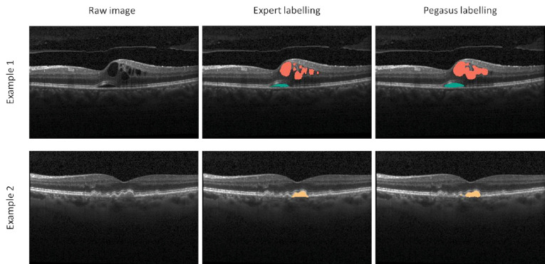
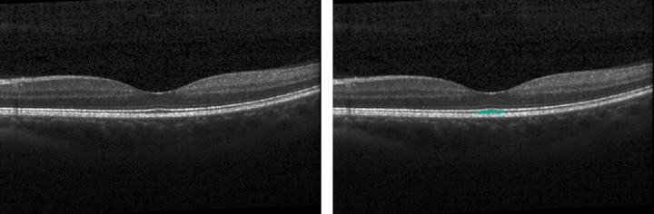
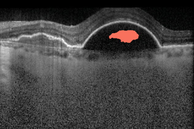
The Pegasus automated fluid segmentation algorithms were applied to three data sets with edematous pathology, comprising 750, 600, and 110 b-scans, respectively. Intraretinal fluid (IRF), sub-retinal fluid (SRF), and pigment epithelial detachment (PED) were automatically segmented by Pegasus-OCT for each b-scan where ground truth from data set owners was available. Detection performance was assessed by calculating sensitivities and specificities, while Dice coefficients were used to assess agreement between the segmentation methods.

RESULTS

For two data sets, IRF detection yielded promising sensitivities (0.98 and 0.94, respectively) and specificities (1.00 and 0.98) but less consistent agreement with the ground truth (dice coefficients 0.81 and 0.59); likewise, SRF detection showed high sensitivity (0.86 and 0.98) and specificity (0.83 and 0.89) but less consistent agreement (0.59 and 0.78). PED detection on the first data set showed moderate agreement (0.66) with high sensitivity (0.97) and specificity (0.98). IRF detection in a third data set yielded less favorable agreement (0.46-0.57) and sensitivity (0.59-0.68), attributed to image quality and ground truth grader discordance.

CONCLUSIONS

The Pegasus automated fluid segmentation algorithms were able to detect IRF, SRF, and PED in SD-OCT b-scans acquired across multiple independent data sets. Dice coefficients and sensitivity and specificity values indicate the potential for application to automated detection and monitoring of retinal diseases such as age-related macular degeneration and diabetic macular edema.

TRANSLATIONAL RELEVANCE

The potential of Pegasus-OCT for automated fluid quantification and differentiation of IRF, SRF, and PED in OCT images has application to both clinical practice and research.

摘要

目的

评估 Pegasus-OCT(Visulytix Ltd)多类别自动流体分割算法在独立的谱域光相干断层扫描数据集上的性能。

方法

将 Pegasus 自动流体分割算法应用于三个包含水肿性病变的数据集,每个数据集分别包含 750、600 和 110 个 B 扫描。在可获得数据集所有者提供的真实数据的情况下,Pegasus-OCT 自动对每个 B 扫描的视网膜内液(IRF)、视网膜下液(SRF)和色素上皮脱离(PED)进行分割。通过计算敏感性和特异性来评估检测性能,而 Dice 系数用于评估分割方法之间的一致性。

结果

对于两个数据集,IRF 检测的敏感性分别为 0.98 和 0.94,特异性分别为 1.00 和 0.98,但与真实数据的一致性不一致(Dice 系数分别为 0.81 和 0.59);同样,SRF 检测的敏感性分别为 0.86 和 0.98,特异性分别为 0.83 和 0.89,但一致性不一致(Dice 系数分别为 0.59 和 0.78)。第一个数据集的 PED 检测与高敏感性(0.97)和特异性(0.98)具有中等一致性(0.66)。第三个数据集的 IRF 检测得到的一致性较差(0.46-0.57)和敏感性(0.59-0.68),这归因于图像质量和真实数据评估员的不一致。

结论

Pegasus 自动流体分割算法能够在多个独立数据集采集的 SD-OCT B 扫描中检测 IRF、SRF 和 PED。Dice 系数以及敏感性和特异性值表明,该算法有可能应用于自动检测和监测年龄相关性黄斑变性和糖尿病性黄斑水肿等视网膜疾病。

翻译

医麦客

https://cdn.ncbi.nlm.nih.gov/pmc/blobs/2c15/9354552/c2d948d66538/tvst-10-1-27-f003.jpg
https://cdn.ncbi.nlm.nih.gov/pmc/blobs/2c15/9354552/7d6903ee6990/tvst-10-1-27-f001.jpg
https://cdn.ncbi.nlm.nih.gov/pmc/blobs/2c15/9354552/08ca1d032c10/tvst-10-1-27-f002.jpg
https://cdn.ncbi.nlm.nih.gov/pmc/blobs/2c15/9354552/c2d948d66538/tvst-10-1-27-f003.jpg
https://cdn.ncbi.nlm.nih.gov/pmc/blobs/2c15/9354552/7d6903ee6990/tvst-10-1-27-f001.jpg
https://cdn.ncbi.nlm.nih.gov/pmc/blobs/2c15/9354552/08ca1d032c10/tvst-10-1-27-f002.jpg
https://cdn.ncbi.nlm.nih.gov/pmc/blobs/2c15/9354552/c2d948d66538/tvst-10-1-27-f003.jpg

相似文献

1
Evaluation of Automated Multiclass Fluid Segmentation in Optical Coherence Tomography Images Using the Pegasus Fluid Segmentation Algorithms.使用 Pegasus 流体分割算法评估光学相干断层扫描图像中的自动多类流体分割。
Transl Vis Sci Technol. 2021 Jan 4;10(1):27. doi: 10.1167/tvst.10.1.27.
2
Evaluation of an Artificial Intelligence-Based Detector of Sub- and Intraretinal Fluid on a Large Set of Optical Coherence Tomography Volumes in Age-Related Macular Degeneration and Diabetic Macular Edema.基于人工智能的视网膜下和视网膜内液检测在大量年龄相关性黄斑变性和糖尿病性黄斑水肿光学相干断层扫描体积上的评估。
Ophthalmologica. 2022;245(6):516-527. doi: 10.1159/000527345. Epub 2022 Oct 10.
3
Automated Segmentation of Lesions Including Subretinal Hyperreflective Material in Neovascular Age-related Macular Degeneration.利用人工智能技术对湿性年龄相关性黄斑变性中包含视网膜下高反射物质的病变进行自动分割。
Am J Ophthalmol. 2018 Jul;191:64-75. doi: 10.1016/j.ajo.2018.04.007. Epub 2018 Apr 12.
4
Spatial Correspondence Between Intraretinal Fluid, Subretinal Fluid, and Pigment Epithelial Detachment in Neovascular Age-Related Macular Degeneration.新生血管性年龄相关性黄斑变性中视网膜内液、视网膜下液与色素上皮脱离之间的空间对应关系
Invest Ophthalmol Vis Sci. 2017 Aug 1;58(10):4039-4048. doi: 10.1167/iovs.16-20201.
5
Quantification of Fluid Resolution and Visual Acuity Gain in Patients With Diabetic Macular Edema Using Deep Learning: A Post Hoc Analysis of a Randomized Clinical Trial.利用深度学习技术对糖尿病性黄斑水肿患者的液体积聚消退和视力提高程度进行定量评估:一项随机临床试验的事后分析。
JAMA Ophthalmol. 2020 Sep 1;138(9):945-953. doi: 10.1001/jamaophthalmol.2020.2457.
6
Fully Automated Detection and Quantification of Macular Fluid in OCT Using Deep Learning.基于深度学习的 OCT 中黄斑区液全自动化检测和定量分析
Ophthalmology. 2018 Apr;125(4):549-558. doi: 10.1016/j.ophtha.2017.10.031. Epub 2017 Dec 8.
7
RetFluidNet: Retinal Fluid Segmentation for SD-OCT Images Using Convolutional Neural Network.RetFluidNet:基于卷积神经网络的 SD-OCT 图像视网膜液分割。
J Digit Imaging. 2021 Jun;34(3):691-704. doi: 10.1007/s10278-021-00459-w. Epub 2021 Jun 2.
8
Validation and Clinical Applicability of Whole-Volume Automated Segmentation of Optical Coherence Tomography in Retinal Disease Using Deep Learning.基于深度学习的全容积自动光学相干断层扫描在视网膜疾病中的验证和临床适用性。
JAMA Ophthalmol. 2021 Sep 1;139(9):964-973. doi: 10.1001/jamaophthalmol.2021.2273.
9
Automated Quantification of Pathological Fluids in Neovascular Age-Related Macular Degeneration, and Its Repeatability Using Deep Learning.利用深度学习对新生血管性年龄相关性黄斑变性病理性液体积分的自动量化及其重复性。
Transl Vis Sci Technol. 2021 Apr 1;10(4):17. doi: 10.1167/tvst.10.4.17.
10
A systematic evaluation of human expert agreement on optical coherence tomography biomarkers using multiple devices.使用多种设备对光学相干断层扫描生物标志物的人类专家一致性进行系统评价。
Eye (Lond). 2023 Aug;37(12):2573-2579. doi: 10.1038/s41433-022-02376-w. Epub 2022 Dec 28.

引用本文的文献

1
Retinal fluid is associated with cytokines of aqueous humor in age-related macular degeneration using automatic 3-dimensional quantification.使用自动三维定量分析,视网膜液与年龄相关性黄斑变性房水中的细胞因子有关。
Front Cell Dev Biol. 2023 Mar 8;11:1157497. doi: 10.3389/fcell.2023.1157497. eCollection 2023.
2
Cyst identification in retinal optical coherence tomography images using hidden Markov model.基于隐马尔可夫模型的视网膜光学相干断层扫描图像中囊泡的识别。
Sci Rep. 2023 Jan 2;13(1):12. doi: 10.1038/s41598-022-27243-2.
3
An Artificial-Intelligence-Based Automated Grading and Lesions Segmentation System for Myopic Maculopathy Based on Color Fundus Photographs.

本文引用的文献

1
DISEASE CLASSIFICATION OF MACULAR OPTICAL COHERENCE TOMOGRAPHY SCANS USING DEEP LEARNING SOFTWARE: Validation on Independent, Multicenter Data.使用深度学习软件对黄斑光学相干断层扫描图像进行疾病分类:独立、多中心数据验证。
Retina. 2020 Aug;40(8):1549-1557. doi: 10.1097/IAE.0000000000002640.
2
Differentiation of Underlying Pathologies of Macular Edema Using Spectral Domain Optical Coherence Tomography (SD-OCT).利用谱域光学相干断层扫描(SD-OCT)对黄斑水肿的潜在病理进行鉴别。
Ocul Immunol Inflamm. 2019;27(3):474-483. doi: 10.1080/09273948.2019.1603313.
3
Deep-learning based multiclass retinal fluid segmentation and detection in optical coherence tomography images using a fully convolutional neural network.
基于彩色眼底照片的人工智能自动分级和近视性黄斑病变分割系统。
Transl Vis Sci Technol. 2022 Jun 1;11(6):16. doi: 10.1167/tvst.11.6.16.
4
Directional analysis of intensity changes for determining the existence of cyst in optical coherence tomography images.用于确定光学相干断层扫描图像中囊肿存在的强度变化的方向分析。
Sci Rep. 2022 Feb 8;12(1):2105. doi: 10.1038/s41598-022-06099-6.
基于深度学习的全卷积神经网络在光学相干断层扫描图像中对多类视网膜液的分割和检测。
Med Image Anal. 2019 May;54:100-110. doi: 10.1016/j.media.2019.02.011. Epub 2019 Feb 22.
4
RETOUCH: The Retinal OCT Fluid Detection and Segmentation Benchmark and Challenge.RETOUCH:视网膜 OCT 流体检测和分割基准及挑战赛。
IEEE Trans Med Imaging. 2019 Aug;38(8):1858-1874. doi: 10.1109/TMI.2019.2901398. Epub 2019 Feb 26.
5
Identifying Medical Diagnoses and Treatable Diseases by Image-Based Deep Learning.基于图像的深度学习识别医学诊断和可治疗疾病。
Cell. 2018 Feb 22;172(5):1122-1131.e9. doi: 10.1016/j.cell.2018.02.010.
6
Fully Automated Detection and Quantification of Macular Fluid in OCT Using Deep Learning.基于深度学习的 OCT 中黄斑区液全自动化检测和定量分析
Ophthalmology. 2018 Apr;125(4):549-558. doi: 10.1016/j.ophtha.2017.10.031. Epub 2017 Dec 8.
7
MEASUREMENTS OF RETINAL FLUID BY OPTICAL COHERENCE TOMOGRAPHY LEAKAGE IN DIABETIC MACULAR EDEMA: A Biomarker of Visual Acuity Response to Treatment.光学相干断层扫描渗漏测量在糖尿病性黄斑水肿中的应用:视力反应治疗的生物标志物。
Retina. 2019 Jan;39(1):52-60. doi: 10.1097/IAE.0000000000001905.
8
Fully-automated segmentation of fluid regions in exudative age-related macular degeneration subjects: Kernel graph cut in neutrosophic domain.渗出性年龄相关性黄斑变性患者中流体区域的全自动分割:中智域中的核图割
PLoS One. 2017 Oct 23;12(10):e0186949. doi: 10.1371/journal.pone.0186949. eCollection 2017.
9
ReLayNet: retinal layer and fluid segmentation of macular optical coherence tomography using fully convolutional networks.ReLayNet:使用全卷积网络对黄斑光学相干断层扫描进行视网膜层和液体分割
Biomed Opt Express. 2017 Jul 13;8(8):3627-3642. doi: 10.1364/BOE.8.003627. eCollection 2017 Aug 1.
10
Deep-learning based, automated segmentation of macular edema in optical coherence tomography.基于深度学习的光学相干断层扫描中黄斑水肿的自动分割
Biomed Opt Express. 2017 Jun 23;8(7):3440-3448. doi: 10.1364/BOE.8.003440. eCollection 2017 Jul 1.