• 文献检索
  • 文档翻译
  • 深度研究
  • 学术资讯
  • Suppr Zotero 插件Zotero 插件
  • 邀请有礼
  • 套餐&价格
  • 历史记录
应用&插件
Suppr Zotero 插件Zotero 插件浏览器插件Mac 客户端Windows 客户端微信小程序
定价
高级版会员购买积分包购买API积分包
服务
文献检索文档翻译深度研究API 文档MCP 服务
关于我们
关于 Suppr公司介绍联系我们用户协议隐私条款
关注我们

Suppr 超能文献

核心技术专利:CN118964589B侵权必究
粤ICP备2023148730 号-1Suppr @ 2026

文献检索

告别复杂PubMed语法,用中文像聊天一样搜索,搜遍4000万医学文献。AI智能推荐,让科研检索更轻松。

立即免费搜索

文件翻译

保留排版,准确专业,支持PDF/Word/PPT等文件格式,支持 12+语言互译。

免费翻译文档

深度研究

AI帮你快速写综述,25分钟生成高质量综述,智能提取关键信息,辅助科研写作。

立即免费体验

基于深度学习的全容积自动光学相干断层扫描在视网膜疾病中的验证和临床适用性。

Validation and Clinical Applicability of Whole-Volume Automated Segmentation of Optical Coherence Tomography in Retinal Disease Using Deep Learning.

机构信息

Google Health, London, United Kingdom.

National Institute for Health Research Biomedical Research Centre for Ophthalmology, Moorfields Eye Hospital NHS (National Health Service) Foundation Trust, London, United Kingdom.

出版信息

JAMA Ophthalmol. 2021 Sep 1;139(9):964-973. doi: 10.1001/jamaophthalmol.2021.2273.

DOI:10.1001/jamaophthalmol.2021.2273
PMID:34236406
原文链接:https://pmc.ncbi.nlm.nih.gov/articles/PMC8444027/
Abstract

IMPORTANCE

Quantitative volumetric measures of retinal disease in optical coherence tomography (OCT) scans are infeasible to perform owing to the time required for manual grading. Expert-level deep learning systems for automatic OCT segmentation have recently been developed. However, the potential clinical applicability of these systems is largely unknown.

OBJECTIVE

To evaluate a deep learning model for whole-volume segmentation of 4 clinically important pathological features and assess clinical applicability.

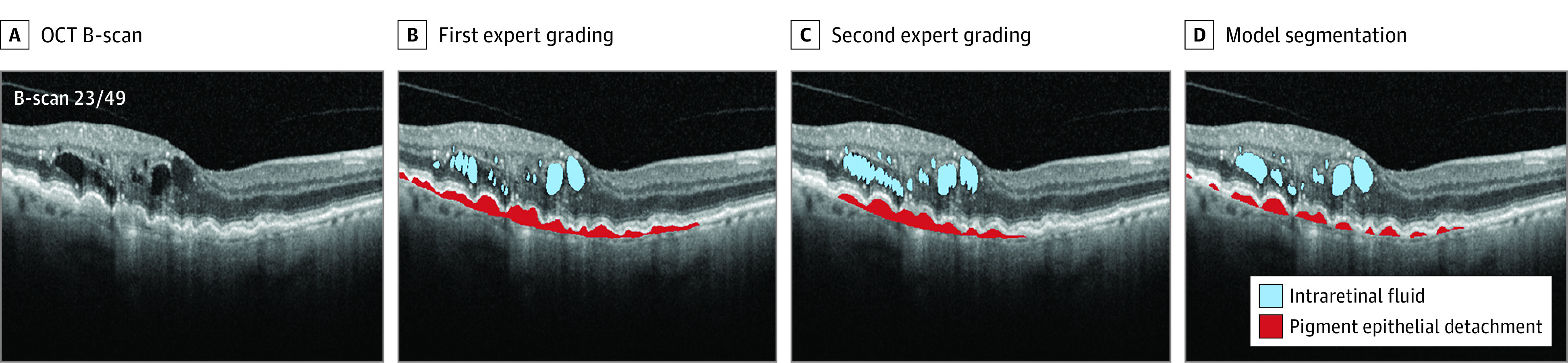
DESIGN, SETTING, PARTICIPANTS: This diagnostic study used OCT data from 173 patients with a total of 15 558 B-scans, treated at Moorfields Eye Hospital. The data set included 2 common OCT devices and 2 macular conditions: wet age-related macular degeneration (107 scans) and diabetic macular edema (66 scans), covering the full range of severity, and from 3 points during treatment. Two expert graders performed pixel-level segmentations of intraretinal fluid, subretinal fluid, subretinal hyperreflective material, and pigment epithelial detachment, including all B-scans in each OCT volume, taking as long as 50 hours per scan. Quantitative evaluation of whole-volume model segmentations was performed. Qualitative evaluation of clinical applicability by 3 retinal experts was also conducted. Data were collected from June 1, 2012, to January 31, 2017, for set 1 and from January 1 to December 31, 2017, for set 2; graded between November 2018 and January 2020; and analyzed from February 2020 to November 2020.

MAIN OUTCOMES AND MEASURES

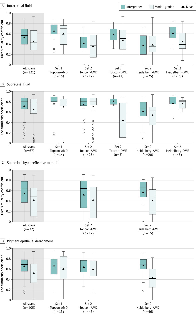
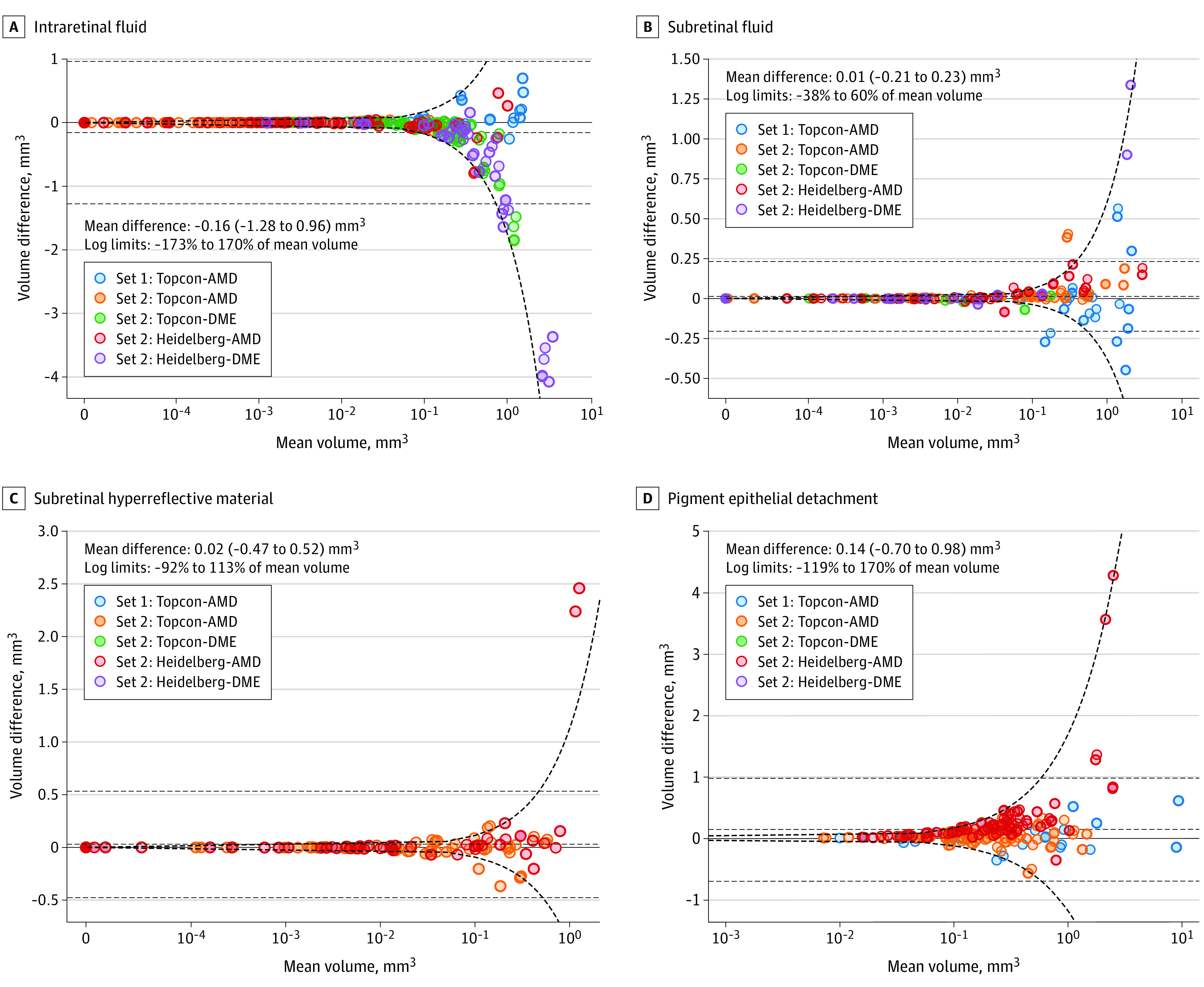
Rating and stack ranking for clinical applicability by retinal specialists, model-grader agreement for voxelwise segmentations, and total volume evaluated using Dice similarity coefficients, Bland-Altman plots, and intraclass correlation coefficients.

RESULTS

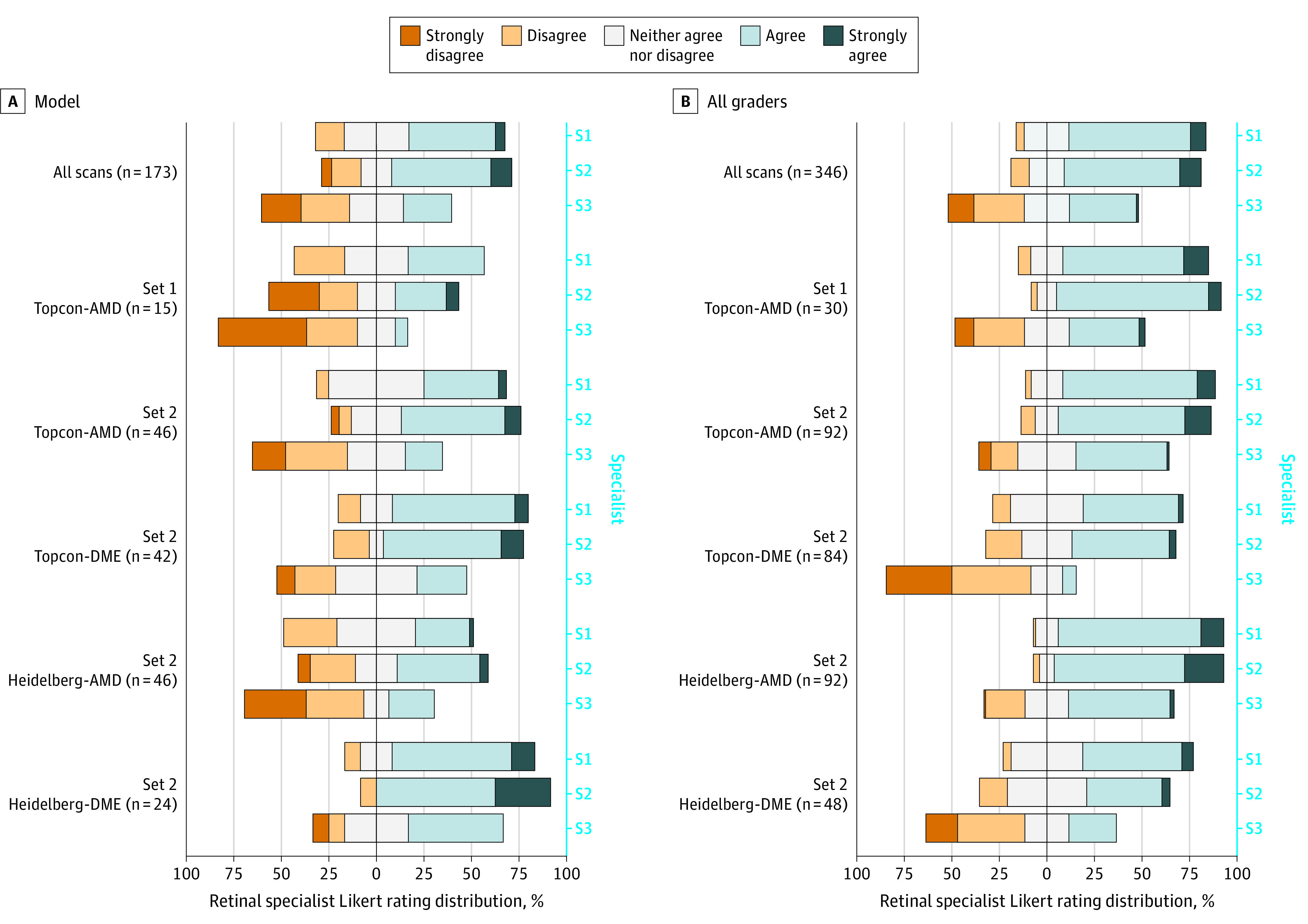
Among the 173 patients included in the analysis (92 [53%] women), qualitative assessment found that automated whole-volume segmentation ranked better than or comparable to at least 1 expert grader in 127 scans (73%; 95% CI, 66%-79%). A neutral or positive rating was given to 135 model segmentations (78%; 95% CI, 71%-84%) and 309 expert gradings (2 per scan) (89%; 95% CI, 86%-92%). The model was rated neutrally or positively in 86% to 92% of diabetic macular edema scans and 53% to 87% of age-related macular degeneration scans. Intraclass correlations ranged from 0.33 (95% CI, 0.08-0.96) to 0.96 (95% CI, 0.90-0.99). Dice similarity coefficients ranged from 0.43 (95% CI, 0.29-0.66) to 0.78 (95% CI, 0.57-0.85).

CONCLUSIONS AND RELEVANCE

This deep learning-based segmentation tool provided clinically useful measures of retinal disease that would otherwise be infeasible to obtain. Qualitative evaluation was additionally important to reveal clinical applicability for both care management and research.

摘要

重要性

由于手动分级所需的时间,光学相干断层扫描(OCT)扫描中视网膜疾病的定量容积测量是不可行的。最近已经开发出用于自动 OCT 分割的专家级深度学习系统。然而,这些系统的潜在临床适用性在很大程度上是未知的。

目的

评估一种用于 4 种临床重要病理特征的全容积分割的深度学习模型,并评估其临床适用性。

设计、设置、参与者:这项诊断研究使用了来自 173 名患者的总共 15558 个 B 扫描的 OCT 数据,这些患者在 Moorfields 眼科医院接受治疗。该数据集包括 2 种常见的 OCT 设备和 2 种黄斑病变:湿性年龄相关性黄斑变性(107 个扫描)和糖尿病性黄斑水肿(66 个扫描),涵盖了所有严重程度,并在治疗过程中的 3 个点进行了扫描。两名专家分级员对视网膜内液、视网膜下液、视网膜下高反射物质和色素上皮脱离进行了像素级分割,包括每个 OCT 容积中的所有 B 扫描,每个扫描的分割时间长达 50 小时。对全容积模型分割进行了定量评估。还由 3 名视网膜专家进行了临床适用性的定性评估。数据于 2012 年 6 月 1 日至 2017 年 1 月 31 日在数据集 1 中收集,于 2017 年 1 月 1 日至 2017 年 12 月 31 日在数据集 2 中收集;于 2018 年 11 月至 2020 年 1 月进行分级;并于 2020 年 2 月至 2020 年 11 月进行分析。

主要结果和测量

视网膜专家的临床适用性评分和堆栈排名、体素分割的模型分级员一致性以及使用 Dice 相似系数、Bland-Altman 图和组内相关系数评估的总容积。

结果

在分析中纳入的 173 名患者中(92[53%]名女性),自动全容积分割在 127 个扫描(73%;95%CI,66%-79%)中被评估为优于或至少与 1 名专家分级员相当。135 个模型分割(78%;95%CI,71%-84%)和 309 个专家分级(每个扫描 2 个)(89%;95%CI,86%-92%)得到了中性或积极的评价。在 86%至 92%的糖尿病性黄斑水肿扫描和 53%至 87%的年龄相关性黄斑变性扫描中,模型得到了中性或积极的评价。组内相关系数范围为 0.33(95%CI,0.08-0.96)至 0.96(95%CI,0.90-0.99)。Dice 相似系数范围为 0.43(95%CI,0.29-0.66)至 0.78(95%CI,0.57-0.85)。

结论和相关性

这种基于深度学习的分割工具提供了临床上有用的视网膜疾病测量方法,否则这些方法是不可行的。定性评估对于揭示护理管理和研究的临床适用性也很重要。

https://cdn.ncbi.nlm.nih.gov/pmc/blobs/8d1a/8444027/3a1e91882f7a/jamaophthalmol-e212273-g004.jpg
https://cdn.ncbi.nlm.nih.gov/pmc/blobs/8d1a/8444027/c26a13b647a6/jamaophthalmol-e212273-g001.jpg
https://cdn.ncbi.nlm.nih.gov/pmc/blobs/8d1a/8444027/55f84b00f93d/jamaophthalmol-e212273-g002.jpg
https://cdn.ncbi.nlm.nih.gov/pmc/blobs/8d1a/8444027/aed721c06a28/jamaophthalmol-e212273-g003.jpg
https://cdn.ncbi.nlm.nih.gov/pmc/blobs/8d1a/8444027/3a1e91882f7a/jamaophthalmol-e212273-g004.jpg

相似文献

1
Validation and Clinical Applicability of Whole-Volume Automated Segmentation of Optical Coherence Tomography in Retinal Disease Using Deep Learning.基于深度学习的全容积自动光学相干断层扫描在视网膜疾病中的验证和临床适用性。
JAMA Ophthalmol. 2021 Sep 1;139(9):964-973. doi: 10.1001/jamaophthalmol.2021.2273.
2
Fully Automated Detection and Quantification of Macular Fluid in OCT Using Deep Learning.基于深度学习的 OCT 中黄斑区液全自动化检测和定量分析
Ophthalmology. 2018 Apr;125(4):549-558. doi: 10.1016/j.ophtha.2017.10.031. Epub 2017 Dec 8.
3
Clinically relevant deep learning for detection and quantification of geographic atrophy from optical coherence tomography: a model development and external validation study.基于深度学习的光学相干断层扫描检测与定量分析脉络膜新生血管的临床研究:模型的建立和外部验证。
Lancet Digit Health. 2021 Oct;3(10):e665-e675. doi: 10.1016/S2589-7500(21)00134-5. Epub 2021 Sep 8.
4
Validation of a deep learning model for automatic detection and quantification of five OCT critical retinal features associated with neovascular age-related macular degeneration.验证一种深度学习模型,用于自动检测和量化与新生血管性年龄相关性黄斑变性相关的五个 OCT 关键视网膜特征。
Br J Ophthalmol. 2024 Sep 20;108(10):1436-1442. doi: 10.1136/bjo-2023-324647.
5
Comparison of Central Macular Fluid Volume With Central Subfield Thickness in Patients With Diabetic Macular Edema Using Optical Coherence Tomography Angiography.应用光学相干断层血管造影术比较糖尿病性黄斑水肿患者的中央视网膜液体积与中央视网膜神经纤维层厚度。
JAMA Ophthalmol. 2021 Jul 1;139(7):734-741. doi: 10.1001/jamaophthalmol.2021.1275.
6
Automated Segmentation of Retinal Fluid Volumes From Structural and Angiographic Optical Coherence Tomography Using Deep Learning.利用深度学习从结构和血管造影光学相干断层扫描中自动分割视网膜液体体积
Transl Vis Sci Technol. 2020 Oct 8;9(2):54. doi: 10.1167/tvst.9.2.54. eCollection 2020 Oct.
7
Simple estimation of clinically relevant lesion volumes using spectral domain-optical coherence tomography in neovascular age-related macular degeneration.使用频域光学相干断层扫描估算新生血管性年龄相关性黄斑变性的临床相关病变体积。
Invest Ophthalmol Vis Sci. 2011 Sep 29;52(10):7792-8. doi: 10.1167/iovs.11-8023.
8
Automated Segmentation of Lesions Including Subretinal Hyperreflective Material in Neovascular Age-related Macular Degeneration.利用人工智能技术对湿性年龄相关性黄斑变性中包含视网膜下高反射物质的病变进行自动分割。
Am J Ophthalmol. 2018 Jul;191:64-75. doi: 10.1016/j.ajo.2018.04.007. Epub 2018 Apr 12.
9
Evaluation of Automated Multiclass Fluid Segmentation in Optical Coherence Tomography Images Using the Pegasus Fluid Segmentation Algorithms.使用 Pegasus 流体分割算法评估光学相干断层扫描图像中的自动多类流体分割。
Transl Vis Sci Technol. 2021 Jan 4;10(1):27. doi: 10.1167/tvst.10.1.27.
10
Optical coherence tomography for age-related macular degeneration and diabetic macular edema: an evidence-based analysis.光学相干断层扫描在年龄相关性黄斑变性和糖尿病性黄斑水肿中的应用:一项基于证据的分析。
Ont Health Technol Assess Ser. 2009;9(13):1-22. Epub 2009 Sep 1.

引用本文的文献

1
Recent Optical Coherence Tomography (OCT) Innovations for Increased Accessibility and Remote Surveillance.近期用于提高可及性和远程监测的光学相干断层扫描(OCT)创新技术。
Bioengineering (Basel). 2025 Apr 23;12(5):441. doi: 10.3390/bioengineering12050441.
2
Artificial intelligence in assessing progression of age-related macular degeneration.人工智能在评估年龄相关性黄斑变性进展中的应用
Eye (Lond). 2025 Feb;39(2):262-273. doi: 10.1038/s41433-024-03460-z. Epub 2024 Nov 18.
3
Artificial intelligence for diagnosing exudative age-related macular degeneration.

本文引用的文献

1
Multiclass semantic segmentation and quantification of traumatic brain injury lesions on head CT using deep learning: an algorithm development and multicentre validation study.基于深度学习的头部 CT 外伤性脑损伤病灶的多类语义分割和定量:一项算法开发和多中心验证研究。
Lancet Digit Health. 2020 Jun;2(6):e314-e322. doi: 10.1016/S2589-7500(20)30085-6. Epub 2020 May 14.
2
Quantitative Analysis of OCT for Neovascular Age-Related Macular Degeneration Using Deep Learning.基于深度学习的 OCT 对新生血管性年龄相关性黄斑变性的定量分析。
Ophthalmology. 2021 May;128(5):693-705. doi: 10.1016/j.ophtha.2020.09.025. Epub 2020 Sep 24.
3
人工智能在渗出性年龄相关性黄斑变性诊断中的应用。
Cochrane Database Syst Rev. 2024 Oct 17;10(10):CD015522. doi: 10.1002/14651858.CD015522.pub2.
4
Artificial intelligence-based analysis of retinal fluid volume dynamics in neovascular age-related macular degeneration and association with vision and atrophy.基于人工智能的新生血管性年龄相关性黄斑变性视网膜液体积聚动力学分析及其与视力和萎缩的关联
Eye (Lond). 2025 Jan;39(1):154-161. doi: 10.1038/s41433-024-03399-1. Epub 2024 Oct 15.
5
Quantifying Changes on OCT in Eyes Receiving Treatment for Neovascular Age-Related Macular Degeneration.量化接受治疗的新生血管性年龄相关性黄斑变性患者眼部的光学相干断层扫描变化。
Ophthalmol Sci. 2024 Jun 28;4(6):100570. doi: 10.1016/j.xops.2024.100570. eCollection 2024 Nov-Dec.
6
Diagnostic decisions of specialist optometrists exposed to ambiguous deep-learning outputs.专科验光师在面对模棱两可的深度学习输出时的诊断决策。
Sci Rep. 2024 Mar 21;14(1):6775. doi: 10.1038/s41598-024-55410-0.
7
A Systematic Prospective Comparison of Fluid Volume Evaluation across OCT Devices Used in Clinical Practice.临床实践中使用的不同光学相干断层扫描(OCT)设备对液体量评估的系统前瞻性比较
Ophthalmol Sci. 2023 Dec 15;4(3):100456. doi: 10.1016/j.xops.2023.100456. eCollection 2024 May-Jun.
8
An artificial intelligence system for the whole process from diagnosis to treatment suggestion of ischemic retinal diseases.一种用于缺血性视网膜疾病从诊断到治疗建议全过程的人工智能系统。
Cell Rep Med. 2023 Oct 17;4(10):101197. doi: 10.1016/j.xcrm.2023.101197. Epub 2023 Sep 20.
9
Deep-Learning-Based Visualization and Volumetric Analysis of Fluid Regions in Optical Coherence Tomography Scans.基于深度学习的光学相干断层扫描中流体区域的可视化与容积分析
Diagnostics (Basel). 2023 Aug 12;13(16):2659. doi: 10.3390/diagnostics13162659.
10
Automated detection and localization of pericardial effusion from point-of-care cardiac ultrasound examination.从即时心脏超声检查中自动检测和定位心包积液。
Med Biol Eng Comput. 2023 Aug;61(8):1947-1959. doi: 10.1007/s11517-023-02855-6. Epub 2023 May 27.
Model-to-Data Approach for Deep Learning in Optical Coherence Tomography Intraretinal Fluid Segmentation.
基于模型到数据的深度学习方法在光学相干断层扫描内视网膜液分割中的应用。
JAMA Ophthalmol. 2020 Oct 1;138(10):1017-1024. doi: 10.1001/jamaophthalmol.2020.2769.
4
Quantification of Fluid Resolution and Visual Acuity Gain in Patients With Diabetic Macular Edema Using Deep Learning: A Post Hoc Analysis of a Randomized Clinical Trial.利用深度学习技术对糖尿病性黄斑水肿患者的液体积聚消退和视力提高程度进行定量评估:一项随机临床试验的事后分析。
JAMA Ophthalmol. 2020 Sep 1;138(9):945-953. doi: 10.1001/jamaophthalmol.2020.2457.
5
Retinal Specialist versus Artificial Intelligence Detection of Retinal Fluid from OCT: Age-Related Eye Disease Study 2: 10-Year Follow-On Study.视网膜专家与人工智能检测 OCT 中的视网膜液:年龄相关性眼病研究 2:10 年随访研究。
Ophthalmology. 2021 Jan;128(1):100-109. doi: 10.1016/j.ophtha.2020.06.038. Epub 2020 Jun 27.
6
Application of Automated Quantification of Fluid Volumes to Anti-VEGF Therapy of Neovascular Age-Related Macular Degeneration.应用液体自动量化技术于抗 VEGF 治疗新生血管性年龄相关性黄斑变性。
Ophthalmology. 2020 Sep;127(9):1211-1219. doi: 10.1016/j.ophtha.2020.03.010. Epub 2020 Mar 16.
7
Quantitative imaging for radiotherapy purposes.放射治疗的定量成像。
Radiother Oncol. 2020 May;146:66-75. doi: 10.1016/j.radonc.2020.01.026. Epub 2020 Feb 27.
8
Deep-learning based multiclass retinal fluid segmentation and detection in optical coherence tomography images using a fully convolutional neural network.基于深度学习的全卷积神经网络在光学相干断层扫描图像中对多类视网膜液的分割和检测。
Med Image Anal. 2019 May;54:100-110. doi: 10.1016/j.media.2019.02.011. Epub 2019 Feb 22.
9
RETOUCH: The Retinal OCT Fluid Detection and Segmentation Benchmark and Challenge.RETOUCH:视网膜 OCT 流体检测和分割基准及挑战赛。
IEEE Trans Med Imaging. 2019 Aug;38(8):1858-1874. doi: 10.1109/TMI.2019.2901398. Epub 2019 Feb 26.
10
Tolerating Subretinal Fluid in Neovascular Age-Related Macular Degeneration Treated with Ranibizumab Using a Treat-and-Extend Regimen: FLUID Study 24-Month Results.采用“治疗即随访”方案的雷珠单抗治疗新生血管性年龄相关性黄斑变性中容忍视网膜下液:FLUID 研究 24 个月结果。
Ophthalmology. 2019 May;126(5):723-734. doi: 10.1016/j.ophtha.2018.11.025. Epub 2018 Nov 29.