Zhang ZhiYuan, Shen LiJun, Wang Yan, Wang Jiazhou, Zhang Hui, Xia Fan, Wan JueFeng, Zhang Zhen
Department of Radiation Oncology, Fudan University Shanghai Cancer Center, Shanghai, China.
Department of Oncology, Shanghai Medical College, Fudan University, Shanghai, China.
Front Oncol. 2021 May 7;11:614052. doi: 10.3389/fonc.2021.614052. eCollection 2021.
Locally advanced rectal cancer (LARC) is a heterogeneous disease with little information about status and image features. The purpose of this study was to analyze the association between T2 magnetic resonance imaging (MRI) radiomics features and status in LARC patients.
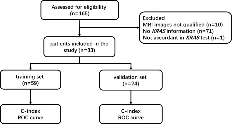
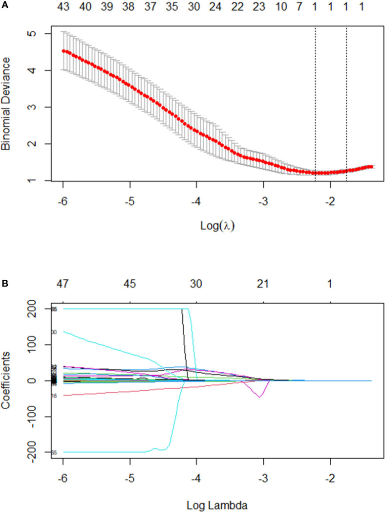
Eighty-three patients with status information and T2 MRI images between 2012.05 and 2019.09 were included. Least absolute shrinkage and selection operator (LASSO) regression was performed to assess the associations between features and gene status. The patients were divided 7:3 into training and validation sets. The C-index and the average area under the receiver operator characteristic curve (AUC) were used for performance evaluation.
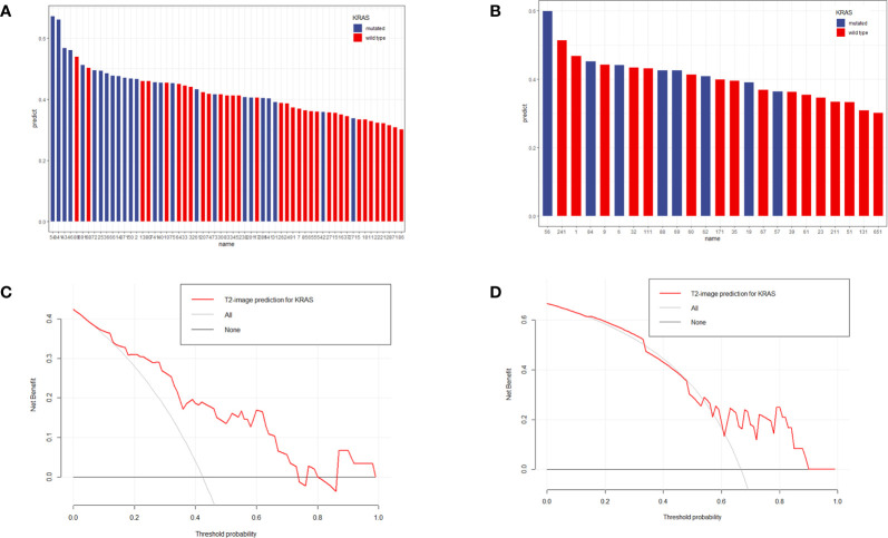
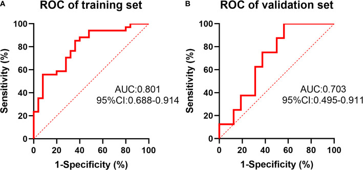
The clinical characteristics of 83 patients in the mutant and wild-type cohorts were balanced. Forty-two (50.6%) patients had mutations, and 41 (49.4%) patients had wild-type . A total of 253 radiomics features were extracted from the T2-MRI images of LARC patients. One radiomic feature named X.LL_scaled_std, a standard deviation value of scaled wavelet-transformed low-pass channel filter, was selected from 253 features (=0.019). The radiomics-based C-index values were 0.801 (95% CI: 0.772-0.830) and 0.703 (95% CI: 0.620-0.786) in the training and validation sets, respectively.
Radiomics features could differentiate status in LARC patients based on T2-MRI images. Further validation in a larger dataset is necessary in the future.
局部进展期直肠癌(LARC)是一种异质性疾病,关于其状态和影像特征的信息较少。本研究旨在分析LARC患者T2加权磁共振成像(MRI)影像组学特征与状态之间的关联。
纳入2012年5月至2019年9月间有状态信息且有T2加权MRI图像的83例患者。采用最小绝对收缩和选择算子(LASSO)回归评估特征与基因状态之间的关联。将患者按7:3比例分为训练集和验证集。采用C指数和受试者操作特征曲线(AUC)下的平均面积进行性能评估。
突变型和野生型队列中83例患者的临床特征均衡。42例(50.6%)患者有突变,41例(49.4%)患者为野生型。从LARC患者的T2加权MRI图像中总共提取了253个影像组学特征。从253个特征中(=0.019)选择了一个名为X.LL_scaled_std的影像组学特征,即小波变换低通通道滤波器的缩放标准差。基于影像组学的训练集和验证集C指数值分别为0.801(95%CI:0.772 - 0.830)和0.703(95%CI:0.620 - 0.786)。
影像组学特征可基于T2加权MRI图像区分LARC患者的状态。未来有必要在更大的数据集中进行进一步验证。