Department of Electrical Engineering in National Taiwan University of Science and Technology, Taipei, 106, Taiwan.
Sci Rep. 2021 May 27;11(1):11174. doi: 10.1038/s41598-021-90599-4.
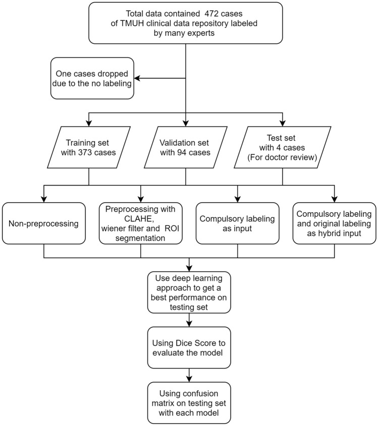
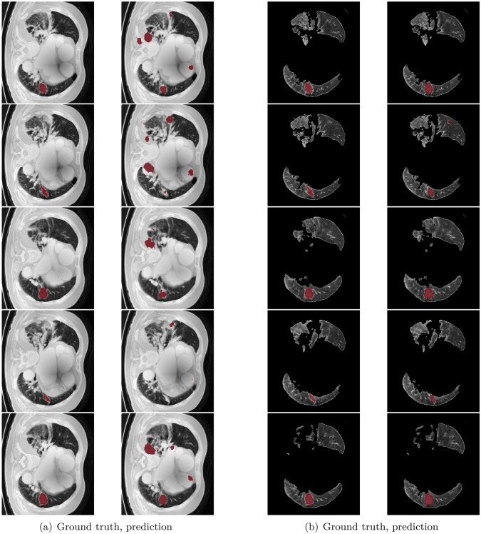
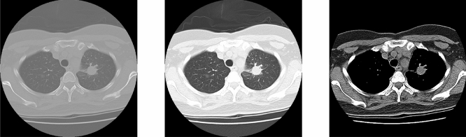
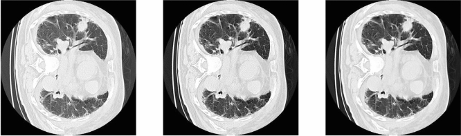
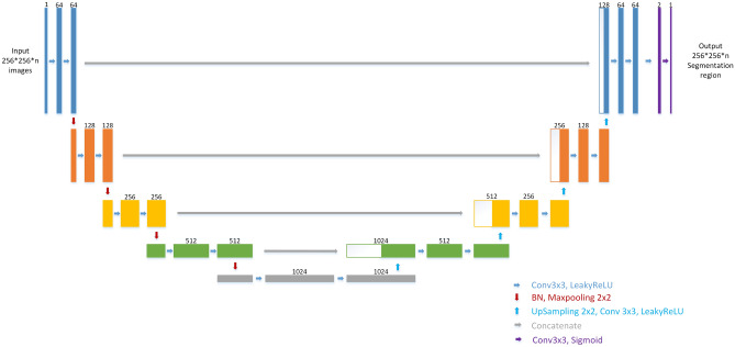
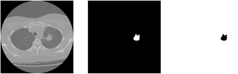
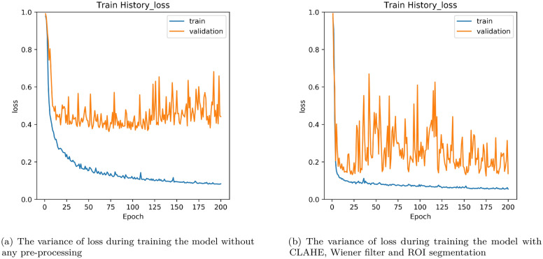
In this study, a novel method with the U-Net-based network architecture, 2D U-Net, is employed to segment the position of lung nodules, which are an early symptom of lung cancer and have a high probability of becoming a carcinoma, especially when a lung nodule is bigger than 15 [Formula: see text]. A serious problem of considering deep learning for all medical images is imbalanced labeling between foreground and background. The lung nodule is the foreground which accounts for a lower percentage in a whole image. The evaluation function adopted in this study is dice coefficient loss, which is usually used in image segmentation tasks. The proposed pre-processing method in this study is to use complementary labeling as the input in U-Net. With this method, the labeling is swapped. The no-nodule position is labeled. And the position of the nodule becomes non-labeled. The result shows that the proposal in this study is efficient in a small quantity of data. This method, complementary labeling could be used in a small data quantity scenario. With the use of ROI segmentation model in the data pre-processing, the results of lung nodule detection can be improved a lot as shown in the experiments.
在这项研究中,采用了一种基于 U-Net 网络架构的新型方法 2D U-Net,用于分割肺结节的位置,肺结节是肺癌的早期症状之一,并且很有可能发展成癌,特别是当肺结节大于 15 [Formula: see text] 时。考虑将深度学习应用于所有医学图像的一个严重问题是前景和背景之间的不平衡标记。肺结节是前景,在整个图像中占比较低。本研究采用的评价函数是骰子系数损失,它通常用于图像分割任务。本研究中提出的预处理方法是将互补标记作为 U-Net 的输入。通过这种方法,对标记进行了交换。对没有结节的位置进行标记,而结节的位置则是非标记的。结果表明,本研究中的方法在少量数据中是有效的。这种方法,互补标记可以用于少量数据的情况。通过在数据预处理中使用 ROI 分割模型,如实验所示,可以大大提高肺结节检测的结果。