Department of Biochemistry and Molecular Biology, Center for Membrane Biology, University of Texas Health Science Center at Houston, Houston, TX, United States; MD Anderson Cancer Center UTHealth Graduate School of Biomedical Sciences, University of Texas Health Science Center at Houston, Houston, TX, United States.
Department of Biochemistry and Molecular Biology, Center for Membrane Biology, University of Texas Health Science Center at Houston, Houston, TX, United States.
Methods Enzymol. 2021;652:193-212. doi: 10.1016/bs.mie.2021.02.005. Epub 2021 Mar 9.
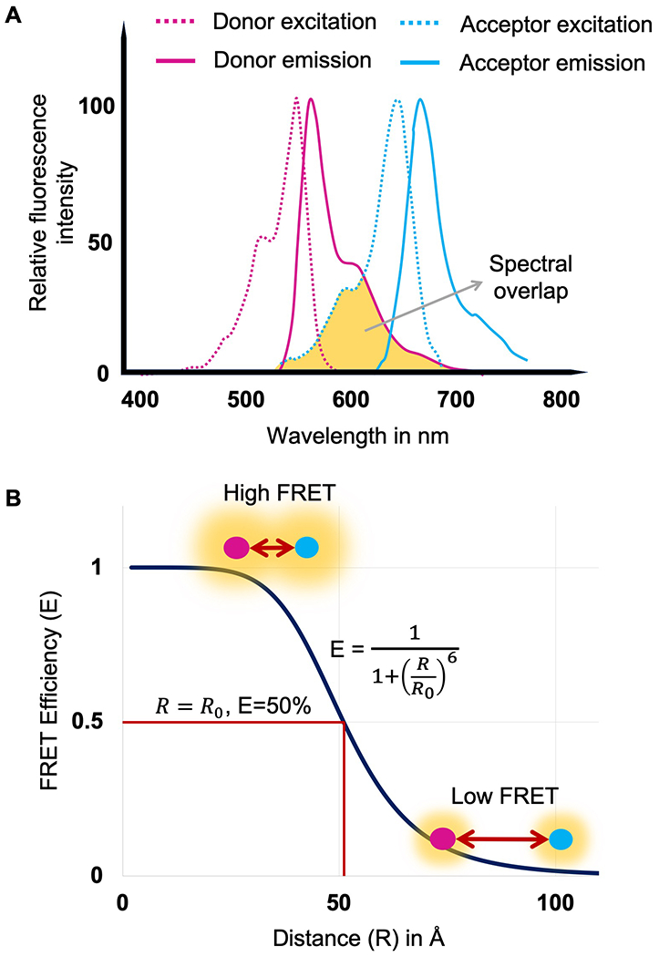
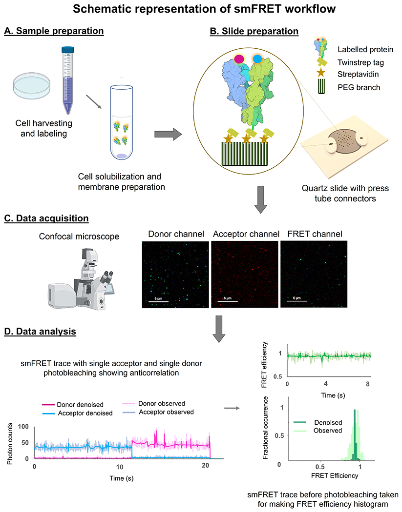
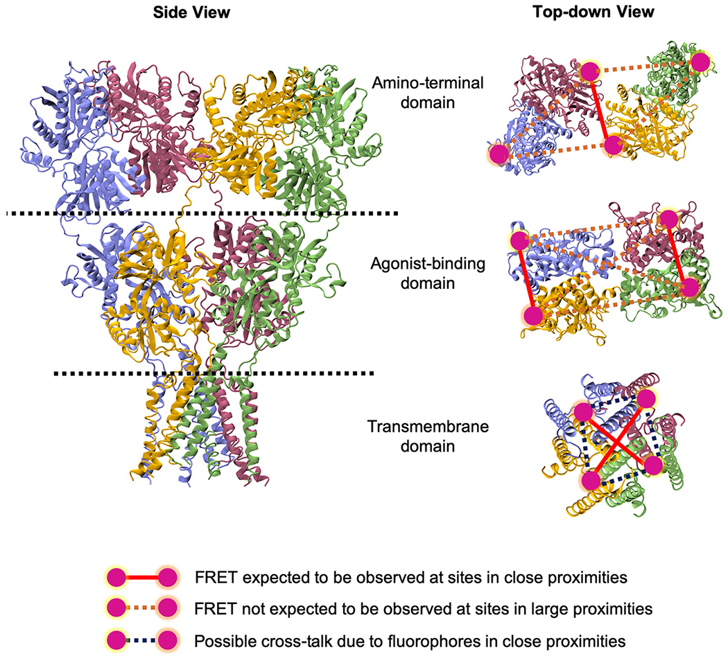
Single molecule Förster Resonance Energy Transfer (smFRET) allows us to measure variation in distances between donor and acceptor fluorophores attached to a protein, providing the conformational landscape of the protein with respect to this specific distance. smFRET can be performed on freely diffusing molecules or on tethered molecules. Here, we describe the tethered method used to study ionotropic glutamate receptors, which allows us to track the changes in FRET as a function of time, thus providing information on the conformations sampled and kinetics of conformational changes in the millisecond to second time scale. Strategies for attaching fluorophores to the proteins, methods for acquiring and analyzing the smFRET trajectories, and limitations are discussed.
单分子Förster 共振能量转移(smFRET)使我们能够测量与蛋白质结合的供体和受体荧光团之间距离的变化,从而提供蛋白质相对于该特定距离的构象景观。smFRET 可在自由扩散的分子或连接的分子上进行。在这里,我们描述了用于研究离子型谷氨酸受体的连接方法,该方法允许我们跟踪 FRET 的变化作为时间的函数,从而提供关于毫秒到秒时间尺度上构象采样和构象变化动力学的信息。讨论了将荧光团连接到蛋白质的策略、获取和分析 smFRET 轨迹的方法以及限制。