• 文献检索
  • 文档翻译
  • 深度研究
  • 学术资讯
  • Suppr Zotero 插件Zotero 插件
  • 邀请有礼
  • 套餐&价格
  • 历史记录
应用&插件
Suppr Zotero 插件Zotero 插件浏览器插件Mac 客户端Windows 客户端微信小程序
定价
高级版会员购买积分包购买API积分包
服务
文献检索文档翻译深度研究API 文档MCP 服务
关于我们
关于 Suppr公司介绍联系我们用户协议隐私条款
关注我们

Suppr 超能文献

核心技术专利:CN118964589B侵权必究
粤ICP备2023148730 号-1Suppr @ 2026

文献检索

告别复杂PubMed语法,用中文像聊天一样搜索,搜遍4000万医学文献。AI智能推荐,让科研检索更轻松。

立即免费搜索

文件翻译

保留排版,准确专业,支持PDF/Word/PPT等文件格式,支持 12+语言互译。

免费翻译文档

深度研究

AI帮你快速写综述,25分钟生成高质量综述,智能提取关键信息,辅助科研写作。

立即免费体验

使用卷积神经网络检测腰椎磁共振图像上的退行性改变:一项可行性研究。

Detection of Degenerative Changes on MR Images of the Lumbar Spine with a Convolutional Neural Network: A Feasibility Study.

作者信息

Lehnen Nils Christian, Haase Robert, Faber Jennifer, Rüber Theodor, Vatter Hartmut, Radbruch Alexander, Schmeel Frederic Carsten

机构信息

Department of Neuroradiology, University Hospital Bonn, Rheinische Friedrich-Wilhelms-Universität Bonn, 53127 Bonn, Germany.

Research Group Clinical Neuroimaging, German Center for Neurodegenerative Diseases, 53127 Bonn, Germany.

出版信息

Diagnostics (Basel). 2021 May 19;11(5):902. doi: 10.3390/diagnostics11050902.

DOI:10.3390/diagnostics11050902
PMID:34069362
原文链接:https://pmc.ncbi.nlm.nih.gov/articles/PMC8158737/
Abstract

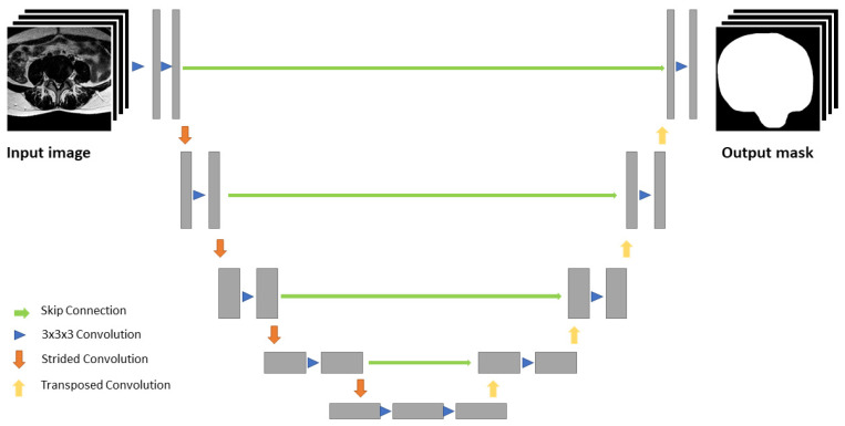
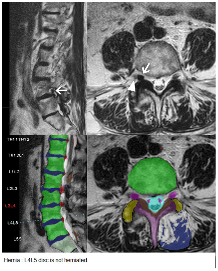
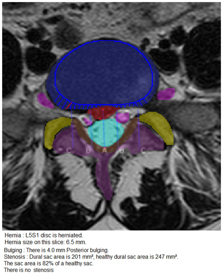
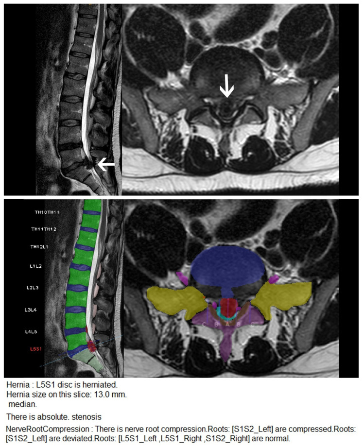
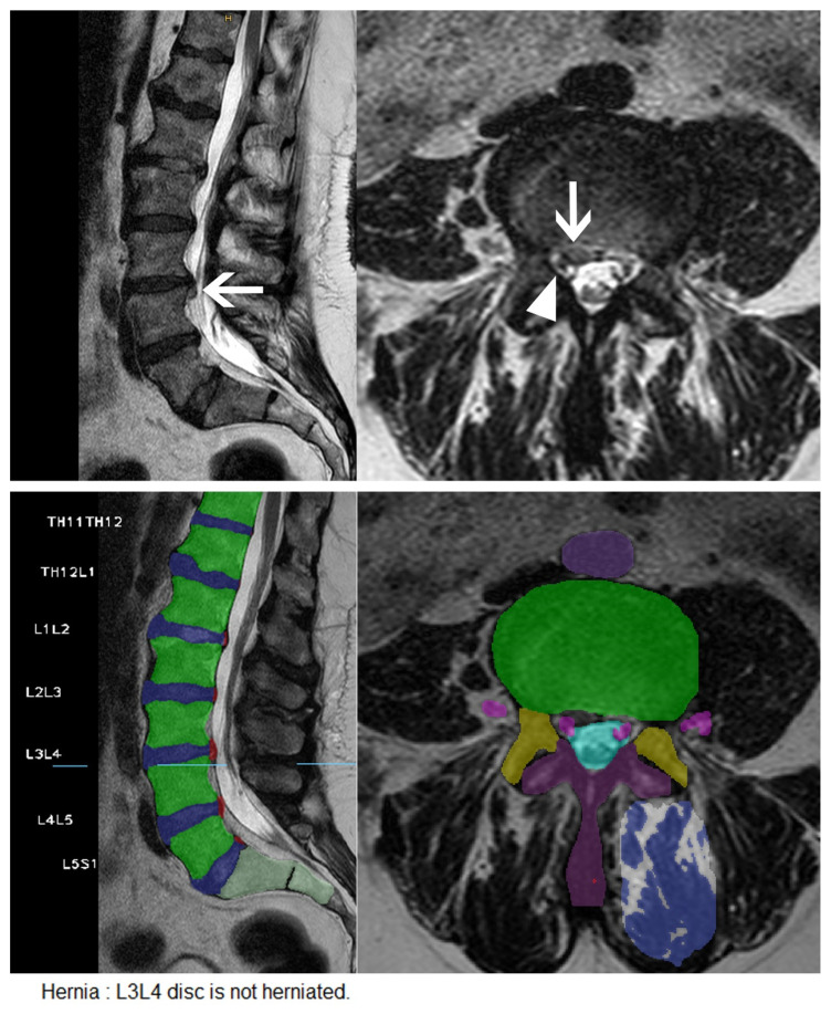
Our objective was to evaluate the diagnostic performance of a convolutional neural network (CNN) trained on multiple MR imaging features of the lumbar spine, to detect a variety of different degenerative changes of the lumbar spine. One hundred and forty-six consecutive patients underwent routine clinical MRI of the lumbar spine including T2-weighted imaging and were retrospectively analyzed using a CNN for detection and labeling of vertebrae, disc segments, as well as presence of disc herniation, disc bulging, spinal canal stenosis, nerve root compression, and spondylolisthesis. The assessment of a radiologist served as the diagnostic reference standard. We assessed the CNN's diagnostic accuracy and consistency using confusion matrices and McNemar's test. In our data, 77 disc herniations (thereof 46 further classified as extrusions), 133 disc bulgings, 35 spinal canal stenoses, 59 nerve root compressions, and 20 segments with spondylolisthesis were present in a total of 888 lumbar spine segments. The CNN yielded a perfect accuracy score for intervertebral disc detection and labeling (100%), and moderate to high diagnostic accuracy for the detection of disc herniations (87%; 95% CI: 0.84, 0.89), extrusions (86%; 95% CI: 0.84, 0.89), bulgings (76%; 95% CI: 0.73, 0.78), spinal canal stenoses (98%; 95% CI: 0.97, 0.99), nerve root compressions (91%; 95% CI: 0.89, 0.92), and spondylolisthesis (87.61%; 95% CI: 85.26, 89.21), respectively. Our data suggest that automatic diagnosis of multiple different degenerative changes of the lumbar spine is feasible using a single comprehensive CNN. The CNN provides high diagnostic accuracy for intervertebral disc labeling and detection of clinically relevant degenerative changes such as spinal canal stenosis and disc extrusion of the lumbar spine.

摘要

我们的目标是评估基于腰椎多种磁共振成像(MR)特征训练的卷积神经网络(CNN)检测腰椎各种不同退变改变的诊断性能。146例连续患者接受了包括T2加权成像在内的腰椎常规临床MRI检查,并使用CNN对椎体、椎间盘节段以及椎间盘突出、椎间盘膨出、椎管狭窄、神经根受压和椎体滑脱的存在情况进行回顾性分析和标注。放射科医生的评估作为诊断参考标准。我们使用混淆矩阵和McNemar检验评估了CNN的诊断准确性和一致性。在我们的数据中,888个腰椎节段中共有77例椎间盘突出(其中46例进一步分类为脱出型)、133例椎间盘膨出、35例椎管狭窄、59例神经根受压和20例椎体滑脱节段。CNN在椎间盘检测和标注方面获得了完美的准确率(100%),在检测椎间盘突出(87%;95%置信区间:0.84,0.89)、脱出型(86%;95%置信区间:0.84,0.89)、膨出(76%;95%置信区间:0.73,0.78)、椎管狭窄(98%;95%置信区间:0.97,0.99)、神经根受压(91%;95%置信区间:0.89,0.92)和椎体滑脱(87.61%;95%置信区间:85.26,89.21)方面具有中度到高度的诊断准确性。我们的数据表明,使用单一综合的CNN对腰椎多种不同退变改变进行自动诊断是可行的。该CNN在椎间盘标注以及检测腰椎临床相关退变改变如椎管狭窄和椎间盘脱出方面具有较高的诊断准确性。

https://cdn.ncbi.nlm.nih.gov/pmc/blobs/389a/8158737/587c96d2413c/diagnostics-11-00902-g005.jpg
https://cdn.ncbi.nlm.nih.gov/pmc/blobs/389a/8158737/67a0ade10ce9/diagnostics-11-00902-g001.jpg
https://cdn.ncbi.nlm.nih.gov/pmc/blobs/389a/8158737/4d5983b9aaf8/diagnostics-11-00902-g002.jpg
https://cdn.ncbi.nlm.nih.gov/pmc/blobs/389a/8158737/bf65c1e7b108/diagnostics-11-00902-g003.jpg
https://cdn.ncbi.nlm.nih.gov/pmc/blobs/389a/8158737/8426ee47981f/diagnostics-11-00902-g004.jpg
https://cdn.ncbi.nlm.nih.gov/pmc/blobs/389a/8158737/587c96d2413c/diagnostics-11-00902-g005.jpg

相似文献

1
Detection of Degenerative Changes on MR Images of the Lumbar Spine with a Convolutional Neural Network: A Feasibility Study.使用卷积神经网络检测腰椎磁共振图像上的退行性改变:一项可行性研究。
Diagnostics (Basel). 2021 May 19;11(5):902. doi: 10.3390/diagnostics11050902.
2
ISSLS PRIZE IN BIOENGINEERING SCIENCE 2017: Automation of reading of radiological features from magnetic resonance images (MRIs) of the lumbar spine without human intervention is comparable with an expert radiologist.2017年国际腰椎研究学会生物工程科学奖:无需人工干预,自动读取腰椎磁共振成像(MRI)的放射学特征,其结果可与放射学专家相媲美。
Eur Spine J. 2017 May;26(5):1374-1383. doi: 10.1007/s00586-017-4956-3. Epub 2017 Feb 6.
3
Deep Learning Model for Automated Detection and Classification of Central Canal, Lateral Recess, and Neural Foraminal Stenosis at Lumbar Spine MRI.深度学习模型在腰椎 MRI 中用于自动检测和分类中央管、侧隐窝和神经孔狭窄
Radiology. 2021 Jul;300(1):130-138. doi: 10.1148/radiol.2021204289. Epub 2021 May 11.
4
Incidental durotomy during lumbar spine surgery: risk factors and anatomic locations: clinical article.腰椎手术中偶然的硬脊膜切开术:危险因素和解剖部位:临床文章。
J Neurosurg Spine. 2013 Feb;18(2):165-9. doi: 10.3171/2012.10.SPINE12271. Epub 2012 Nov 30.
5
Feasibility of Deep Learning Algorithms for Reporting in Routine Spine Magnetic Resonance Imaging.深度学习算法用于常规脊柱磁共振成像报告的可行性
Int J Spine Surg. 2020 Dec;14(s3):S86-S97. doi: 10.14444/7131.
6
Artificial Intelligence Comparison of the Radiologist Report With Endoscopic Predictors of Successful Transforaminal Decompression for Painful Conditions of the Lumber Spine: Application of Deep Learning Algorithm Interpretation of Routine Lumbar Magnetic Resonance Imaging Scan.人工智能将放射科医生报告与腰椎疼痛性疾病经椎间孔减压成功的内镜预测指标进行比较:深度学习算法在常规腰椎磁共振成像扫描解读中的应用
Int J Spine Surg. 2020 Dec;14(s3):S75-S85. doi: 10.14444/7130. Epub 2020 Nov 18.
7
Deep learning-based detection of lumbar spinal canal stenosis using convolutional neural networks.基于卷积神经网络的腰椎椎管狭窄症深度学习检测。
Spine J. 2024 Nov;24(11):2086-2101. doi: 10.1016/j.spinee.2024.06.009. Epub 2024 Jun 22.
8
[Adjacent segment degeneration after lumbosacral fusion in spondylolisthesis: a retrospective radiological and clinical analysis].腰椎滑脱症腰骶融合术后相邻节段退变:一项回顾性影像学及临床分析
Acta Chir Orthop Traumatol Cech. 2010 Apr;77(2):124-30.
9
What types of degenerative lumbar pathologies respond to nerve root injection? A retrospective review of six hundred and forty one cases.哪些类型的退行性腰椎疾病对神经根注射有反应?对641例病例的回顾性研究。
Int Orthop. 2015 Jul;39(7):1379-82. doi: 10.1007/s00264-015-2761-3. Epub 2015 Apr 16.
10
Reliability Analysis of Deep Learning Algorithms for Reporting of Routine Lumbar MRI Scans.用于常规腰椎磁共振成像扫描报告的深度学习算法的可靠性分析
Int J Spine Surg. 2020 Dec;14(s3):S98-S107. doi: 10.14444/7132. Epub 2020 Oct 29.

引用本文的文献

1
Quantification and classification of lumbar disc herniation on axial magnetic resonance images using deep learning models.使用深度学习模型对腰椎间盘突出症进行轴向磁共振图像的定量和分类
Radiol Med. 2025 Mar 24. doi: 10.1007/s11547-025-01996-y.
2
MRI to digital medicine diagnosis: integrating deep learning into clinical decision-making for lumbar degenerative diseases.从磁共振成像到数字医学诊断:将深度学习整合到腰椎退行性疾病的临床决策中
Front Surg. 2025 Jan 6;11:1424716. doi: 10.3389/fsurg.2024.1424716. eCollection 2024.
3
Machine Learning and Deep Learning for Diagnosis of Lumbar Spinal Stenosis: Systematic Review and Meta-Analysis.

本文引用的文献

1
Improving Breast Cancer Detection Accuracy of Mammography with the Concurrent Use of an Artificial Intelligence Tool.同时使用人工智能工具提高乳腺钼靶检查的乳腺癌检测准确性。
Radiol Artif Intell. 2020 Nov 4;2(6):e190208. doi: 10.1148/ryai.2020190208. eCollection 2020 Nov.
2
Labeling Vertebrae with Two-dimensional Reformations of Multidetector CT Images: An Adversarial Approach for Incorporating Prior Knowledge of Spine Anatomy.利用多排螺旋CT图像的二维重建标记椎骨:一种融入脊柱解剖先验知识的对抗方法
Radiol Artif Intell. 2020 Mar 25;2(2):e190074. doi: 10.1148/ryai.2020190074. eCollection 2020 Mar.
3
Quantitative Analysis of Neural Foramina in the Lumbar Spine: An Imaging Informatics and Machine Learning Study.
用于诊断腰椎管狭窄症的机器学习与深度学习:系统评价与荟萃分析
J Med Internet Res. 2024 Dec 23;26:e54676. doi: 10.2196/54676.
4
Lumbar Spinal Stenosis Grading in Multiple Level Magnetic Resonance Imaging Using Deep Convolutional Neural Networks.使用深度卷积神经网络对多节段磁共振成像中的腰椎管狭窄进行分级
Global Spine J. 2025 May;15(4):2309-2317. doi: 10.1177/21925682241299332. Epub 2024 Nov 1.
5
A machine learning based quantification system for automated diagnosis of lumbar spondylolisthesis on spinal X-rays.一种基于机器学习的腰椎滑脱症脊柱X光自动诊断量化系统。
Heliyon. 2024 Sep 4;10(17):e37418. doi: 10.1016/j.heliyon.2024.e37418. eCollection 2024 Sep 15.
6
Artificial Intelligence in Spinal Imaging and Patient Care: A Review of Recent Advances.脊柱成像与患者护理中的人工智能:近期进展综述
Neurospine. 2024 Jun;21(2):474-486. doi: 10.14245/ns.2448388.194. Epub 2024 Jun 30.
7
Artificial intelligence: a new cutting-edge tool in spine surgery.人工智能:脊柱外科领域的一种新型前沿工具。
Asian Spine J. 2024 Jun;18(3):458-471. doi: 10.31616/asj.2023.0382. Epub 2024 Jun 25.
8
Artificial intelligence in musculoskeletal imaging: realistic clinical applications in the next decade.人工智能在肌肉骨骼成像中的应用:未来十年的现实临床应用。
Skeletal Radiol. 2024 Sep;53(9):1849-1868. doi: 10.1007/s00256-024-04684-6. Epub 2024 Jun 20.
9
How AI May Transform Musculoskeletal Imaging.人工智能如何改变肌肉骨骼成像。
Radiology. 2024 Jan;310(1):e230764. doi: 10.1148/radiol.230764.
10
A neural network model for detection and classification of lumbar spinal stenosis on MRI.一种基于 MRI 的腰椎管狭窄症检测和分类的神经网络模型。
Eur Spine J. 2024 Mar;33(3):941-948. doi: 10.1007/s00586-023-08089-2. Epub 2023 Dec 27.
腰椎神经孔的定量分析:一项影像信息学与机器学习研究
Radiol Artif Intell. 2019 Mar 6;1(2):180037. doi: 10.1148/ryai.2019180037. eCollection 2019 Mar.
4
Systematic Review and Meta-Analysis of Vesical Imaging-Reporting and Data System (VI-RADS) Inter-Observer Reliability: An Added Value for Muscle Invasive Bladder Cancer Detection.膀胱影像报告和数据系统(VI-RADS)观察者间可靠性的系统评价和Meta分析:对肌层浸润性膀胱癌检测的附加价值
Cancers (Basel). 2020 Oct 15;12(10):2994. doi: 10.3390/cancers12102994.
5
Global burden of 369 diseases and injuries in 204 countries and territories, 1990-2019: a systematic analysis for the Global Burden of Disease Study 2019.204 个国家和地区 1990-2019 年 369 种疾病和伤害导致的全球负担:2019 年全球疾病负担研究的系统分析。
Lancet. 2020 Oct 17;396(10258):1204-1222. doi: 10.1016/S0140-6736(20)30925-9.
6
Preoperative detection of Vesical Imaging-Reporting and Data System (VI-RADS) score 5 reliably identifies extravesical extension of urothelial carcinoma of the urinary bladder and predicts significant delayed time to cystectomy: time to reconsider the need for primary deep transurethral resection of bladder tumour in cases of locally advanced disease?术前膀胱成像报告和数据系统(VI-RADS)评分 5 可可靠识别膀胱癌的膀胱外延伸,并预测显著的膀胱切除术延迟时间:是否需要重新考虑局部进展性疾病患者行原发性经尿道膀胱肿瘤深度切除术?
BJU Int. 2020 Nov;126(5):610-619. doi: 10.1111/bju.15188. Epub 2020 Aug 17.
7
Classification in Brief: The Meyerding Classification System of Spondylolisthesis.简要分类:腰椎滑脱的迈耶丁分类系统
Clin Orthop Relat Res. 2020 May;478(5):1125-1130. doi: 10.1097/CORR.0000000000001153.
8
Spinal Stenosis Grading in Magnetic Resonance Imaging Using Deep Convolutional Neural Networks.基于深度卷积神经网络的磁共振成像中脊柱狭窄分级。
Spine (Phila Pa 1976). 2020 Jun 15;45(12):804-812. doi: 10.1097/BRS.0000000000003377.
9
Prospective Assessment of Vesical Imaging Reporting and Data System (VI-RADS) and Its Clinical Impact on the Management of High-risk Non-muscle-invasive Bladder Cancer Patients Candidate for Repeated Transurethral Resection.前瞻性评估膀胱影像报告和数据系统(VI-RADS)及其对高危非肌肉浸润性膀胱癌患者重复经尿道切除术候选者管理的临床影响。
Eur Urol. 2020 Jan;77(1):101-109. doi: 10.1016/j.eururo.2019.09.029. Epub 2019 Nov 5.
10
Fully automated radiological analysis of spinal disorders and deformities: a deep learning approach.脊柱疾病和畸形的全自动放射学分析:深度学习方法。
Eur Spine J. 2019 May;28(5):951-960. doi: 10.1007/s00586-019-05944-z. Epub 2019 Mar 12.