Takatsuna Masafumi, Azumi Rie, Mizusawa Takeshi, Sato Hiroki, Mizuno Ken-Ichi, Kato Takashi, Yokoyama Junji, Ajioka Yoichi, Terai Shuji
Division of Gastroenterology and Hepatology, Graduate School of Medical and Dental Sciences, Niigata University, Niigata, Japan.
Division of Molecular and Diagnostic Pathology, Graduate School of Medical and Dental Sciences, Niigata University, Niigata, Japan.
Endosc Int Open. 2021 Jun;9(6):E863-E866. doi: 10.1055/a-1396-3854. Epub 2021 May 27.
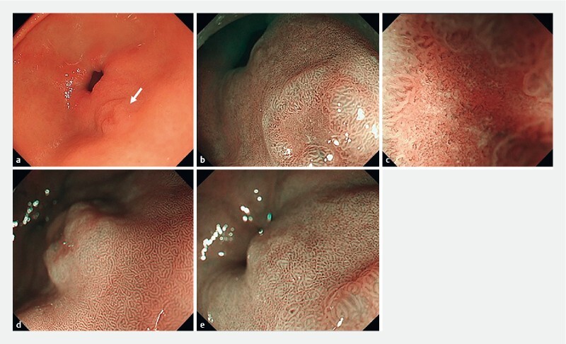
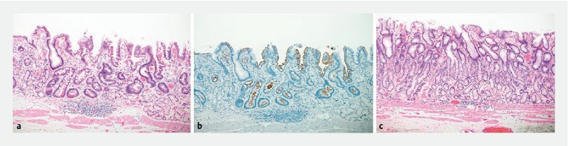
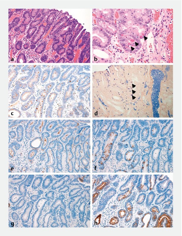
A 40-year-old man with slightly depressed (0-IIc) type gastric cancer of the pyloric anterior gastric area underwent pre-operative screening for tetralogy of Fallot and endoscopic submucosal dissection (ESD) and was tested for antigens and antibodies. Both tests were negative. He did not have a history of eradication. Pathological diagnosis of ESD showed a well-differentiated adenocarcinoma. The tumor was CD10-positive, MUC5AC-negative, and MUC6-confocal positive; it showed differentiation with gastrointestinal phenotype. Moreover, the tumor cells were lysozyme-positive, resembling Paneth cells. Mucosal glands exhibited intestinal metaplasia on the anal side of the tumor lesion. On the oral side of the tumor, metaplasia was non-existent, with normal pyloric glands present in the mucosal layer. The patient was not infected with ; however, intestinal metaplasia existed around the early gastric cancer. This suggested that the intestinal metaplasia occurred due to bile reflux, and the gastric neoplasia arose with the metaplasia without an infection. This case may potentially help explain gastric cancer development in the absence of infection.
一名40岁男性,患有幽门前胃区轻度凹陷型(0-IIc型)胃癌,接受了法洛四联症的术前筛查和内镜黏膜下剥离术(ESD),并进行了抗原和抗体检测。两项检测均为阴性。他没有根除治疗史。ESD的病理诊断显示为高分化腺癌。肿瘤CD10阳性,MUC5AC阴性,MUC6共聚焦阳性;表现出胃肠道表型分化。此外,肿瘤细胞溶菌酶阳性,类似潘氏细胞。肿瘤病变肛门侧的黏膜腺体呈现肠化生。在肿瘤的口腔侧,不存在化生,黏膜层有正常的幽门腺。该患者未感染 ;然而,早期胃癌周围存在肠化生。这表明肠化生是由胆汁反流引起的,胃肿瘤是在化生的基础上发生的,没有 感染。该病例可能有助于解释在没有 感染的情况下胃癌的发生发展。