Department of Cellular and Molecular Biophysics, Max Planck Institute of Biochemistry, Martinsried, Germany.
Graduate School for Quantitative Biosciences (QBM), Ludwig-Maximillians-University, Munich, Germany.
Nat Commun. 2021 Jun 3;12(1):3310. doi: 10.1038/s41467-021-23387-3.
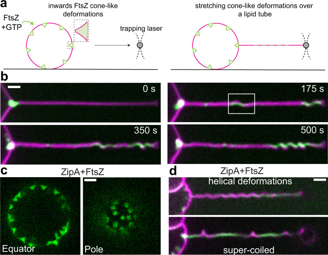
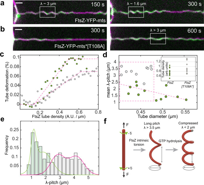
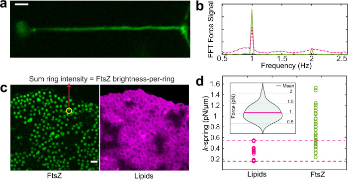
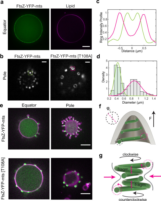
FtsZ is a key component in bacterial cell division, being the primary protein of the presumably contractile Z ring. In vivo and in vitro, it shows two distinctive features that could so far, however, not be mechanistically linked: self-organization into directionally treadmilling vortices on solid supported membranes, and shape deformation of flexible liposomes. In cells, circumferential treadmilling of FtsZ was shown to recruit septum-building enzymes, but an active force production remains elusive. To gain mechanistic understanding of FtsZ dependent membrane deformations and constriction, we design an in vitro assay based on soft lipid tubes pulled from FtsZ decorated giant lipid vesicles (GUVs) by optical tweezers. FtsZ filaments actively transform these tubes into spring-like structures, where GTPase activity promotes spring compression. Operating the optical tweezers in lateral vibration mode and assigning spring constants to FtsZ coated tubes, the directional forces that FtsZ-YFP-mts rings exert upon GTP hydrolysis can be estimated to be in the pN range. They are sufficient to induce membrane budding with constricting necks on both, giant vesicles and E.coli cells devoid of their cell walls. We hypothesize that these forces result from torsional stress in a GTPase activity dependent manner.
FtsZ 是细菌细胞分裂的关键组成部分,是假定可收缩 Z 环的主要蛋白质。在体内和体外,它表现出两个独特的特征,但迄今为止,这两个特征还不能在机制上联系起来:在固体支撑膜上自我组织成定向的履带式涡旋,以及柔性脂质体的形状变形。在细胞中,FtsZ 的圆周履带式运动被证明可以招募隔膜构建酶,但产生主动力的机制仍不清楚。为了深入了解 FtsZ 依赖性膜变形和收缩的机制,我们设计了一种基于软脂质管的体外测定方法,该方法通过光学镊子从 FtsZ 修饰的巨大脂质体 (GUV) 中拉出脂质管。FtsZ 丝体将这些管主动转化为弹簧状结构,其中 GTPase 活性促进弹簧压缩。在横向振动模式下操作光学镊子,并为 FtsZ 涂层管分配弹簧常数,可以估计 FtsZ-YFP-mts 环在 GTP 水解时对管施加的定向力在皮牛范围内。这些力足以诱导没有细胞壁的巨大囊泡和 E. coli 细胞的膜出芽,并形成收缩的颈部。我们假设这些力是由 GTPase 活性依赖性的扭转应力产生的。