Salivary Gland Disease Center and Beijing Key Laboratory of Tooth Regeneration and Function Reconstruction, Capital Medical University School of Stomatology, Beijing, China.
Department of Oral and Maxillofacial and Head and Neck Oncology, Capital Medical University School of Stomatology, Beijing, China.
Cell Prolif. 2021 Jul;54(7):e13078. doi: 10.1111/cpr.13078. Epub 2021 Jun 7.
Salivary gland regeneration is closely related to the parasympathetic nerve; however, the mechanism behind this relationship is still unclear. The aim of this study was to evaluate the relationship between the parasympathetic nerve and morphological differences during salivary gland regeneration.
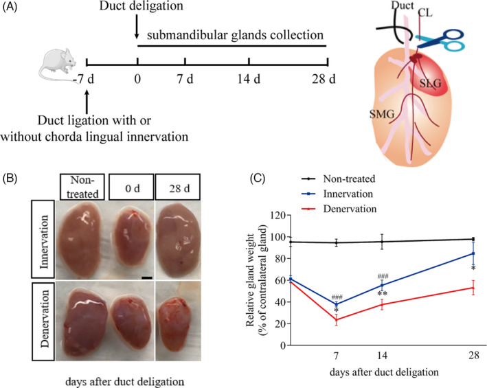
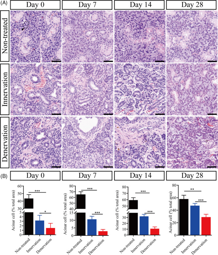
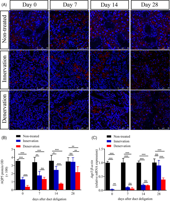
We used a duct ligation/deligation-induced submandibular gland regeneration model of Sprague-Dawley (SD) rats. The regenerated submandibular gland with or without chorda lingual (CL) innervation was detected by haematoxylin-eosin staining, real-time PCR (RT-PCR), immunohistochemistry and Western blotting. We counted the number of Ki67-positive cells to reveal the proliferation process that occurs during gland regeneration. Finally, we examined the expression of the following markers: aquaporin 5, cytokeratin 7, neural cell adhesion molecule (NCAM) and polysialyltransferases.
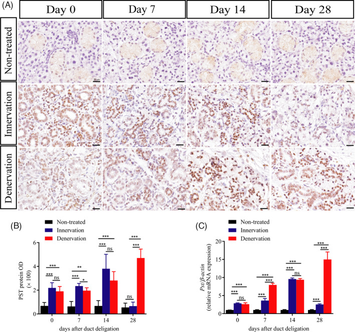
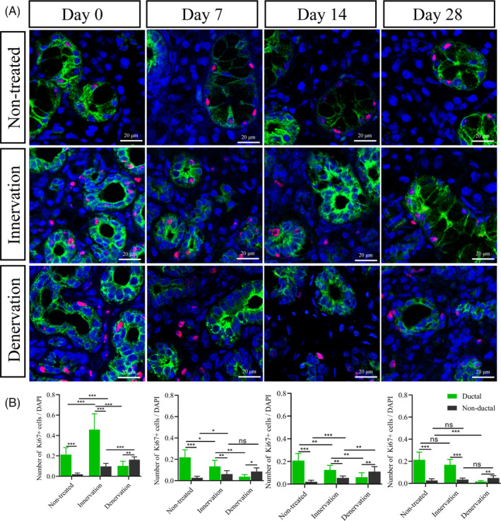
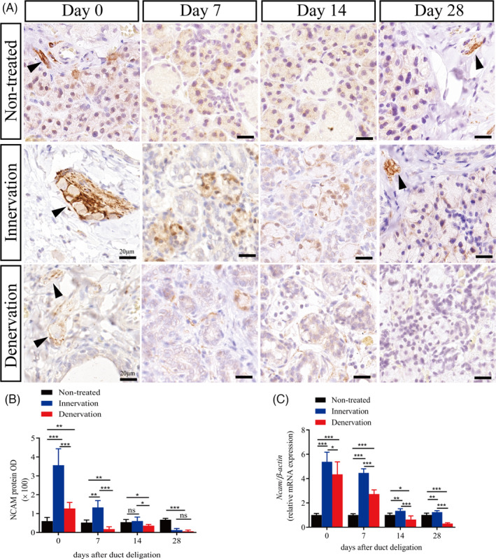
Intact parasympathetic innervation promoted submandibular gland regeneration. The process of gland regeneration was significantly repressed by cutting off the CL nerve. During gland regeneration, Ki67-positive cells were mainly found in the ductal structures. Moreover, the expression of NCAM and polysialyltransferases-1 (PST) expression in the innervation group was significantly increased during early regeneration and decreased in the late stages. In the denervated submandibular glands, the expression of NCAM decreased during regeneration.
Our findings revealed that the regeneration of submandibular glands with intact parasympathetic innervation was associated with duct cell proliferation and the increased expression of PST and NCAM.
唾液腺再生与副交感神经密切相关,但两者之间的关系机制尚不清楚。本研究旨在评估副交感神经与唾液腺再生过程中形态差异之间的关系。
我们使用了 Sprague-Dawley(SD)大鼠的导管结扎/松解诱导的下颌下腺再生模型。通过苏木精-伊红染色、实时 PCR(RT-PCR)、免疫组织化学和 Western blot 检测具有或不具有舌下神经(CL)支配的再生下颌下腺。我们计数 Ki67 阳性细胞的数量,以揭示在腺体再生过程中发生的增殖过程。最后,我们检查了以下标记物的表达:水通道蛋白 5、细胞角蛋白 7、神经细胞黏附分子(NCAM)和多涎酸转移酶。
完整的副交感神经支配促进了下颌下腺的再生。切断 CL 神经会显著抑制腺体的再生过程。在腺体再生过程中,Ki67 阳性细胞主要存在于导管结构中。此外,在神经支配组中,NCAM 和多涎酸转移酶-1(PST)的表达在早期再生时显著增加,在后期则减少。在去神经支配的下颌下腺中,NCAM 的表达在再生过程中减少。
我们的研究结果表明,具有完整副交感神经支配的下颌下腺再生与导管细胞增殖以及 PST 和 NCAM 的表达增加有关。