• 文献检索
  • 文档翻译
  • 深度研究
  • 学术资讯
  • Suppr Zotero 插件Zotero 插件
  • 邀请有礼
  • 套餐&价格
  • 历史记录
应用&插件
Suppr Zotero 插件Zotero 插件浏览器插件Mac 客户端Windows 客户端微信小程序
定价
高级版会员购买积分包购买API积分包
服务
文献检索文档翻译深度研究API 文档MCP 服务
关于我们
关于 Suppr公司介绍联系我们用户协议隐私条款
关注我们

Suppr 超能文献

核心技术专利:CN118964589B侵权必究
粤ICP备2023148730 号-1Suppr @ 2026

文献检索

告别复杂PubMed语法,用中文像聊天一样搜索,搜遍4000万医学文献。AI智能推荐,让科研检索更轻松。

立即免费搜索

文件翻译

保留排版,准确专业,支持PDF/Word/PPT等文件格式,支持 12+语言互译。

免费翻译文档

深度研究

AI帮你快速写综述,25分钟生成高质量综述,智能提取关键信息,辅助科研写作。

立即免费体验

对肺部和心脏区域的特征进行加权,用于胸科疾病分类。

Weighing features of lung and heart regions for thoracic disease classification.

机构信息

School of Computer Science and Technology, Harbin Institute of Technology, Harbin, China.

Department of Computer Science and Engineering, Southern University of Science and Technology, Shenzhen, China.

出版信息

BMC Med Imaging. 2021 Jun 10;21(1):99. doi: 10.1186/s12880-021-00627-y.

DOI:10.1186/s12880-021-00627-y
PMID:34112095
原文链接:https://pmc.ncbi.nlm.nih.gov/articles/PMC8194196/
Abstract

BACKGROUND

Chest X-rays are the most commonly available and affordable radiological examination for screening thoracic diseases. According to the domain knowledge of screening chest X-rays, the pathological information usually lay on the lung and heart regions. However, it is costly to acquire region-level annotation in practice, and model training mainly relies on image-level class labels in a weakly supervised manner, which is highly challenging for computer-aided chest X-ray screening. To address this issue, some methods have been proposed recently to identify local regions containing pathological information, which is vital for thoracic disease classification. Inspired by this, we propose a novel deep learning framework to explore discriminative information from lung and heart regions.

RESULT

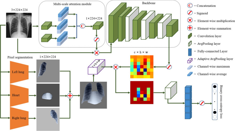
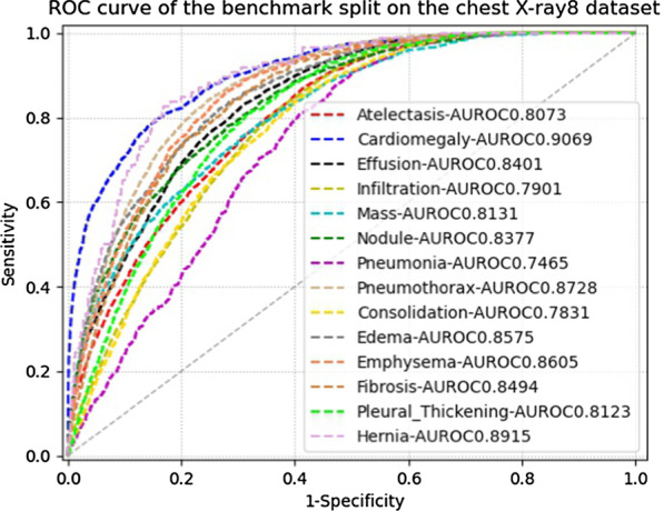
We design a feature extractor equipped with a multi-scale attention module to learn global attention maps from global images. To exploit disease-specific cues effectively, we locate lung and heart regions containing pathological information by a well-trained pixel-wise segmentation model to generate binarization masks. By introducing element-wise logical AND operator on the learned global attention maps and the binarization masks, we obtain local attention maps in which pixels are are 1 for lung and heart region and 0 for other regions. By zeroing features of non-lung and heart regions in attention maps, we can effectively exploit their disease-specific cues in lung and heart regions. Compared to existing methods fusing global and local features, we adopt feature weighting to avoid weakening visual cues unique to lung and heart regions. Our method with pixel-wise segmentation can help overcome the deviation of locating local regions. Evaluated by the benchmark split on the publicly available chest X-ray14 dataset, the comprehensive experiments show that our method achieves superior performance compared to the state-of-the-art methods.

CONCLUSION

We propose a novel deep framework for the multi-label classification of thoracic diseases in chest X-ray images. The proposed network aims to effectively exploit pathological regions containing the main cues for chest X-ray screening. Our proposed network has been used in clinic screening to assist the radiologists. Chest X-ray accounts for a significant proportion of radiological examinations. It is valuable to explore more methods for improving performance.

摘要

背景

胸部 X 光检查是最常用和最经济的胸部疾病筛查放射学检查。根据筛查胸部 X 光的领域知识,病理信息通常位于肺部和心脏区域。然而,在实践中获取区域级别的注释是昂贵的,并且模型训练主要依赖于图像级别的弱监督类标签,这对计算机辅助胸部 X 光筛查具有很大的挑战性。为了解决这个问题,最近提出了一些方法来识别包含病理信息的局部区域,这对胸部疾病分类至关重要。受此启发,我们提出了一种新的深度学习框架,从肺部和心脏区域中探索有区别的信息。

结果

我们设计了一个特征提取器,配备了一个多尺度注意力模块,从全局图像中学习全局注意力图。为了有效地利用疾病特异性线索,我们通过训练有素的像素级分割模型定位包含病理信息的肺部和心脏区域,生成二值化掩模。通过在学习到的全局注意力图和二值化掩模上引入元素级逻辑与操作,我们得到了局部注意力图,其中肺部和心脏区域的像素值为 1,其他区域的像素值为 0。通过在注意力图中对非肺部和心脏区域的特征进行置零,可以有效地利用它们在肺部和心脏区域中的疾病特异性线索。与融合全局和局部特征的现有方法相比,我们采用特征加权来避免削弱肺部和心脏区域特有的视觉线索。我们的方法使用像素级分割,可以帮助克服定位局部区域的偏差。在公开的胸部 X 射线 14 数据集的基准分割上进行评估,综合实验表明,与最先进的方法相比,我们的方法表现出优越的性能。

结论

我们提出了一种新的深度学习框架,用于胸部 X 射线图像中的多标签胸部疾病分类。所提出的网络旨在有效地利用包含胸部 X 光筛查主要线索的病理区域。我们提出的网络已用于临床筛查,以协助放射科医生。胸部 X 光检查在放射学检查中占有很大比例。探索更多提高性能的方法是有价值的。

https://cdn.ncbi.nlm.nih.gov/pmc/blobs/6453/8194196/b594919c29e4/12880_2021_627_Fig4_HTML.jpg
https://cdn.ncbi.nlm.nih.gov/pmc/blobs/6453/8194196/c9acc93b124c/12880_2021_627_Fig1_HTML.jpg
https://cdn.ncbi.nlm.nih.gov/pmc/blobs/6453/8194196/a29bc581a0c0/12880_2021_627_Fig2_HTML.jpg
https://cdn.ncbi.nlm.nih.gov/pmc/blobs/6453/8194196/ffd9f9c3fa01/12880_2021_627_Fig3_HTML.jpg
https://cdn.ncbi.nlm.nih.gov/pmc/blobs/6453/8194196/b594919c29e4/12880_2021_627_Fig4_HTML.jpg

相似文献

1
Weighing features of lung and heart regions for thoracic disease classification.对肺部和心脏区域的特征进行加权,用于胸科疾病分类。
BMC Med Imaging. 2021 Jun 10;21(1):99. doi: 10.1186/s12880-021-00627-y.
2
Triple attention learning for classification of 14 thoracic diseases using chest radiography.基于胸部 X 光的 14 种胸部疾病分类的三重注意学习。
Med Image Anal. 2021 Jan;67:101846. doi: 10.1016/j.media.2020.101846. Epub 2020 Oct 16.
3
SDFN: Segmentation-based deep fusion network for thoracic disease classification in chest X-ray images.基于分割的深度融合网络在胸部 X 射线图像中的胸部疾病分类。
Comput Med Imaging Graph. 2019 Jul;75:66-73. doi: 10.1016/j.compmedimag.2019.05.005. Epub 2019 May 28.
4
Lesion Location Attention Guided Network for Multi-Label Thoracic Disease Classification in Chest X-Rays.基于病灶位置注意引导网络的胸部 X 射线多病种分类方法。
IEEE J Biomed Health Inform. 2020 Jul;24(7):2016-2027. doi: 10.1109/JBHI.2019.2952597. Epub 2019 Nov 11.
5
Contralaterally Enhanced Networks for Thoracic Disease Detection.用于胸部疾病检测的对侧增强网络。
IEEE Trans Med Imaging. 2021 Sep;40(9):2428-2438. doi: 10.1109/TMI.2021.3077913. Epub 2021 Aug 31.
6
PCAN: Pixel-wise classification and attention network for thoracic disease classification and weakly supervised localization.PCAN:用于胸部疾病分类和弱监督定位的像素级分类和注意力网络。
Comput Med Imaging Graph. 2022 Dec;102:102137. doi: 10.1016/j.compmedimag.2022.102137. Epub 2022 Oct 18.
7
Improving Computer-Aided Thoracic Disease Diagnosis through Comparative Analysis Using Chest X-ray Images Taken at Different Times.通过对不同时间拍摄的胸部 X 光图像进行比较分析,提高计算机辅助胸部疾病诊断水平。
Sensors (Basel). 2024 Feb 24;24(5):1478. doi: 10.3390/s24051478.
8
CheXNet and feature pyramid network: a fusion deep learning architecture for multilabel chest X-Ray clinical diagnoses classification.CheXNet 和特征金字塔网络:一种融合深度学习架构,用于多标签胸部 X 射线临床诊断分类。
Int J Cardiovasc Imaging. 2024 Apr;40(4):709-722. doi: 10.1007/s10554-023-03039-x. Epub 2023 Dec 27.
9
Automatic Localization and Identification of Thoracic Diseases from Chest X-rays with Deep Learning.基于深度学习的胸部X光片上胸部疾病自动定位与识别
Curr Med Imaging. 2022;18(13):1416-1425. doi: 10.2174/1573405618666220518110113.
10
Centralized contrastive loss with weakly supervised progressive feature extraction for fine-grained common thorax disease retrieval in chest x-ray.基于集中对比损失和弱监督渐进式特征提取的胸部 X 射线细粒度常见胸部疾病检索方法。
Med Phys. 2023 Jun;50(6):3560-3572. doi: 10.1002/mp.16144. Epub 2023 Jan 11.

引用本文的文献

1
Machine Learning Augmented Interpretation of Chest X-rays: A Systematic Review.机器学习辅助的胸部X光解读:一项系统综述
Diagnostics (Basel). 2023 Feb 15;13(4):743. doi: 10.3390/diagnostics13040743.
2
A Powerful Paradigm for Cardiovascular Risk Stratification Using Multiclass, Multi-Label, and Ensemble-Based Machine Learning Paradigms: A Narrative Review.一种使用多类、多标签和基于集成的机器学习范式进行心血管风险分层的强大范式:叙述性综述。
Diagnostics (Basel). 2022 Mar 16;12(3):722. doi: 10.3390/diagnostics12030722.

本文引用的文献

1
PadChest: A large chest x-ray image dataset with multi-label annotated reports.PadChest:一个大型胸部 X 射线图像数据集,带有多标签注释报告。
Med Image Anal. 2020 Dec;66:101797. doi: 10.1016/j.media.2020.101797. Epub 2020 Aug 20.
2
CoroNet: A deep neural network for detection and diagnosis of COVID-19 from chest x-ray images.CoroNet:一种用于从胸部 X 光图像中检测和诊断 COVID-19 的深度神经网络。
Comput Methods Programs Biomed. 2020 Nov;196:105581. doi: 10.1016/j.cmpb.2020.105581. Epub 2020 Jun 5.
3
MIMIC-CXR, a de-identified publicly available database of chest radiographs with free-text reports.
MIMIC-CXR,一个去标识化的、公开可用的、包含自由文本报告的胸部 X 光数据库。
Sci Data. 2019 Dec 12;6(1):317. doi: 10.1038/s41597-019-0322-0.
4
Thorax-Net: An Attention Regularized Deep Neural Network for Classification of Thoracic Diseases on Chest Radiography.Thorax-Net:一种基于注意力正则化的深度学习神经网络,用于胸部 X 射线影像中胸部疾病的分类。
IEEE J Biomed Health Inform. 2020 Feb;24(2):475-485. doi: 10.1109/JBHI.2019.2928369. Epub 2019 Jul 12.
5
SDFN: Segmentation-based deep fusion network for thoracic disease classification in chest X-ray images.基于分割的深度融合网络在胸部 X 射线图像中的胸部疾病分类。
Comput Med Imaging Graph. 2019 Jul;75:66-73. doi: 10.1016/j.compmedimag.2019.05.005. Epub 2019 May 28.
6
Attention gated networks: Learning to leverage salient regions in medical images.注意门控网络:学习利用医学图像中的显著区域。
Med Image Anal. 2019 Apr;53:197-207. doi: 10.1016/j.media.2019.01.012. Epub 2019 Feb 5.
7
Discrepancy and error in radiology: concepts, causes and consequences.放射学中的差异与误差:概念、成因及后果。
Ulster Med J. 2012 Jan;81(1):3-9.
8
Segmentation of anatomical structures in chest radiographs using supervised methods: a comparative study on a public database.使用监督方法对胸部X光片中的解剖结构进行分割:在一个公共数据库上的比较研究
Med Image Anal. 2006 Feb;10(1):19-40. doi: 10.1016/j.media.2005.02.002.
9
Development of a digital image database for chest radiographs with and without a lung nodule: receiver operating characteristic analysis of radiologists' detection of pulmonary nodules.有和无肺结节的胸部X光数字图像数据库的开发:放射科医生对肺结节检测的接受者操作特征分析
AJR Am J Roentgenol. 2000 Jan;174(1):71-4. doi: 10.2214/ajr.174.1.1740071.