• 文献检索
  • 文档翻译
  • 深度研究
  • 学术资讯
  • Suppr Zotero 插件Zotero 插件
  • 邀请有礼
  • 套餐&价格
  • 历史记录
应用&插件
Suppr Zotero 插件Zotero 插件浏览器插件Mac 客户端Windows 客户端微信小程序
定价
高级版会员购买积分包购买API积分包
服务
文献检索文档翻译深度研究API 文档MCP 服务
关于我们
关于 Suppr公司介绍联系我们用户协议隐私条款
关注我们

Suppr 超能文献

核心技术专利:CN118964589B侵权必究
粤ICP备2023148730 号-1Suppr @ 2026

文献检索

告别复杂PubMed语法,用中文像聊天一样搜索,搜遍4000万医学文献。AI智能推荐,让科研检索更轻松。

立即免费搜索

文件翻译

保留排版,准确专业,支持PDF/Word/PPT等文件格式,支持 12+语言互译。

免费翻译文档

深度研究

AI帮你快速写综述,25分钟生成高质量综述,智能提取关键信息,辅助科研写作。

立即免费体验

肝细胞的干性通过 YAP1 的激活由高水平的脂多糖维持。

The stemness of hepatocytes is maintained by high levels of lipopolysaccharide via YAP1 activation.

机构信息

Tumor Immunology and Gene Therapy Center, Third Affiliated Hospital of Second Military Medical University, Shanghai, 200438, China.

Institute of Translational Medicine, Shanghai University, Shanghai, 200444, China.

出版信息

Stem Cell Res Ther. 2021 Jun 10;12(1):342. doi: 10.1186/s13287-021-02421-7.

DOI:10.1186/s13287-021-02421-7
PMID:34112239
原文链接:https://pmc.ncbi.nlm.nih.gov/articles/PMC8193885/
Abstract

BACKGROUND

The liver possesses a powerful regeneration ability, which is correlated with the stemness of hepatocytes in the portal vein (PV). However, the mechanism underlying the maintenance of hepatocyte stemness has not been elucidated. Here, we hypothesized that high levels of lipopolysaccharide from the portal vein might maintain the stemness of hepatocytes in the PV area.

METHODS

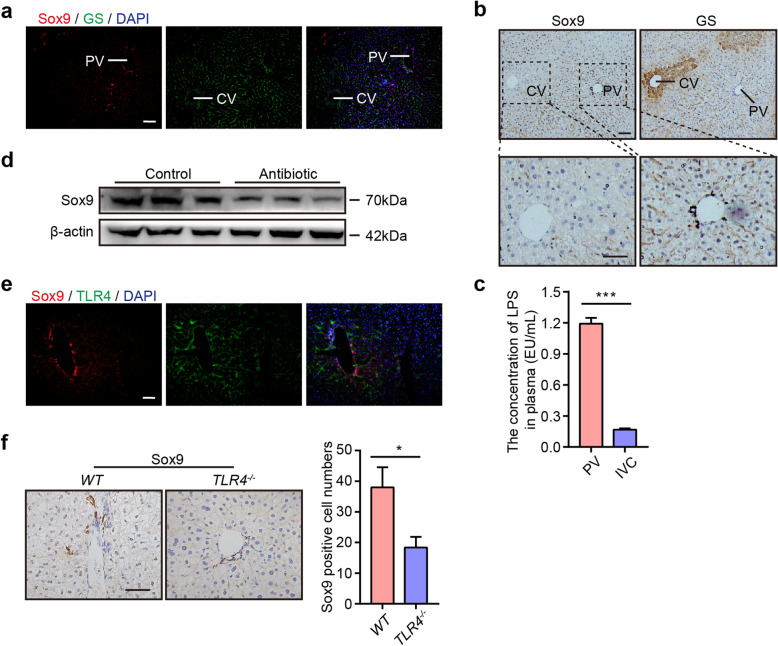
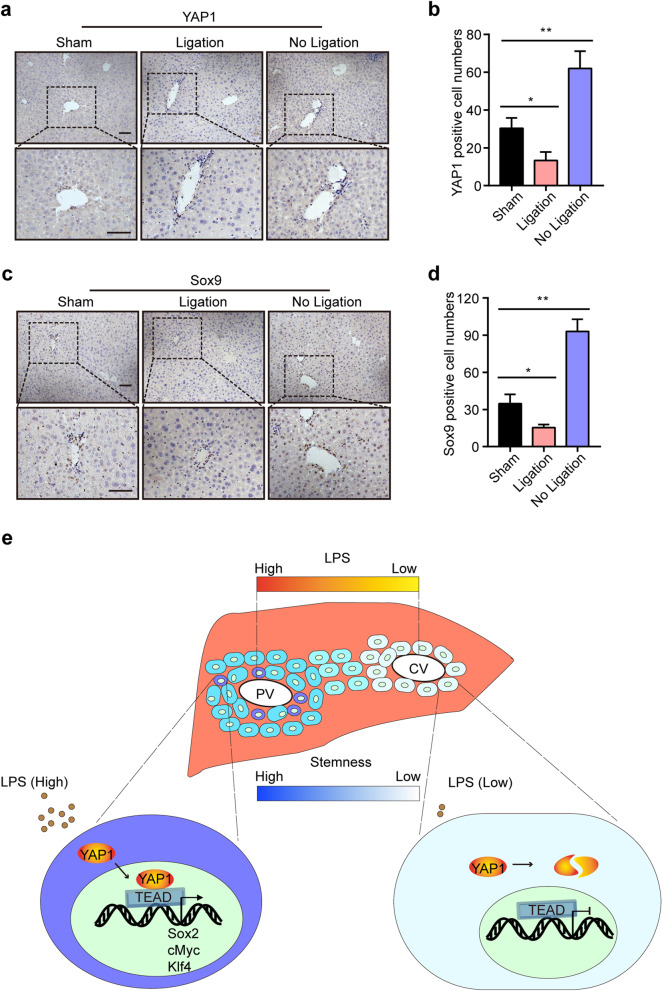
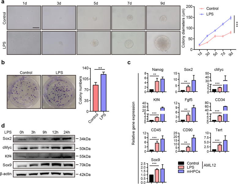
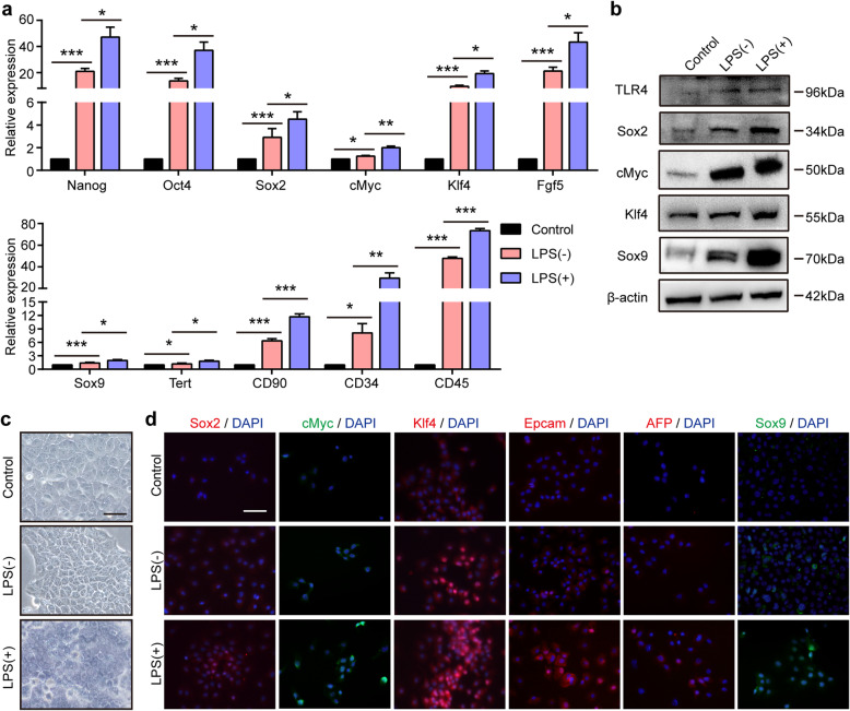
First, we examined the location of hepatic stem cells and the concentration of lipopolysaccharide (LPS) in the portal vein and inferior vena cava. Then, we assessed the effect of LPS on stemness maintenance in mice by using antibiotics to eliminate LPS and knocking out the LPS receptor, TLR4. In vitro, the effect of LPS on the stemness of hepatocytes was investigated by colony and sphere formation assays and assessment of pluripotent and stem cell marker expression. Furthermore, we studied the mechanism by which LPS regulates the stemness of hepatocytes. Finally, we ligated the portal vein branch to further verify the effect of LPS.

RESULTS

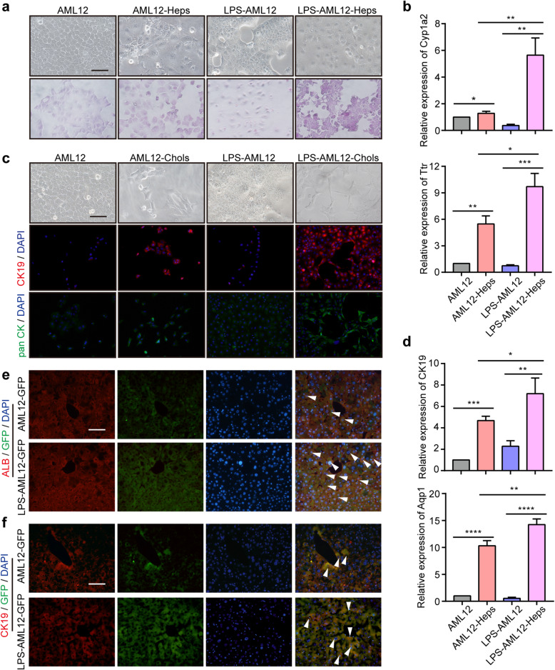
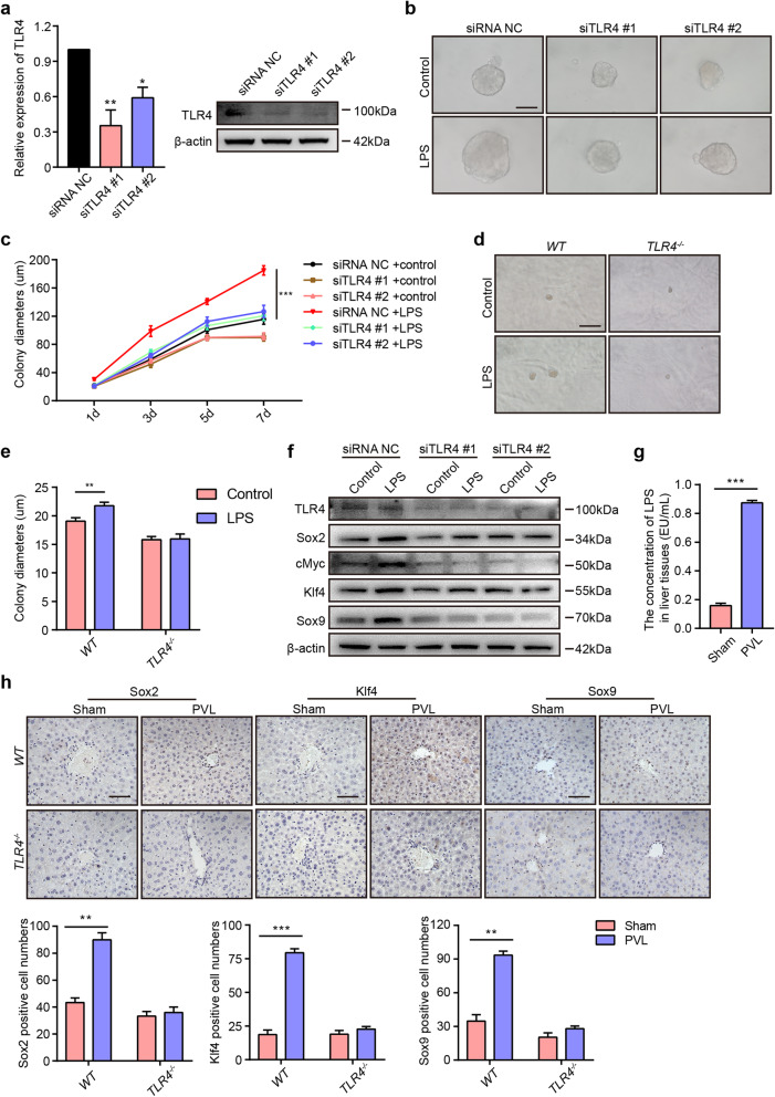
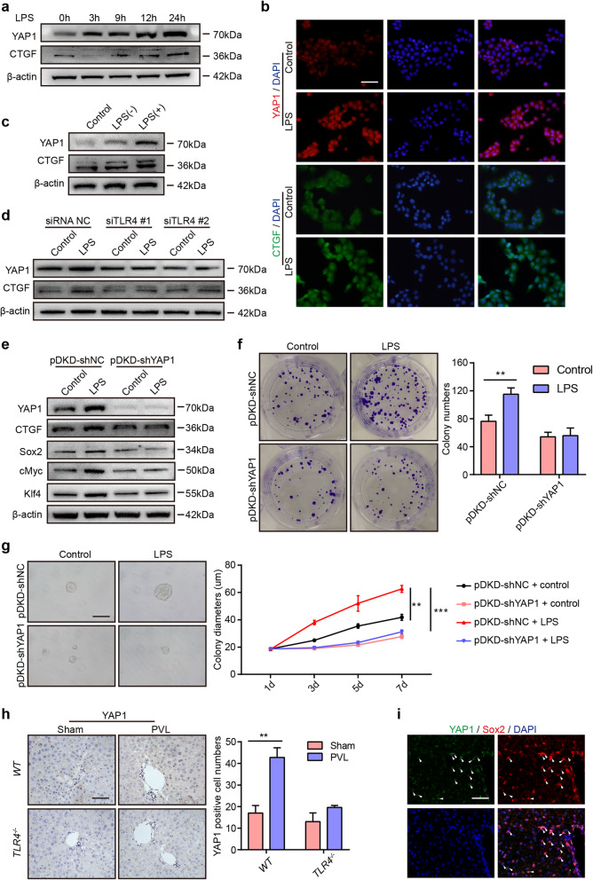
We found that a high level of LPS from the portal vein was correlated with the location of hepatic stem cells in the PV area, and elimination of LPS by antibiotics inhibited the expression of the stemness marker. LPS promoted colony and sphere formation and induced the upregulation of pluripotent and stem cell markers in AML12 cells. Furthermore, in the reprogramming medium, LPS facilitated the dedifferentiation of mature hepatocytes into hepatic progenitor-like cells, which exhibited a bipotent differentiation capacity in vivo and in vitro. Mechanistically, LPS bound TLR4 to regulate stemness of hepatocytes via the activation of YAP1 signaling, and blockade of YAP1 abolished the LPS-induced cell stemness and upregulation of pluripotent markers.

CONCLUSIONS

Our study implies a correlation between LPS/TLR4/YAP1 signaling and cell stemness, and LPS was shown to be involved in stemness maintenance of hepatocytes in the PV area. LPS might be used to induce the dedifferentiation of mature hepatocytes into progenitor-like cells for repair of liver injury.

摘要

背景

肝脏具有强大的再生能力,这与门静脉(PV)中的肝细胞的干性有关。然而,维持肝细胞干性的机制尚未阐明。在这里,我们假设门静脉中的高浓度脂多糖可能维持 PV 区域肝细胞的干性。

方法

首先,我们检查了肝干细胞的位置和门静脉及下腔静脉中的脂多糖(LPS)浓度。然后,我们通过使用抗生素消除 LPS 和敲除 LPS 受体 TLR4 来评估 LPS 对小鼠干性维持的影响。在体外,通过集落和球体形成测定以及多能和干细胞标志物表达评估来研究 LPS 对肝细胞干性的影响。此外,我们研究了 LPS 调节肝细胞干性的机制。最后,我们结扎门静脉分支以进一步验证 LPS 的作用。

结果

我们发现门静脉中的高 LPS 水平与 PV 区域肝干细胞的位置相关,抗生素消除 LPS 抑制了干性标志物的表达。LPS 促进集落和球体形成,并诱导 AML12 细胞中多能和干细胞标志物的上调。此外,在重编程培养基中,LPS 促进成熟肝细胞向肝祖细胞样细胞的去分化,在体内和体外均表现出双潜能分化能力。在机制上,LPS 通过 TLR4 结合来调节肝细胞的干性,通过 YAP1 信号的激活,LPS 结合 TLR4 调节肝细胞的干性,而 YAP1 信号的阻断则消除了 LPS 诱导的细胞干性和多能标志物的上调。

结论

我们的研究表明 LPS/TLR4/YAP1 信号与细胞干性之间存在相关性,并且 LPS 参与了 PV 区域肝细胞干性的维持。LPS 可能用于诱导成熟肝细胞向祖细胞样细胞的去分化,以修复肝损伤。

https://cdn.ncbi.nlm.nih.gov/pmc/blobs/e27d/8193885/a336f33ad18a/13287_2021_2421_Fig7_HTML.jpg
https://cdn.ncbi.nlm.nih.gov/pmc/blobs/e27d/8193885/ac0e95f6c40f/13287_2021_2421_Fig1_HTML.jpg
https://cdn.ncbi.nlm.nih.gov/pmc/blobs/e27d/8193885/444dab5e3fab/13287_2021_2421_Fig2_HTML.jpg
https://cdn.ncbi.nlm.nih.gov/pmc/blobs/e27d/8193885/10f72946b249/13287_2021_2421_Fig3_HTML.jpg
https://cdn.ncbi.nlm.nih.gov/pmc/blobs/e27d/8193885/1edbce72ca6e/13287_2021_2421_Fig4_HTML.jpg
https://cdn.ncbi.nlm.nih.gov/pmc/blobs/e27d/8193885/bbdae4b25df4/13287_2021_2421_Fig5_HTML.jpg
https://cdn.ncbi.nlm.nih.gov/pmc/blobs/e27d/8193885/58faa4653844/13287_2021_2421_Fig6_HTML.jpg
https://cdn.ncbi.nlm.nih.gov/pmc/blobs/e27d/8193885/a336f33ad18a/13287_2021_2421_Fig7_HTML.jpg

相似文献

1
The stemness of hepatocytes is maintained by high levels of lipopolysaccharide via YAP1 activation.肝细胞的干性通过 YAP1 的激活由高水平的脂多糖维持。
Stem Cell Res Ther. 2021 Jun 10;12(1):342. doi: 10.1186/s13287-021-02421-7.
2
Ligustrazine disrupts lipopolysaccharide-activated NLRP3 inflammasome pathway associated with inhibition of Toll-like receptor 4 in hepatocytes.川芎嗪通过抑制肝细胞中Toll样受体4,破坏脂多糖激活的NLRP3炎性小体途径。
Biomed Pharmacother. 2016 Mar;78:204-209. doi: 10.1016/j.biopha.2016.01.018. Epub 2016 Feb 2.
3
Toll-like receptor 4 mediates inflammatory signaling by bacterial lipopolysaccharide in human hepatic stellate cells.Toll样受体4介导细菌脂多糖在人肝星状细胞中的炎症信号传导。
Hepatology. 2003 May;37(5):1043-55. doi: 10.1053/jhep.2003.50182.
4
Lipopolysaccharide promotes tumorigenicity of hepatic progenitor cells by promoting proliferation and blocking normal differentiation.脂多糖通过促进增殖和阻断正常分化来促进肝祖细胞的致瘤性。
Cancer Lett. 2017 Feb 1;386:35-46. doi: 10.1016/j.canlet.2016.10.044. Epub 2016 Nov 15.
5
Lipopolysaccharide induces the differentiation of hepatic progenitor cells into myofibroblasts via activation of the Hedgehog signaling pathway.脂多糖通过激活刺猬信号通路诱导肝祖细胞分化为肌成纤维细胞。
Cell Cycle. 2017 Jul 18;16(14):1357-1365. doi: 10.1080/15384101.2017.1325976. Epub 2017 May 31.
6
Small molecule-mediated reprogramming of human hepatocytes into bipotent progenitor cells.小分子介导的人肝细胞重编程为双能祖细胞。
J Hepatol. 2019 Jan;70(1):97-107. doi: 10.1016/j.jhep.2018.09.007. Epub 2018 Sep 19.
7
Failure of the gut barrier system enhances liver injury in rats: protection of hepatocytes by gut-derived hepatocyte growth factor.肠道屏障系统功能障碍会加重大鼠肝脏损伤:肠道源性肝细胞生长因子对肝细胞的保护作用。
Eur J Gastroenterol Hepatol. 2004 Oct;16(10):1017-25. doi: 10.1097/00042737-200410000-00011.
8
Role of toll-like receptors in changes in gene expression and NF-kappa B activation in mouse hepatocytes stimulated with lipopolysaccharide.Toll样受体在脂多糖刺激的小鼠肝细胞基因表达变化及核因子κB激活中的作用
Infect Immun. 2002 Jul;70(7):3433-42. doi: 10.1128/IAI.70.7.3433-3442.2002.
9
Hepatocyte toll-like receptor 4 mediates lipopolysaccharide-induced hepcidin expression.肝细胞 Toll 样受体 4 介导脂多糖诱导的铁调素表达。
Exp Mol Med. 2017 Dec 8;49(12):e408. doi: 10.1038/emm.2017.207.
10
NADPH oxidase 4 modulates hepatic responses to lipopolysaccharide mediated by Toll-like receptor-4.NADPH 氧化酶 4 通过 Toll 样受体-4 调节脂多糖介导的肝反应。
Sci Rep. 2017 Oct 30;7(1):14346. doi: 10.1038/s41598-017-14574-8.

引用本文的文献

1
Advances in liver organoids: replicating hepatic complexity for toxicity assessment and disease modeling.肝脏类器官的进展:复制肝脏复杂性用于毒性评估和疾病建模。
Stem Cell Res Ther. 2025 Jan 26;16(1):27. doi: 10.1186/s13287-025-04139-2.
2
In Vitro Generated Equine Hepatic-Like Progenitor Cells as a Novel Potent Cell Pool for Equine Metabolic Syndrome (EMS) Treatment.体外生成的马源类肝祖细胞作为治疗马代谢综合征(EMS)的新型有效细胞池。
Stem Cell Rev Rep. 2023 May;19(4):1124-1134. doi: 10.1007/s12015-023-10507-3. Epub 2023 Jan 20.
3
Commensal gut microbiota-based strategies for oral delivery of therapeutic proteins.

本文引用的文献

1
Cell Plasticity in Liver Regeneration.肝再生中的细胞可塑性。
Trends Cell Biol. 2020 Apr;30(4):329-338. doi: 10.1016/j.tcb.2020.01.007. Epub 2020 Feb 11.
2
Epigenetic induction of tumor stemness via the lipopolysaccharide-TET3-HOXB2 signaling axis in esophageal squamous cell carcinoma.通过 LPS-TET3-HOXB2 信号轴在食管鳞状细胞癌中诱导肿瘤干性的表观遗传。
Cell Commun Signal. 2020 Feb 3;18(1):17. doi: 10.1186/s12964-020-0510-8.
3
Understanding the marvels behind liver regeneration.了解肝脏再生背后的奇迹。
基于共生肠道菌群的治疗性蛋白口服递送策略。
Trends Pharmacol Sci. 2022 Dec;43(12):1004-1013. doi: 10.1016/j.tips.2022.08.002. Epub 2022 Aug 31.
4
The gut-liver axis: host microbiota interactions shape hepatocarcinogenesis.肠-肝轴:宿主微生物群相互作用塑造肝癌发生。
Trends Cancer. 2022 Jul;8(7):583-597. doi: 10.1016/j.trecan.2022.02.009. Epub 2022 Mar 21.
Wiley Interdiscip Rev Dev Biol. 2019 May;8(3):e340. doi: 10.1002/wdev.340. Epub 2019 Mar 28.
4
Lineage Tracing Reveals the Bipotency of SOX9 Hepatocytes during Liver Regeneration.谱系追踪揭示了 SOX9 肝细胞在肝再生过程中的双能性。
Stem Cell Reports. 2019 Mar 5;12(3):624-638. doi: 10.1016/j.stemcr.2019.01.010. Epub 2019 Feb 14.
5
Inflammatory Cytokine TNFα Promotes the Long-Term Expansion of Primary Hepatocytes in 3D Culture.炎症细胞因子 TNFα 促进原代肝细胞在 3D 培养中的长期扩增。
Cell. 2018 Nov 29;175(6):1607-1619.e15. doi: 10.1016/j.cell.2018.11.012.
6
Cell Aggregation Culture Induces Functional Differentiation of Induced Hepatocyte-like Cells through Activation of Hippo Signaling.细胞聚集培养通过激活 Hippo 信号诱导诱导的肝细胞样细胞的功能分化。
Cell Rep. 2018 Oct 2;25(1):183-198. doi: 10.1016/j.celrep.2018.09.010.
7
Small molecule-mediated reprogramming of human hepatocytes into bipotent progenitor cells.小分子介导的人肝细胞重编程为双能祖细胞。
J Hepatol. 2019 Jan;70(1):97-107. doi: 10.1016/j.jhep.2018.09.007. Epub 2018 Sep 19.
8
The Hippo Signaling Network and Its Biological Functions.Hippo 信号通路及其生物学功能。
Annu Rev Genet. 2018 Nov 23;52:65-87. doi: 10.1146/annurev-genet-120417-031621. Epub 2018 Sep 5.
9
Glucagon contributes to liver zonation.胰高血糖素有助于肝脏分区。
Proc Natl Acad Sci U S A. 2018 Apr 24;115(17):E4111-E4119. doi: 10.1073/pnas.1721403115. Epub 2018 Mar 19.
10
Ecological Balance of Oral Microbiota Is Required to Maintain Oral Mesenchymal Stem Cell Homeostasis.口腔微生物组的生态平衡对于维持口腔间充质干细胞的内稳态至关重要。
Stem Cells. 2018 Apr;36(4):551-561. doi: 10.1002/stem.2762. Epub 2018 Jan 4.