BRAIN Centre (Biomarker Research and Imaging for Neuroscience), Department of Neuroimaging, King's College London, The James Black Centre, 125 Coldharbour Lane, London, SE5 9NU, UK.
Department of Life, Health and Environmental Sciences, University of L'Aquila, Piazzale Salvatore Tommasi 1, 67100, L'Aquila, Italy.
Sci Rep. 2021 Jun 14;11(1):12419. doi: 10.1038/s41598-021-91899-5.
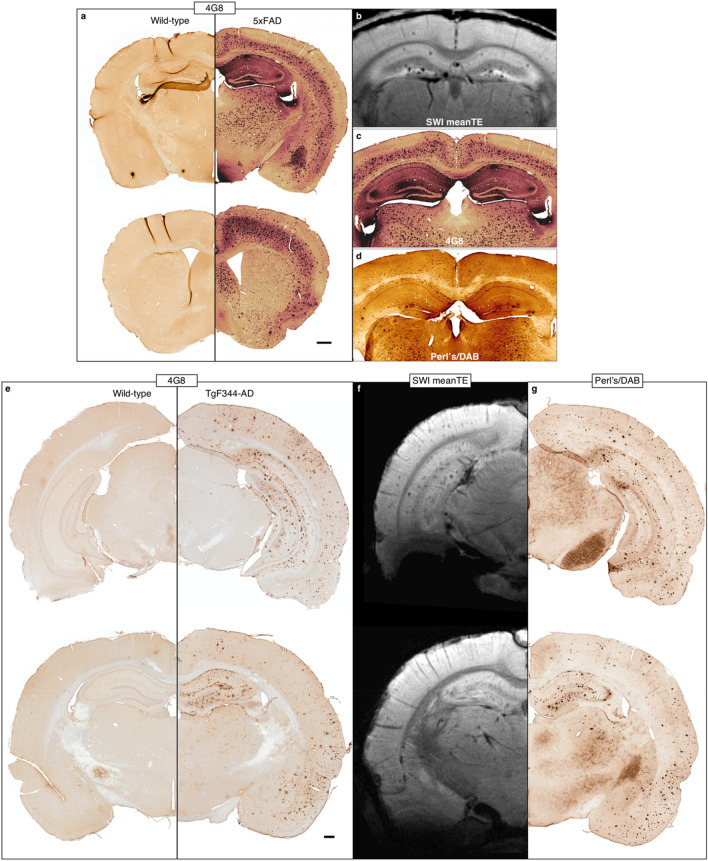
Amyloid plaques are a hallmark of Alzheimer's disease (AD) that develop in its earliest stages. Thus, non-invasive detection of these plaques would be invaluable for diagnosis and the development and monitoring of treatments, but this remains a challenge due to their small size. Here, we investigated the utility of manganese-enhanced MRI (MEMRI) for visualizing plaques in transgenic rodent models of AD across two species: 5xFAD mice and TgF344-AD rats. Animals were given subcutaneous injections of MnCl and imaged in vivo using a 9.4 T Bruker scanner. MnCl improved signal-to-noise ratio but was not necessary to detect plaques in high-resolution images. Plaques were visible in all transgenic animals and no wild-types, and quantitative susceptibility mapping showed that they were more paramagnetic than the surrounding tissue. This, combined with beta-amyloid and iron staining, indicate that plaque MR visibility in both animal models was driven by plaque size and iron load. Longitudinal relaxation rate mapping revealed increased manganese uptake in brain regions of high plaque burden in transgenic animals compared to their wild-type littermates. This was limited to the rhinencephalon in the TgF344-AD rats, while it was most significantly increased in the cortex of the 5xFAD mice. Alizarin Red staining suggests that manganese bound to plaques in 5xFAD mice but not in TgF344-AD rats. Multi-parametric MEMRI is a simple, viable method for detecting amyloid plaques in rodent models of AD. Manganese-induced signal enhancement can enable higher-resolution imaging, which is key to visualizing these small amyloid deposits. We also present the first in vivo evidence of manganese as a potential targeted contrast agent for imaging plaques in the 5xFAD model of AD.
淀粉样斑块是阿尔茨海默病(AD)的一个标志,在疾病的早期阶段就会出现。因此,非侵入性地检测这些斑块对于诊断以及治疗方法的开发和监测将是非常有价值的,但由于其体积小,这仍然是一个挑战。在这里,我们研究了锰增强磁共振成像(MEMRI)在两种 AD 转基因啮齿动物模型中的斑块可视化的效用:5xFAD 小鼠和 TgF344-AD 大鼠。动物接受皮下注射 MnCl 后,在 Bruker 9.4T 扫描仪上进行体内成像。MnCl 提高了信噪比,但对于在高分辨率图像中检测斑块并非必需。在所有转基因动物中都可见斑块,而在野生型动物中则不可见,定量磁化率映射显示它们比周围组织具有更强的顺磁性。这一点,再加上β-淀粉样蛋白和铁染色,表明两种动物模型中的斑块 MR 可见性是由斑块大小和铁负荷驱动的。纵向弛豫率映射显示,与野生型同窝仔相比,转基因动物高斑块负担的脑区中锰摄取增加。在 TgF344-AD 大鼠中,这种情况仅限于边缘叶,而在 5xFAD 小鼠中则最为显著增加。茜素红染色表明,锰与 5xFAD 小鼠中的斑块结合,但与 TgF344-AD 大鼠中的斑块不结合。多参数 MEMRI 是一种简单、可行的方法,可用于检测 AD 转基因啮齿动物模型中的淀粉样斑块。锰诱导的信号增强可以实现更高分辨率的成像,这是可视化这些小淀粉样沉积物的关键。我们还首次提供了体内证据,表明锰可能是 AD 的 5xFAD 模型中成像斑块的潜在靶向对比剂。