Idilman Ilkay S, Low Hsien Min, Gidener Tolga, Philbrick Kenneth, Mounajjed Taofic, Li Jiahui, Allen Alina M, Yin Meng, Venkatesh Sudhakar K
Division of Abdominal Imaging, Department of Radiology, Mayo Clinic, Rochester, MN 55905, USA.
Department of Radiology, School of Medicine, Hacettepe University, Ankara 06100, Turkey.
J Clin Med. 2021 Jun 10;10(12):2565. doi: 10.3390/jcm10122565.
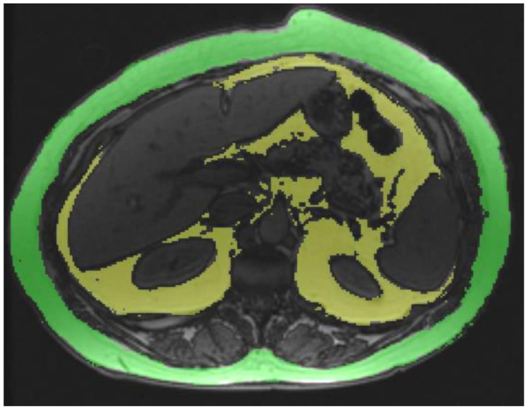
(1) Purpose: To determine the association between visceral adipose tissue (VAT) and proton density fat fraction (PDFF) with magnetic resonance imaging (MRI), and hepatic steatosis (HS), non-alcoholic steatohepatitis (NASH) and hepatic fibrosis (HF) in patients with known or suspected non-alcoholic fatty liver disease (NAFLD). (2) Methods: 135 subjects that had a liver biopsy performed within 3 months (bariatric cohort) or 1 month (NAFLD cohort) of an MRI exam formed the study group. VAT volume was quantified at L2-L3 level on opposed-phase images with signal intensity-based painting using a semi-quantitative software. Liver PDFF and pancreas PDFF were calculated on fat fraction maps. Liver volume (Lvol) and spleen volume (Svol) were also calculated using a semi-automated 3D volume tool available on PACS. A histological analysis was performed by an expert hepatopathologist blinded to imaging findings. (3) Results: The mean Lvol, Svol, liver PDFF, pancreas PDFF and VAT of the study population were 2492.2 mL, 381.6 mL, 13.2%, 12.7% and 120.6 mL, respectively. VAT showed moderate correlation with liver PDFF (r = 0.41, < 0.001) and weak correlation with Lvol (r = 0.38, < 0.001), Svol (r = 0.20, = 0.025) and pancreas PDFF (r = 0.29, = 0.001). VAT, Lvol and liver PDFF were significantly higher in patients with HS ( < 0.001), NASH ( < 0.05) and HF ( < 0.05). VAT was also significantly higher in the presence of lobular inflammation ( = 0.019) and hepatocyte ballooning ( = 0.001). The cut-off VAT volumes for predicting HS, NASH and HF were 101.8 mL (AUC, 0.7), 111.8 mL (AUC, 0.64) and 111.6 mL (AUC, 0.66), respectively. (4) Conclusion: The MRI determined VAT can be used for predicting the presence of HS, NASH and HF in patients with known or suspected NAFLD.
(1)目的:利用磁共振成像(MRI)确定已知或疑似非酒精性脂肪性肝病(NAFLD)患者的内脏脂肪组织(VAT)和质子密度脂肪分数(PDFF)与肝脂肪变性(HS)、非酒精性脂肪性肝炎(NASH)及肝纤维化(HF)之间的关联。(2)方法:135名在MRI检查后3个月内(减重手术队列)或1个月内(NAFLD队列)接受肝活检的受试者组成研究组。使用基于信号强度绘制的半定量软件,在L2-L3水平的反相位图像上对VAT体积进行定量。在脂肪分数图上计算肝脏PDFF和胰腺PDFF。还使用PACS上可用的半自动3D体积工具计算肝脏体积(Lvol)和脾脏体积(Svol)。由对影像结果不知情的专业肝病病理学家进行组织学分析。(3)结果:研究人群的平均Lvol、Svol、肝脏PDFF、胰腺PDFF和VAT分别为2492.2 mL、381.6 mL、13.2%、12.7%和120.6 mL。VAT与肝脏PDFF呈中度相关(r = 0.41,P < 0.001),与Lvol呈弱相关(r = 0.38,P < 0.001),与Svol呈弱相关(r = 0.20,P = 0.025),与胰腺PDFF呈弱相关(r = 0.29,P = 0.001)。HS(P < 0.001)、NASH(P < 0.05)和HF(P < 0.05)患者的VAT、Lvol和肝脏PDFF显著更高。在存在小叶炎症(P = 0.019)和肝细胞气球样变(P = 0.001)时,VAT也显著更高。预测HS、NASH和HF的VAT体积截断值分别为101.8 mL(AUC,0.7)、111.8 mL(AUC,0.64)和111.6 mL(AUC,0.66)。(4)结论:MRI测定的VAT可用于预测已知或疑似NAFLD患者中HS、NASH和HF的存在。