• 文献检索
  • 文档翻译
  • 深度研究
  • 学术资讯
  • Suppr Zotero 插件Zotero 插件
  • 邀请有礼
  • 套餐&价格
  • 历史记录
应用&插件
Suppr Zotero 插件Zotero 插件浏览器插件Mac 客户端Windows 客户端微信小程序
定价
高级版会员购买积分包购买API积分包
服务
文献检索文档翻译深度研究API 文档MCP 服务
关于我们
关于 Suppr公司介绍联系我们用户协议隐私条款
关注我们

Suppr 超能文献

核心技术专利:CN118964589B侵权必究
粤ICP备2023148730 号-1Suppr @ 2026

文献检索

告别复杂PubMed语法,用中文像聊天一样搜索,搜遍4000万医学文献。AI智能推荐,让科研检索更轻松。

立即免费搜索

文件翻译

保留排版,准确专业,支持PDF/Word/PPT等文件格式,支持 12+语言互译。

免费翻译文档

深度研究

AI帮你快速写综述,25分钟生成高质量综述,智能提取关键信息,辅助科研写作。

立即免费体验

具有不同毒性、结构和种子潜力的亨廷顿纤维可以相互转化。

Huntingtin fibrils with different toxicity, structure, and seeding potential can be interconverted.

机构信息

Department of Physiology & Neuroscience, Zilkha Neurogenetic Institute, Keck School of Medicine, University of Southern California, Los Angeles, CA, USA.

Department of Emergency Medicine, Regions Hospital, St. Paul, MN, USA.

出版信息

Nat Commun. 2021 Jul 13;12(1):4272. doi: 10.1038/s41467-021-24411-2.

DOI:10.1038/s41467-021-24411-2
PMID:34257293
原文链接:https://pmc.ncbi.nlm.nih.gov/articles/PMC8277859/
Abstract

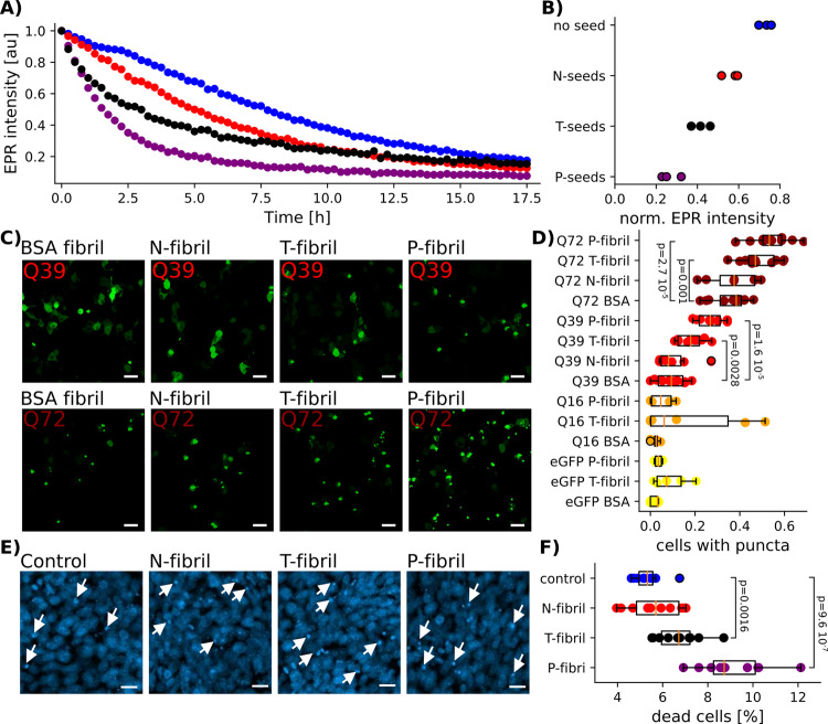
The first exon of the huntingtin protein (HTTex1) important in Huntington's disease (HD) can form cross-β fibrils of varying toxicity. We find that the difference between these fibrils is the degree of entanglement and dynamics of the C-terminal proline-rich domain (PRD) in a mechanism analogous to polyproline film formation. In contrast to fibril strains found for other cross-β fibrils, these HTTex1 fibril types can be interconverted. This is because the structure of their polyQ fibril core remains unchanged. Further, we find that more toxic fibrils of low entanglement have higher affinities for protein interactors and are more effective seeds for recombinant HTTex1 and HTTex1 in cells. Together these data show how the structure of a framing sequence at the surface of a fibril can modulate seeding, protein-protein interactions, and thereby toxicity in neurodegenerative disease.

摘要

亨廷顿病(HD)中重要的亨廷顿蛋白(HTTex1)的第一个外显子可以形成具有不同毒性的交叉β纤维。我们发现,这些纤维之间的差异在于 C 端脯氨酸丰富结构域(PRD)的缠结程度和动力学,这一机制类似于多聚脯氨酸膜的形成。与其他交叉β纤维的纤维菌株不同,这些 HTTex1 纤维类型可以相互转换。这是因为它们的多聚 Q 纤维核心结构保持不变。此外,我们发现缠结程度较低的毒性纤维具有更高的蛋白相互作用亲和力,并且对于重组 HTTex1 和 HTTex1 在细胞中的成核作用更有效。这些数据共同表明,纤维表面框架序列的结构如何调节成核、蛋白-蛋白相互作用,从而影响神经退行性疾病的毒性。

https://cdn.ncbi.nlm.nih.gov/pmc/blobs/8b22/8277859/4d81cb37c22e/41467_2021_24411_Fig8_HTML.jpg
https://cdn.ncbi.nlm.nih.gov/pmc/blobs/8b22/8277859/b9b877144c73/41467_2021_24411_Fig1_HTML.jpg
https://cdn.ncbi.nlm.nih.gov/pmc/blobs/8b22/8277859/314cf0428190/41467_2021_24411_Fig2_HTML.jpg
https://cdn.ncbi.nlm.nih.gov/pmc/blobs/8b22/8277859/0d32c4111130/41467_2021_24411_Fig3_HTML.jpg
https://cdn.ncbi.nlm.nih.gov/pmc/blobs/8b22/8277859/3ec69cd9f283/41467_2021_24411_Fig4_HTML.jpg
https://cdn.ncbi.nlm.nih.gov/pmc/blobs/8b22/8277859/341a325ab573/41467_2021_24411_Fig5_HTML.jpg
https://cdn.ncbi.nlm.nih.gov/pmc/blobs/8b22/8277859/bc2c58ebb89d/41467_2021_24411_Fig6_HTML.jpg
https://cdn.ncbi.nlm.nih.gov/pmc/blobs/8b22/8277859/e98c77c21cf4/41467_2021_24411_Fig7_HTML.jpg
https://cdn.ncbi.nlm.nih.gov/pmc/blobs/8b22/8277859/4d81cb37c22e/41467_2021_24411_Fig8_HTML.jpg

相似文献

1
Huntingtin fibrils with different toxicity, structure, and seeding potential can be interconverted.具有不同毒性、结构和种子潜力的亨廷顿纤维可以相互转化。
Nat Commun. 2021 Jul 13;12(1):4272. doi: 10.1038/s41467-021-24411-2.
2
Formation and Structure of Wild Type Huntingtin Exon-1 Fibrils.野生型亨廷顿蛋白外显子1纤维的形成与结构
Biochemistry. 2017 Jul 18;56(28):3579-3586. doi: 10.1021/acs.biochem.7b00138. Epub 2017 Jul 7.
3
Amplification of neurotoxic HTTex1 assemblies in human neurons.在人神经元中神经毒性 HTTex1 组装物的扩增。
Neurobiol Dis. 2021 Nov;159:105517. doi: 10.1016/j.nbd.2021.105517. Epub 2021 Sep 24.
4
The 17-residue-long N terminus in huntingtin controls stepwise aggregation in solution and on membranes via different mechanisms.亨廷顿蛋白的 17 个氨基酸长的 N 端通过不同的机制控制在溶液中和膜上的逐步聚集。
J Biol Chem. 2018 Feb 16;293(7):2597-2605. doi: 10.1074/jbc.M117.813667. Epub 2017 Dec 27.
5
The polyglutamine domain is the primary driver of seeding in huntingtin aggregation.多聚谷氨酰胺结构域是亨廷顿聚集物中引发聚集的主要驱动因素。
PLoS One. 2024 Mar 14;19(3):e0298323. doi: 10.1371/journal.pone.0298323. eCollection 2024.
6
TDP43 and huntingtin Exon-1 undergo a conformationally specific interaction that strongly alters the fibril formation of both proteins.TDP43 和亨廷顿外显子 1 发生构象特异性相互作用,强烈改变这两种蛋白质的纤维形成。
J Biol Chem. 2024 Sep;300(9):107660. doi: 10.1016/j.jbc.2024.107660. Epub 2024 Aug 13.
7
Structural Model of the Proline-Rich Domain of Huntingtin Exon-1 Fibrils.亨廷顿蛋白外显子 1 纤维中脯氨酸丰富结构域的结构模型。
Biophys J. 2020 Nov 17;119(10):2019-2028. doi: 10.1016/j.bpj.2020.10.010. Epub 2020 Oct 20.
8
Monomeric Huntingtin Exon 1 Has Similar Overall Structural Features for Wild-Type and Pathological Polyglutamine Lengths.单体 Huntingtin 外显子 1 具有相似的整体结构特征,无论野生型还是病理性聚谷氨酰胺长度。
J Am Chem Soc. 2017 Oct 18;139(41):14456-14469. doi: 10.1021/jacs.7b06659. Epub 2017 Oct 9.
9
Generation of Native, Untagged Huntingtin Exon1 Monomer and Fibrils Using a SUMO Fusion Strategy.使用SUMO融合策略生成天然、无标签的亨廷顿蛋白外显子1单体和原纤维。
J Vis Exp. 2018 Jun 27(136):57506. doi: 10.3791/57506.
10
Toxic effects of mutant huntingtin in axons are mediated by its proline-rich domain.突变型亨廷顿蛋白在轴突中的毒性作用是由其脯氨酸丰富结构域介导的。
Brain. 2024 Jun 3;147(6):2098-2113. doi: 10.1093/brain/awad280.

引用本文的文献

1
Probing How Anti-huntingtin Antibodies Bind the Fibrillar Fuzzy Coat Using Solid-State NMR.利用固态核磁共振探究抗亨廷顿蛋白抗体如何结合纤维状模糊外壳。
Chemistry. 2025 Aug 13;31(45):e01676. doi: 10.1002/chem.202501676. Epub 2025 Jul 24.
2
Direct Observation of Secondary Nucleation in Huntingtin Amyloid Formation by High-Speed Atomic Force Microscopy.通过高速原子力显微镜直接观察亨廷顿蛋白淀粉样蛋白形成过程中的二次成核
J Am Chem Soc. 2025 Jun 25;147(25):21973-21984. doi: 10.1021/jacs.5c05571. Epub 2025 Jun 12.
3
Multiscale simulations elucidate the mechanism of polyglutamine aggregation and the role of flanking domains in fibril polymorphism.

本文引用的文献

1
Structural Model of the Proline-Rich Domain of Huntingtin Exon-1 Fibrils.亨廷顿蛋白外显子 1 纤维中脯氨酸丰富结构域的结构模型。
Biophys J. 2020 Nov 17;119(10):2019-2028. doi: 10.1016/j.bpj.2020.10.010. Epub 2020 Oct 20.
2
Dynamics of the Proline-Rich C-Terminus of Huntingtin Exon-1 Fibrils.亨廷顿蛋白外显子 1 纤维中脯氨酸丰富的 C 末端的动力学。
J Phys Chem B. 2018 Oct 18;122(41):9507-9515. doi: 10.1021/acs.jpcb.8b09213. Epub 2018 Oct 4.
3
Identification of distinct conformations associated with monomers and fibril assemblies of mutant huntingtin.
多尺度模拟阐明了多聚谷氨酰胺聚集的机制以及侧翼结构域在纤维多态性中的作用。
bioRxiv. 2025 May 23:2025.05.19.654960. doi: 10.1101/2025.05.19.654960.
4
mTOR inhibition in Q175 Huntington's disease model mice facilitates neuronal autophagy and mutant huntingtin clearance.在Q175亨廷顿舞蹈病模型小鼠中抑制mTOR可促进神经元自噬及突变型亨廷顿蛋白的清除。
Elife. 2025 May 20;14:RP104979. doi: 10.7554/eLife.104979.
5
Inhibitor-based modulation of huntingtin aggregation mechanisms mitigates fibril-induced cellular stress.基于抑制剂对亨廷顿蛋白聚集机制的调节可减轻原纤维诱导的细胞应激。
Nat Commun. 2025 Apr 15;16(1):3588. doi: 10.1038/s41467-025-58691-9.
6
Integrative determination of atomic structure of mutant huntingtin exon 1 fibrils implicated in Huntington disease.与亨廷顿病相关的突变型亨廷顿蛋白外显子1原纤维原子结构的综合测定。
Nat Commun. 2024 Dec 30;15(1):10793. doi: 10.1038/s41467-024-55062-8.
7
Cryo-EM and solid state NMR together provide a more comprehensive structural investigation of protein fibrils.低温电子显微镜和固态核磁共振技术共同为蛋白质纤维的结构研究提供了更全面的方法。
Protein Sci. 2024 Oct;33(10):e5168. doi: 10.1002/pro.5168.
8
Solid-state nuclear magnetic resonance in the structural study of polyglutamine aggregation.固态核磁共振在聚谷氨酰胺聚集结构研究中的应用
Biochem Soc Trans. 2024 Apr 24;52(2):719-731. doi: 10.1042/BST20230731.
9
The polyglutamine domain is the primary driver of seeding in huntingtin aggregation.多聚谷氨酰胺结构域是亨廷顿聚集物中引发聚集的主要驱动因素。
PLoS One. 2024 Mar 14;19(3):e0298323. doi: 10.1371/journal.pone.0298323. eCollection 2024.
10
Fluorescent protein tagging promotes phase separation and alters the aggregation pathway of huntingtin exon-1.荧光蛋白标记促进相分离,并改变亨廷顿外显子 1 的聚集途径。
J Biol Chem. 2024 Jan;300(1):105585. doi: 10.1016/j.jbc.2023.105585. Epub 2023 Dec 21.
鉴定与突变型亨廷顿蛋白单体和纤维组装相关的不同构象。
Hum Mol Genet. 2018 Jul 1;27(13):2330-2343. doi: 10.1093/hmg/ddy141.
4
Profilin reduces aggregation and phase separation of huntingtin N-terminal fragments by preferentially binding to soluble monomers and oligomers.丝状肌动蛋白结合蛋白通过优先结合可溶性单体和低聚物,减少 huntingtin N 端片段的聚集和相分离。
J Biol Chem. 2018 Mar 9;293(10):3734-3746. doi: 10.1074/jbc.RA117.000357. Epub 2018 Jan 22.
5
The 17-residue-long N terminus in huntingtin controls stepwise aggregation in solution and on membranes via different mechanisms.亨廷顿蛋白的 17 个氨基酸长的 N 端通过不同的机制控制在溶液中和膜上的逐步聚集。
J Biol Chem. 2018 Feb 16;293(7):2597-2605. doi: 10.1074/jbc.M117.813667. Epub 2017 Dec 27.
6
ImageJ2: ImageJ for the next generation of scientific image data.ImageJ2:面向下一代科学图像数据的ImageJ。
BMC Bioinformatics. 2017 Nov 29;18(1):529. doi: 10.1186/s12859-017-1934-z.
7
In Situ Architecture and Cellular Interactions of PolyQ Inclusions.多聚谷氨酰胺包涵体的原位结构和细胞相互作用。
Cell. 2017 Sep 21;171(1):179-187.e10. doi: 10.1016/j.cell.2017.08.009. Epub 2017 Sep 7.
8
Formation and Structure of Wild Type Huntingtin Exon-1 Fibrils.野生型亨廷顿蛋白外显子1纤维的形成与结构
Biochemistry. 2017 Jul 18;56(28):3579-3586. doi: 10.1021/acs.biochem.7b00138. Epub 2017 Jul 7.
9
Fibril polymorphism affects immobilized non-amyloid flanking domains of huntingtin exon1 rather than its polyglutamine core.纤维多态性影响的是不溶性的亨廷顿外显子 1 中非淀粉样侧翼结构域,而不是其多聚谷氨酰胺核心。
Nat Commun. 2017 May 24;8:15462. doi: 10.1038/ncomms15462.
10
Huntingtin Inclusions Trigger Cellular Quiescence, Deactivate Apoptosis, and Lead to Delayed Necrosis.亨廷顿蛋白包涵体引发细胞静止,使细胞凋亡失活,并导致延迟性坏死。
Cell Rep. 2017 May 2;19(5):919-927. doi: 10.1016/j.celrep.2017.04.029.