Tian Xuan, Cao Huan, Wu Longlong, Zheng Weiping, Yuan Mengshu, Li Xiang, Song Hongli, Shen Zhongyang
School of Medicine, Nankai University, Tianjin, China.
Tianjin First Central Hospital Clinic Institute, Tianjin Medical University, Tianjin 300070, China.
Stem Cells Int. 2021 Jul 1;2021:9935370. doi: 10.1155/2021/9935370. eCollection 2021.
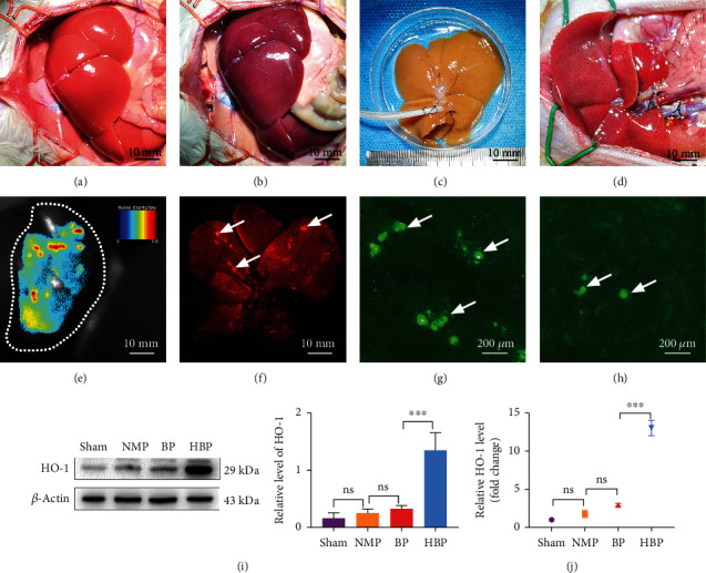
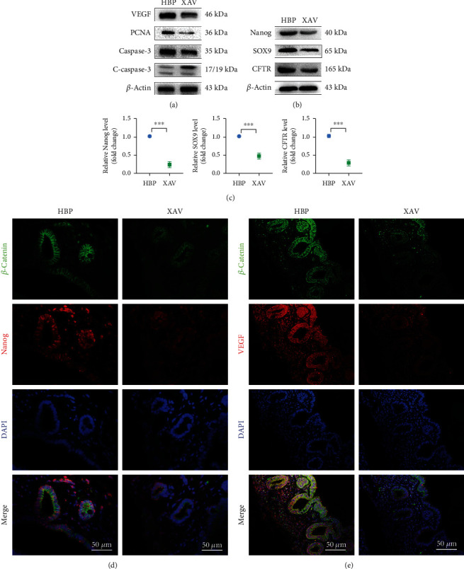
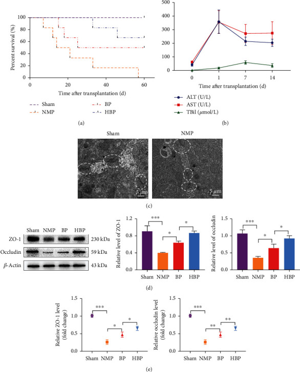
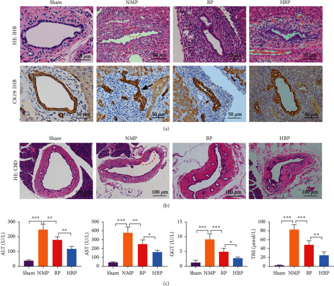
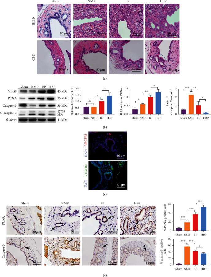
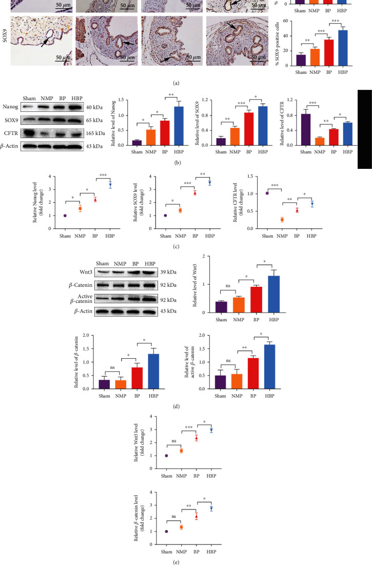
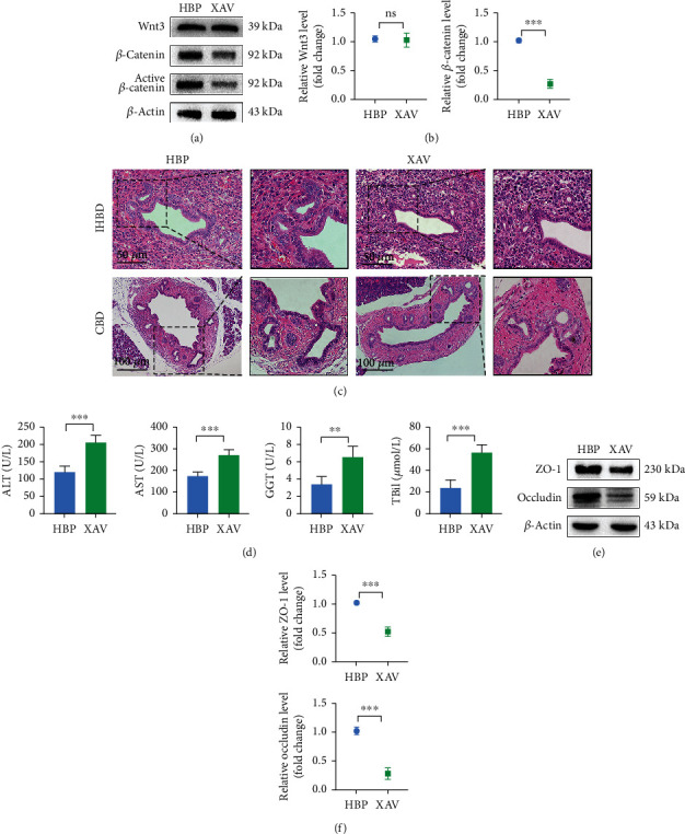
Livers from donors after circulatory death (DCD) are inevitably exposed to a longer warm ischemic period, which might increase the incidence of postoperative bile duct complications. Bone marrow mesenchymal stem cells (BMMSCs) have tissue repair properties. The present study was aimed at exploring the repair effect of heme oxygenase-1- (HO-1-) modified BMMSCs (HO-1/BMMSCs) combined with normothermic machine perfusion (NMP) on bile duct injury after DCD liver transplantation and at revealing the underlying mechanisms. Rat livers were exposed to warm ischemia for 30 min; then, NMP was performed through the portal vein for 4 h with BMMSCs, HO-1/BMMSCs, or neither before implantation. Obvious bile duct histological damage and liver functional damage were observed postoperatively. In the group treated with HO-1/BMMSCs combined with NMP (HBP group), liver functions and bile duct histology were improved; meanwhile, cell apoptosis was reduced and cell proliferation was active. A large number of regenerative cells appeared at the injured site, and the defective bile duct epithelium was restored. Dilatation of peribiliary glands (PBGs), proliferation of PBG cells, high expression of vascular endothelial growth factor (VEGF), and increased proportion of bile duct progenitor cells with stem/progenitor cells biomarkers were observed. Blocking Wnt signaling significantly inhibited the repair effect of HO-1/BMMSCs on bile duct injury. In conclusion, HO-1/BMMSCs combined with NMP were relevant to the activation of biliary progenitor cells in PBGs which repaired bile duct injury in DCD liver transplantation via the Wnt signaling pathway. Proliferation and differentiation of PBG cells were involved in the renewal of the injured biliary epithelium.
循环死亡后供体(DCD)的肝脏不可避免地会经历更长的热缺血期,这可能会增加术后胆管并发症的发生率。骨髓间充质干细胞(BMMSCs)具有组织修复特性。本研究旨在探讨血红素加氧酶-1(HO-1)修饰的BMMSCs(HO-1/BMMSCs)联合常温机器灌注(NMP)对DCD肝移植术后胆管损伤的修复作用,并揭示其潜在机制。将大鼠肝脏暴露于热缺血30分钟;然后,在植入前通过门静脉进行4小时的NMP,分别加入BMMSCs、HO-1/BMMSCs或不加任何细胞。术后观察到明显的胆管组织学损伤和肝功能损害。在HO-1/BMMSCs联合NMP治疗组(HBP组)中,肝功能和胆管组织学得到改善;同时,细胞凋亡减少,细胞增殖活跃。损伤部位出现大量再生细胞,受损的胆管上皮得以恢复。观察到胆管周围腺体(PBGs)扩张、PBG细胞增殖、血管内皮生长因子(VEGF)高表达以及具有干/祖细胞生物标志物的胆管祖细胞比例增加。阻断Wnt信号通路显著抑制了HO-1/BMMSCs对胆管损伤的修复作用。总之,HO-1/BMMSCs联合NMP与激活PBGs中的胆管祖细胞有关,这些祖细胞通过Wnt信号通路修复DCD肝移植中的胆管损伤。PBG细胞的增殖和分化参与了受损胆管上皮的更新。