Suzuki Godai, Saito Yutaka, Seki Motoaki, Evans-Yamamoto Daniel, Negishi Mikiko, Kakoi Kentaro, Kawai Hiroki, Landry Christian R, Yachie Nozomu, Mitsuyama Toutai
Artificial Intelligence Research Center, National Institute of Advanced Industrial Science and Technology (AIST), Tokyo, 135-0064, Japan.
AIST-Waseda University Computational Bio Big-Data Open Innovation Laboratory (CBBD-OIL), Tokyo, 169-8555, Japan.
NPJ Syst Biol Appl. 2021 Jul 21;7(1):31. doi: 10.1038/s41540-021-00190-w.
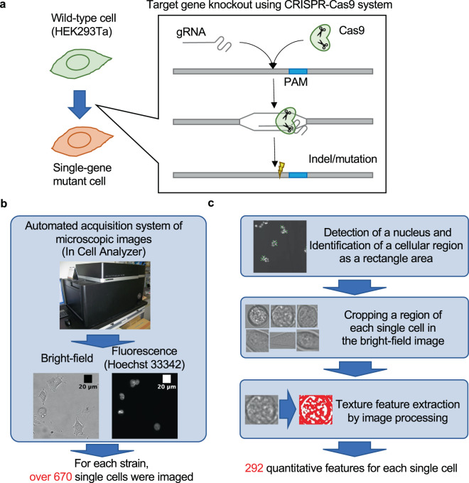
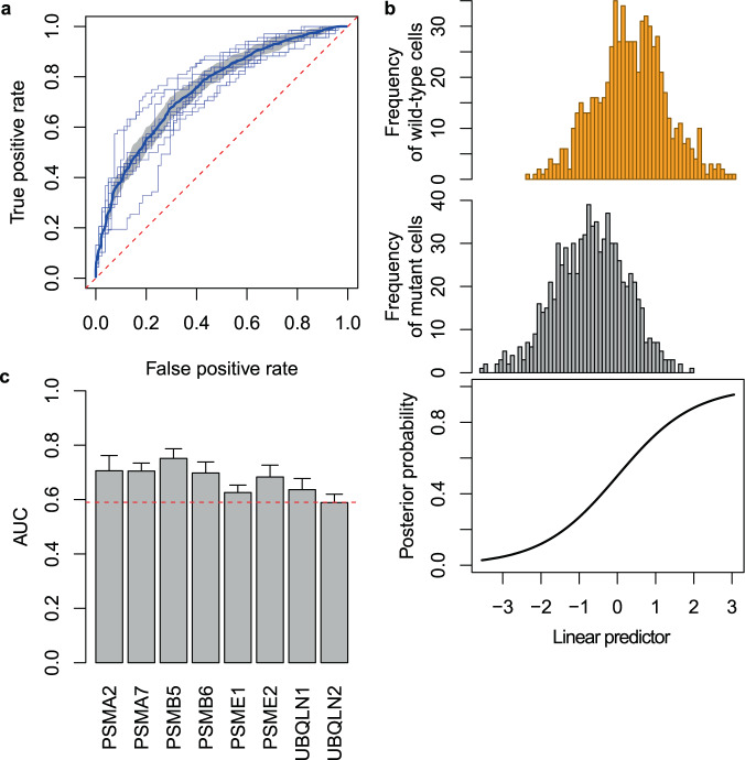
Morphological profiling is a combination of established optical microscopes and cutting-edge machine vision technologies, which stacks up successful applications in high-throughput phenotyping. One major question is how much information can be extracted from an image to identify genetic differences between cells. While fluorescent microscopy images of specific organelles have been broadly used for single-cell profiling, the potential ability of bright-field (BF) microscopy images of label-free cells remains to be tested. Here, we examine whether single-gene perturbation can be discriminated based on BF images of label-free cells using a machine learning approach. We acquired hundreds of BF images of single-gene mutant cells, quantified single-cell profiles consisting of texture features of cellular regions, and constructed a machine learning model to discriminate mutant cells from wild-type cells. Interestingly, the mutants were successfully discriminated from the wild type (area under the receiver operating characteristic curve = 0.773). The features that contributed to the discrimination were identified, and they included those related to the morphology of structures that appeared within cellular regions. Furthermore, functionally close gene pairs showed similar feature profiles of the mutant cells. Our study reveals that single-gene mutant cells can be discriminated from wild-type cells based on BF images, suggesting the potential as a useful tool for mutant cell profiling.
形态学分析是将成熟的光学显微镜与前沿的机器视觉技术相结合,在高通量表型分析中积累了成功的应用。一个主要问题是从图像中可以提取多少信息来识别细胞之间的遗传差异。虽然特定细胞器的荧光显微镜图像已广泛用于单细胞分析,但无标记细胞的明场(BF)显微镜图像的潜在能力仍有待测试。在这里,我们使用机器学习方法研究是否可以基于无标记细胞的BF图像区分单基因扰动。我们获取了数百张单基因突变体细胞的BF图像,量化了由细胞区域纹理特征组成的单细胞轮廓,并构建了一个机器学习模型来区分突变细胞和野生型细胞。有趣的是,突变体与野生型成功区分(受试者工作特征曲线下面积 = 0.773)。确定了有助于区分的特征,其中包括与细胞区域内出现的结构形态相关的特征。此外,功能相近的基因对显示出突变体细胞的相似特征谱。我们的研究表明,可以基于BF图像区分单基因突变体细胞和野生型细胞,这表明其作为突变体细胞分析有用工具的潜力。