Ghaffarinovin Zeinab, Soltaninia Omid, Mortazavi Yousef, Esmaeilzadeh Abdolreza, Nadri Samad
Department of Medical Biotechnology, School of Medicine, Zanjan University of Medical Sciences, Zanjan, Iran.
Department of Oral & Maxillofacial Surgery, Hamadan University of Medical Sciences, Hamadan, Iran.
Bioimpacts. 2021;11(3):209-217. doi: 10.34172/bi.2021.28. Epub 2020 Jul 8.
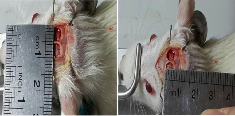
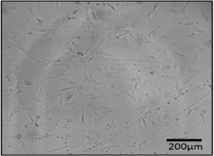
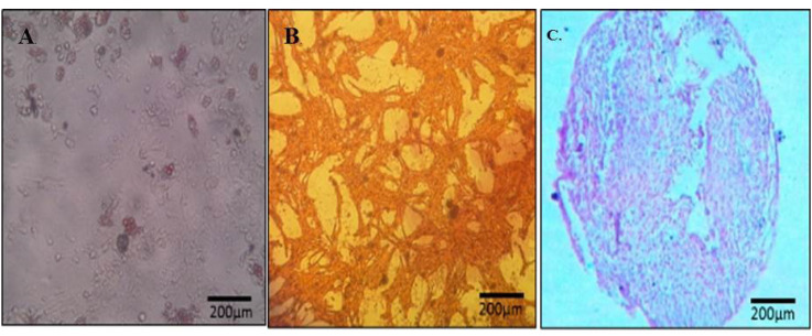
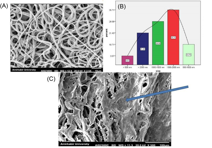
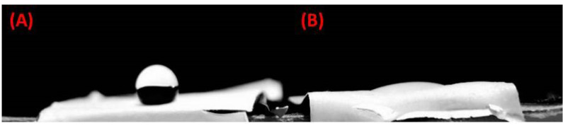
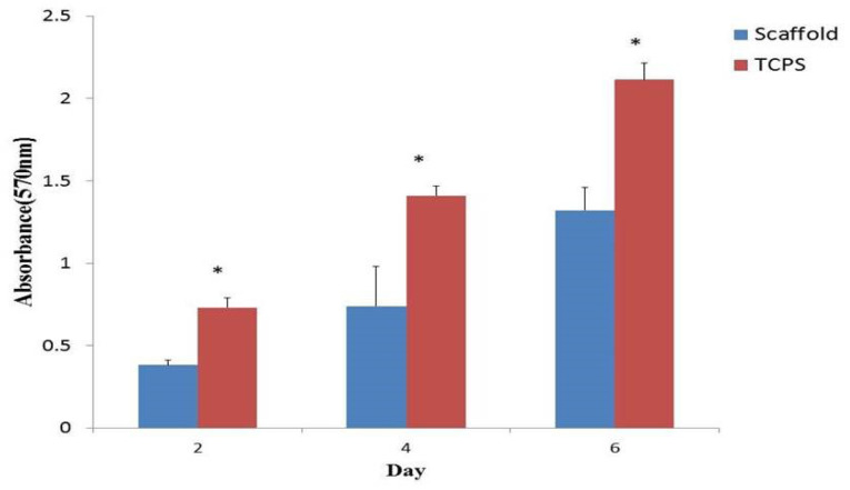
Tissue regenerative medicine strategies, as a promising alternative has become of major interest to the reconstruction of critical size bone defects. This study evaluated the effects of the simultaneous application of polycaprolactone (PCL), amniotic fluid mesenchymal stem cells (AF-MSCs) and platelet-rich plasma (PRP) on the repair of rat cranial bone defects. The AF-MSCs were isolated at the end of the second week of pregnancy in rats. PRP obtained from rat blood and the random PCL fibrous scaffolds were prepared using the electrospinning method. Circular full thickness (5 mm) bone defects were developed on both sides of the parietal bones (animal number=24) and the scaffolds containing AF-MSCs and PRP were implanted in the right lesions. Thereafter, after eight weeks the histological and immunohistochemistry studies were performed to evaluate the bone formation and collagen type I expression. The spindle-shaped mesenchymal stem cells were isolated and the electron microscope images indicated the preparation of a random PCL scaffold. Immunohistochemical findings showed that collagen type I was expressed by AF-MSCs cultured on the scaffold. The results of hematoxylin and eosin (H&E) staining indicated the formation of blood vessels in the presence of PRP. Additionally, immunofluorescence findings suggested that PRP had a positive effect on collagen type I expression. The simultaneous application of fibrous scaffold + AF-MSCs + PRP has positive effects on bone regeneration. This study showed that PRP can affect the formation of new blood vessels in the scaffold transplanted in the bone defect.
组织再生医学策略作为一种有前景的替代方法,已成为修复临界尺寸骨缺损的主要研究热点。本研究评估了聚己内酯(PCL)、羊水间充质干细胞(AF-MSCs)和富血小板血浆(PRP)联合应用对大鼠颅骨缺损修复的影响。在大鼠妊娠第二周结束时分离AF-MSCs。从大鼠血液中获取PRP,并采用静电纺丝法制备随机排列的PCL纤维支架。在顶骨两侧制造圆形全层(5毫米)骨缺损(动物数量=24),将含有AF-MSCs和PRP的支架植入右侧缺损处。此后,八周后进行组织学和免疫组织化学研究,以评估骨形成和I型胶原表达。分离出纺锤形间充质干细胞,电子显微镜图像显示制备了随机排列的PCL支架。免疫组织化学结果表明,在支架上培养的AF-MSCs表达I型胶原。苏木精和伊红(H&E)染色结果表明,在PRP存在的情况下形成了血管。此外,免疫荧光结果表明,PRP对I型胶原表达有积极影响。纤维支架+AF-MSCs+PRP联合应用对骨再生有积极作用。本研究表明,PRP可影响移植到骨缺损处的支架中新血管的形成。