• 文献检索
  • 文档翻译
  • 深度研究
  • 学术资讯
  • Suppr Zotero 插件Zotero 插件
  • 邀请有礼
  • 套餐&价格
  • 历史记录
应用&插件
Suppr Zotero 插件Zotero 插件浏览器插件Mac 客户端Windows 客户端微信小程序
定价
高级版会员购买积分包购买API积分包
服务
文献检索文档翻译深度研究API 文档MCP 服务
关于我们
关于 Suppr公司介绍联系我们用户协议隐私条款
关注我们

Suppr 超能文献

核心技术专利:CN118964589B侵权必究
粤ICP备2023148730 号-1Suppr @ 2026

文献检索

告别复杂PubMed语法,用中文像聊天一样搜索,搜遍4000万医学文献。AI智能推荐,让科研检索更轻松。

立即免费搜索

文件翻译

保留排版,准确专业,支持PDF/Word/PPT等文件格式,支持 12+语言互译。

免费翻译文档

深度研究

AI帮你快速写综述,25分钟生成高质量综述,智能提取关键信息,辅助科研写作。

立即免费体验

巨噬细胞样细胞密度在增殖性糖尿病性视网膜病变中增加,其特征为光相干断层扫描血管造影。

Macrophage-Like Cell Density Is Increased in Proliferative Diabetic Retinopathy Characterized by Optical Coherence Tomography Angiography.

机构信息

Department of Ophthalmology, Feinberg School of Medicine, Northwestern University, Chicago, Illinois, United States.

出版信息

Invest Ophthalmol Vis Sci. 2021 Aug 2;62(10):2. doi: 10.1167/iovs.62.10.2.

DOI:10.1167/iovs.62.10.2
PMID:34338748
原文链接:https://pmc.ncbi.nlm.nih.gov/articles/PMC8340655/
Abstract

PURPOSE

To quantitatively characterize macrophage-like cells (MLCs) at the vitreoretinal interface in different severity stages of diabetic retinopathy (DR) using optical coherence tomography angiography (OCTA).

METHODS

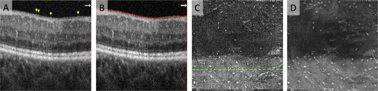
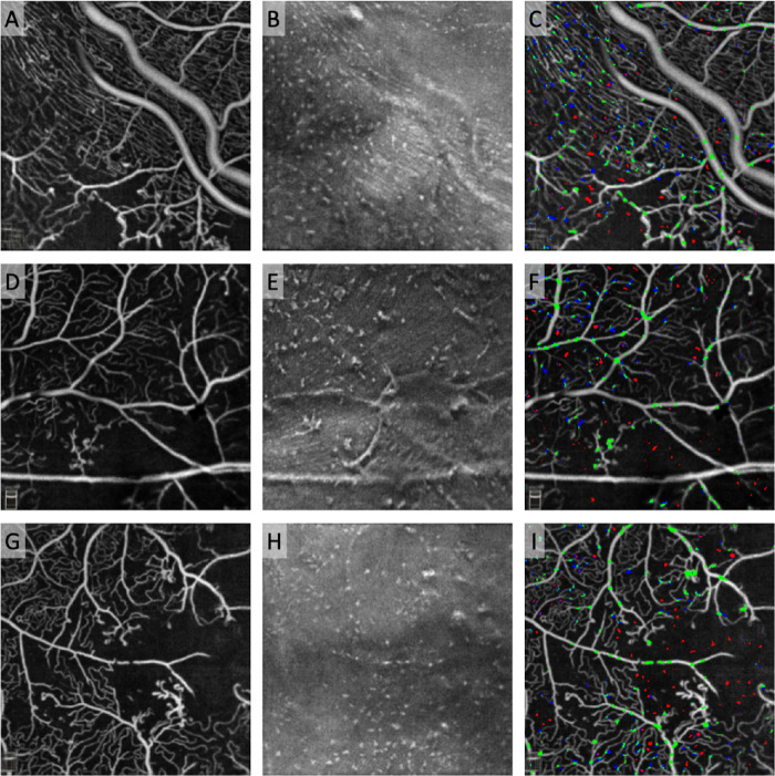
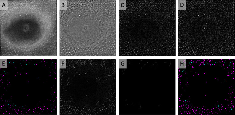
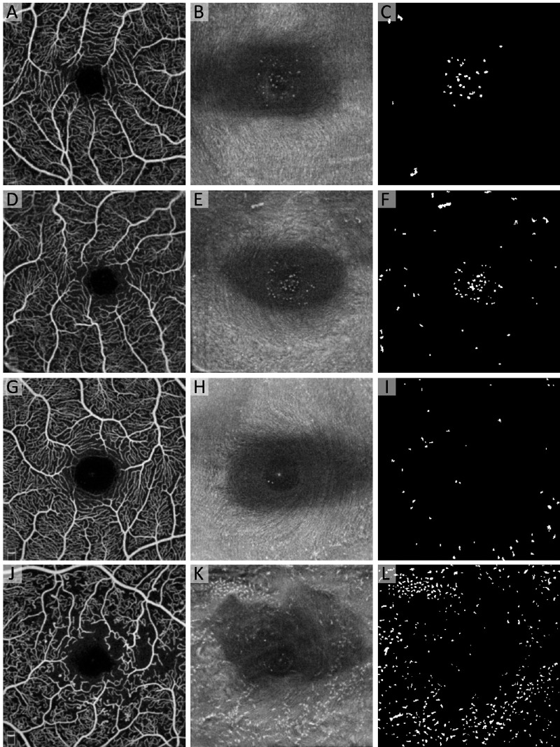
The study included 72 eyes of 72 subjects: 18 healthy controls, 22 diabetes mellitus (DM) without DR, 17 nonproliferative DR (NPDR), and 15 proliferative DR (PDR). We obtained repeated (average, 6.5; range, 3-10) macular OCTA scans for each eye. We registered and averaged the 3-µm OCT slab above the vitreoretinal interface to visualize MLCs. Using a semiautomated method, we binarized and quantified MLCs and compared MLC densities among groups. We also evaluated MLC distribution relative to underlying superficial capillary plexus vasculature and quantified MLCs overlying blood vessels within the perivascular 30-µm watershed region and within ischemic zones (defined as >30 µm from the nearest vessel).

RESULTS

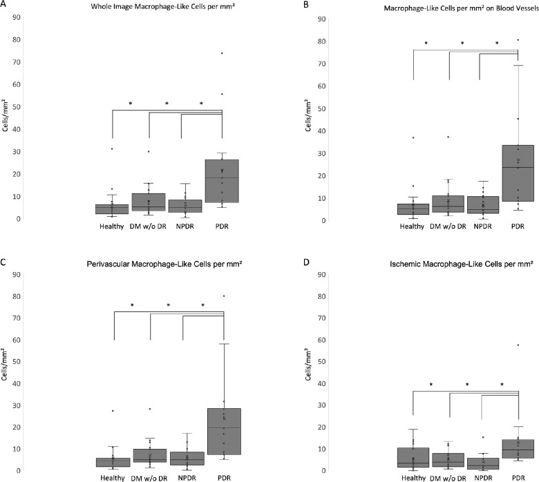
MLC density was 2.8- to 3.8-fold higher in PDR compared with all other groups (P < 0.05 for all). MLC density in PDR was most increased in perivascular areas (3.3- to 4.2-fold; P < 0.05 vs. all) and on blood vessels (3.0- to 4.0-fold; P < 0.05 vs. all), and elevated to a lesser extent in ischemic areas (2.3- to 3.4-fold; P < 0.05 vs. all). MLCs were more likely to localize on blood vessels in DM without DR, NPDR, and PDR (P < 0.05 for all), but not healthy eyes.

CONCLUSIONS

MLC density was significantly increased in PDR. MLCs clustered on blood vessels in diabetic but not in healthy eyes. Further studies are needed to confirm the origin, identity, and function of MLCs during DR.

摘要

目的

利用光相干断层扫描血管造影术(OCTA)定量描述不同严重程度糖尿病视网膜病变(DR)患者玻璃体视网膜界面处的巨噬细胞样细胞(MLCs)。

方法

该研究纳入了 72 只眼的 72 名受试者:18 名健康对照者、22 名无 DR 的糖尿病患者、17 名非增生性 DR(NPDR)患者和 15 名增生性 DR(PDR)患者。我们对每只眼进行了重复(平均 6.5 次;范围 3-10 次)黄斑 OCTA 扫描。我们在玻璃体视网膜界面上方注册并平均了 3µm OCT 切片以可视化 MLCs。使用半自动方法,我们对 MLCs 进行二值化并进行量化,并比较各组之间的 MLC 密度。我们还评估了 MLC 分布与下方浅层毛细血管丛血管的关系,并量化了血管周围 30µm 流域区域和缺血区域(定义为距离最近血管 >30µm)内的 MLCs。

结果

与所有其他组相比,PDR 中的 MLC 密度高 2.8-3.8 倍(所有 P<0.05)。PDR 中的 MLC 密度在血管周围区域(3.3-4.2 倍;与所有组相比 P<0.05)和血管上(3.0-4.0 倍;与所有组相比 P<0.05)增加最多,在缺血区域增加较少(2.3-3.4 倍;与所有组相比 P<0.05)。在无 DR 的糖尿病患者、NPDR 患者和 PDR 患者中,MLCs 更有可能定位于血管上(所有 P<0.05),而不是健康眼中。

结论

在 PDR 中,MLC 密度显著增加。在糖尿病患者中,MLCs 聚集在血管上,但在健康眼中则不然。需要进一步的研究来确认 DR 期间 MLC 的来源、身份和功能。

https://cdn.ncbi.nlm.nih.gov/pmc/blobs/c0ce/8340655/83947167148c/iovs-62-10-2-f007.jpg
https://cdn.ncbi.nlm.nih.gov/pmc/blobs/c0ce/8340655/0ee4d394520b/iovs-62-10-2-f001.jpg
https://cdn.ncbi.nlm.nih.gov/pmc/blobs/c0ce/8340655/e2c89748ec06/iovs-62-10-2-f002.jpg
https://cdn.ncbi.nlm.nih.gov/pmc/blobs/c0ce/8340655/f49b1667d8e8/iovs-62-10-2-f003.jpg
https://cdn.ncbi.nlm.nih.gov/pmc/blobs/c0ce/8340655/a75236d04fd3/iovs-62-10-2-f004.jpg
https://cdn.ncbi.nlm.nih.gov/pmc/blobs/c0ce/8340655/473ce640f0f1/iovs-62-10-2-f005.jpg
https://cdn.ncbi.nlm.nih.gov/pmc/blobs/c0ce/8340655/5f98cfc5bb47/iovs-62-10-2-f006.jpg
https://cdn.ncbi.nlm.nih.gov/pmc/blobs/c0ce/8340655/83947167148c/iovs-62-10-2-f007.jpg

相似文献

1
Macrophage-Like Cell Density Is Increased in Proliferative Diabetic Retinopathy Characterized by Optical Coherence Tomography Angiography.巨噬细胞样细胞密度在增殖性糖尿病性视网膜病变中增加,其特征为光相干断层扫描血管造影。
Invest Ophthalmol Vis Sci. 2021 Aug 2;62(10):2. doi: 10.1167/iovs.62.10.2.
2
Proliferative diabetic retinopathy and diabetic macular edema are two factors that increase macrophage-like cell density characterized by en face optical coherence tomography.增生性糖尿病视网膜病变和糖尿病性黄斑水肿是两种会增加细胞密度的因素,其特征可以通过光学相干断层扫描术进行检测。
BMC Ophthalmol. 2023 Feb 1;23(1):46. doi: 10.1186/s12886-023-02794-8.
3
Association between macrophage-like cell density and ischemia metrics in diabetic eyes.糖尿病性眼病中巨噬细胞样细胞密度与缺血指标之间的关联。
Exp Eye Res. 2023 Dec;237:109703. doi: 10.1016/j.exer.2023.109703. Epub 2023 Oct 28.
4
Interaction Between the Distribution of Diabetic Retinopathy Lesions and the Association of Optical Coherence Tomography Angiography Scans With Diabetic Retinopathy Severity.糖尿病视网膜病变病变分布与光学相干断层扫描血管造影扫描与糖尿病视网膜病变严重程度的相关性分析。
JAMA Ophthalmol. 2020 Dec 1;138(12):1291-1297. doi: 10.1001/jamaophthalmol.2020.4516.
5
Elevated number and density of macrophage-like cell as a novel inflammation biomarker in diabetic macular edema.巨噬细胞样细胞数量和密度升高可作为糖尿病性黄斑水肿的新型炎症生物标志物。
Sci Rep. 2023 Mar 31;13(1):5320. doi: 10.1038/s41598-023-32455-1.
6
Macular Capillary Perfusion in Chinese Patients With Diabetic Retinopathy Obtained With Optical Coherence Tomography Angiography.光学相干断层扫描血管造影术检测中国糖尿病视网膜病变患者的黄斑区毛细血管灌注情况
Ophthalmic Surg Lasers Imaging Retina. 2019 Apr 1;50(4):e88-e95. doi: 10.3928/23258160-20190401-12.
7
Linking Retinal Microvasculature Features With Severity of Diabetic Retinopathy Using Optical Coherence Tomography Angiography.使用光学相干断层扫描血管造影术将视网膜微血管特征与糖尿病性视网膜病变的严重程度相联系。
Invest Ophthalmol Vis Sci. 2016 Jul 1;57(9):OCT519-25. doi: 10.1167/iovs.15-18901.
8
QUANTIFICATION OF RETINAL VESSEL TORTUOSITY IN DIABETIC RETINOPATHY USING OPTICAL COHERENCE TOMOGRAPHY ANGIOGRAPHY.应用光学相干断层扫描血管造影术定量分析糖尿病性视网膜病变中的视网膜血管迂曲。
Retina. 2018 May;38(5):976-985. doi: 10.1097/IAE.0000000000001618.
9
Deep Capillary Macular Perfusion Indices Obtained with OCT Angiography Correlate with Degree of Nonproliferative Diabetic Retinopathy.通过光学相干断层扫描血管造影获得的深部黄斑毛细血管灌注指数与非增殖性糖尿病视网膜病变程度相关。
Eur J Ophthalmol. 2017 Nov 8;27(6):716-729. doi: 10.5301/ejo.5000948.
10
Choriocapillaris and retinal vascular plexus density of diabetic eyes using split-spectrum amplitude decorrelation spectral-domain optical coherence tomography angiography.应用谱域光相干断层扫描血管成像术分析糖尿病患者脉络膜毛细血管和视网膜血管丛密度。
Br J Ophthalmol. 2019 Apr;103(4):452-456. doi: 10.1136/bjophthalmol-2018-311903. Epub 2018 May 23.

引用本文的文献

1
Elevated hyperreflective foci as a novel characteristic in idiopathic epiretinal membrane by optical coherence tomography angiography.光学相干断层扫描血管造影显示,高反射灶增强是特发性视网膜前膜的一种新特征。
BMC Ophthalmol. 2025 Feb 25;25(1):89. doi: 10.1186/s12886-025-03929-9.
2
Association and multimodal model of retinal and blood-based biomarkers for detection of preclinical Alzheimer's disease.用于检测临床前阿尔茨海默病的视网膜和血液生物标志物的关联及多模态模型
Alzheimers Res Ther. 2025 Jan 10;17(1):19. doi: 10.1186/s13195-024-01668-5.
3
Retinal perivascular macrophages regulate immune cell infiltration during neuroinflammation in mouse models of ocular disease.

本文引用的文献

1
Label-free adaptive optics imaging of human retinal macrophage distribution and dynamics.无标记自适应光学成像技术对人视网膜巨噬细胞分布和动态的研究。
Proc Natl Acad Sci U S A. 2020 Dec 1;117(48):30661-30669. doi: 10.1073/pnas.2010943117. Epub 2020 Nov 9.
2
Geometric Perfusion Deficits: A Novel OCT Angiography Biomarker for Diabetic Retinopathy Based on Oxygen Diffusion.几何性灌注缺损:基于氧扩散的糖尿病视网膜病变新型 OCT 血管造影生物标志物
Am J Ophthalmol. 2021 Feb;222:256-270. doi: 10.1016/j.ajo.2020.09.007. Epub 2020 Sep 9.
3
Glycolysis links reciprocal activation of myeloid cells and endothelial cells in the retinal angiogenic niche.
视网膜血管周巨噬细胞在眼部疾病小鼠模型的神经炎症期间调节免疫细胞浸润。
J Clin Invest. 2024 Aug 29;134(20):e180904. doi: 10.1172/JCI180904.
4
Redefining the ontogeny of hyalocytes as yolk sac-derived tissue-resident macrophages of the vitreous body.重新定义玻璃体内的原纤维细胞为卵黄囊来源的组织驻留巨噬细胞。
J Neuroinflammation. 2024 Jul 3;21(1):168. doi: 10.1186/s12974-024-03110-x.
5
Vogt-Koyanagi-Harada Disease: A Narrative Review.伏格特-小柳-原田病:一篇叙述性综述
Cureus. 2024 Apr 23;16(4):e58867. doi: 10.7759/cureus.58867. eCollection 2024 Apr.
6
Vitreoretinal Interface Cells Correlate In Vivo With Uveitis Activity and Decrease With Anti-Inflammatory Treatment.玻璃体视网膜界面细胞与葡萄膜炎活动相关,并随抗炎治疗而减少。
Transl Vis Sci Technol. 2024 May 1;13(5):15. doi: 10.1167/tvst.13.5.15.
7
Quantifying Putative Retinal Gliosis in Preclinical Alzheimer's Disease.定量临床前阿尔茨海默病中的疑似视网膜神经胶质增生。
Invest Ophthalmol Vis Sci. 2024 May 1;65(5):5. doi: 10.1167/iovs.65.5.5.
8
Association between macrophage-like cell density and ischemia metrics in diabetic eyes.糖尿病性眼病中巨噬细胞样细胞密度与缺血指标之间的关联。
Exp Eye Res. 2023 Dec;237:109703. doi: 10.1016/j.exer.2023.109703. Epub 2023 Oct 28.
9
Visualizing features with wide-field volumetric OCT angiography.宽场 OCT 血管造影术可视化特征。
Opt Express. 2024 Mar 11;32(6):10329-10347. doi: 10.1364/OE.510640.
10
Hyalocytes-guardians of the vitreoretinal interface.玻璃体细胞——视网膜界面的守护者。
Graefes Arch Clin Exp Ophthalmol. 2024 Sep;262(9):2765-2784. doi: 10.1007/s00417-024-06448-3. Epub 2024 Apr 3.
糖酵解在视网膜血管生成微环境中连接了髓样细胞和内皮细胞的相互激活。
Sci Transl Med. 2020 Aug 5;12(555). doi: 10.1126/scitranslmed.aay1371.
4
Imaging of Macrophage-Like Cells in Living Human Retina Using Clinical OCT.临床 OCT 对活体人视网膜内类巨噬细胞成像。
Invest Ophthalmol Vis Sci. 2020 Jun 3;61(6):48. doi: 10.1167/iovs.61.6.48.
5
Glial cells of the human fovea.人黄斑区胶质细胞。
Mol Vis. 2020 Apr 1;26:235-245. eCollection 2020.
6
Assessing the Use of Incorrectly Scaled Optical Coherence Tomography Angiography Images in Peer-Reviewed Studies: A Systematic Review.评估同行评审研究中使用错误缩放光学相干断层扫描血管造影图像的情况:系统评价。
JAMA Ophthalmol. 2020 Jan 1;138(1):86-94. doi: 10.1001/jamaophthalmol.2019.4821.
7
Mo-derived perivascular macrophage recruitment protects against endothelial cell death in retinal vein occlusion.Mo 衍生的血管周巨噬细胞募集可防止视网膜静脉阻塞中的内皮细胞死亡。
J Neuroinflammation. 2019 Jul 27;16(1):157. doi: 10.1186/s12974-019-1547-8.
8
Influence of Axial Length on Parafoveal and Peripapillary Metrics from Swept Source Optical Coherence Tomography Angiography.扫频源光学相干断层血管造影术(SS-OCTA)中眼轴长度对视盘旁和旁中心凹区参数的影响。
Curr Eye Res. 2019 Sep;44(9):980-986. doi: 10.1080/02713683.2019.1607393. Epub 2019 Apr 29.
9
Microglial Function Is Distinct in Different Anatomical Locations during Retinal Homeostasis and Degeneration.在视网膜稳态和变性过程中,不同解剖位置的小胶质细胞功能不同。
Immunity. 2019 Mar 19;50(3):723-737.e7. doi: 10.1016/j.immuni.2019.02.007. Epub 2019 Mar 5.
10
Hemodynamic Response of the Three Macular Capillary Plexuses in Dark Adaptation and Flicker Stimulation Using Optical Coherence Tomography Angiography.利用光相干断层扫描血管造影术观察暗适应和闪烁刺激下三个黄斑毛细血管丛的血流动力学反应。
Invest Ophthalmol Vis Sci. 2019 Feb 1;60(2):694-703. doi: 10.1167/iovs.18-25478.