• 文献检索
  • 文档翻译
  • 深度研究
  • 学术资讯
  • Suppr Zotero 插件Zotero 插件
  • 邀请有礼
  • 套餐&价格
  • 历史记录
应用&插件
Suppr Zotero 插件Zotero 插件浏览器插件Mac 客户端Windows 客户端微信小程序
定价
高级版会员购买积分包购买API积分包
服务
文献检索文档翻译深度研究API 文档MCP 服务
关于我们
关于 Suppr公司介绍联系我们用户协议隐私条款
关注我们

Suppr 超能文献

核心技术专利:CN118964589B侵权必究
粤ICP备2023148730 号-1Suppr @ 2026

文献检索

告别复杂PubMed语法,用中文像聊天一样搜索,搜遍4000万医学文献。AI智能推荐,让科研检索更轻松。

立即免费搜索

文件翻译

保留排版,准确专业,支持PDF/Word/PPT等文件格式,支持 12+语言互译。

免费翻译文档

深度研究

AI帮你快速写综述,25分钟生成高质量综述,智能提取关键信息,辅助科研写作。

立即免费体验

基于 U-Net 的深度学习自动分割扩散加权图像中的直肠肿瘤。

Automatic segmentation of rectal tumor on diffusion-weighted images by deep learning with U-Net.

机构信息

Department of Radiology, Key Laboratory of Carcinogenesis and Translational Research (Ministry of Education/Beijing), Peking University Cancer Hospital & Institute, Beijing, China.

出版信息

J Appl Clin Med Phys. 2021 Sep;22(9):324-331. doi: 10.1002/acm2.13381. Epub 2021 Aug 3.

DOI:10.1002/acm2.13381
PMID:34343402
原文链接:https://pmc.ncbi.nlm.nih.gov/articles/PMC8425941/
Abstract

PURPOSE

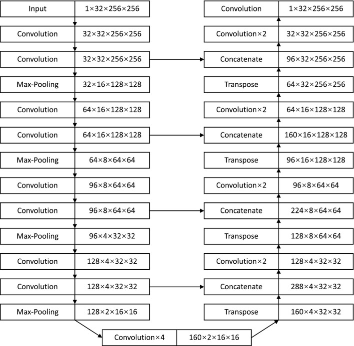
Manual delineation of a rectal tumor on a volumetric image is time-consuming and subjective. Deep learning has been used to segment rectal tumors automatically on T2-weighted images, but automatic segmentation on diffusion-weighted imaging is challenged by noise, artifact, and low resolution. In this study, a volumetric U-shaped neural network (U-Net) is proposed to automatically segment rectal tumors on diffusion-weighted images.

METHODS

Three hundred patients of locally advanced rectal cancer were enrolled in this study and divided into a training group, a validation group, and a test group. The region of rectal tumor was delineated on the diffusion-weighted images by experienced radiologists as the ground truth. A U-Net was designed with a volumetric input of the diffusion-weighted images and an output of segmentation with the same size. A semi-automatic segmentation method was used for comparison by manually choosing a threshold of gray level and automatically selecting the largest connected region. Dice similarity coefficient (DSC) was calculated to evaluate the methods.

RESULTS

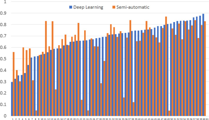
On the test group, deep learning method (DSC = 0.675 ± 0.144, median DSC is 0.702, maximum DSC is 0.893, and minimum DSC is 0.297) showed higher segmentation accuracy than the semi-automatic method (DSC = 0.614 ± 0.225, median DSC is 0.685, maximum DSC is 0.869, and minimum DSC is 0.047). Paired t-test shows significant difference (T = 2.160, p = 0.035) in DSC between the deep learning method and the semi-automatic method in the test group.

CONCLUSION

Volumetric U-Net can automatically segment rectal tumor region on DWI images of locally advanced rectal cancer.

摘要

目的

在容积图像上手动勾画直肠肿瘤费时且主观。深度学习已被用于在 T2 加权图像上自动分割直肠肿瘤,但在扩散加权成像上的自动分割受到噪声、伪影和低分辨率的挑战。在本研究中,提出了一种用于在扩散加权图像上自动分割直肠肿瘤的容积 U 形神经网络(U-Net)。

方法

本研究纳入了 300 例局部进展期直肠癌患者,将其分为训练组、验证组和测试组。由经验丰富的放射科医生在扩散加权图像上勾画直肠肿瘤区域作为金标准。设计了一个具有扩散加权图像容积输入和相同大小分割输出的 U-Net。通过手动选择灰度级阈值和自动选择最大连通区域,比较了一种半自动分割方法。计算了 Dice 相似系数(DSC)以评估方法。

结果

在测试组中,深度学习方法(DSC=0.675±0.144,中位数 DSC 为 0.702,最大 DSC 为 0.893,最小 DSC 为 0.297)的分割准确性高于半自动方法(DSC=0.614±0.225,中位数 DSC 为 0.685,最大 DSC 为 0.869,最小 DSC 为 0.047)。配对 t 检验显示,在测试组中,深度学习方法与半自动方法在 DSC 上的差异具有统计学意义(T=2.160,p=0.035)。

结论

容积 U-Net 可自动分割局部进展期直肠癌 DWI 图像上的直肠肿瘤区域。

https://cdn.ncbi.nlm.nih.gov/pmc/blobs/dbe4/8425941/5a17aadfdb35/ACM2-22-324-g001.jpg
https://cdn.ncbi.nlm.nih.gov/pmc/blobs/dbe4/8425941/3debc3193a89/ACM2-22-324-g002.jpg
https://cdn.ncbi.nlm.nih.gov/pmc/blobs/dbe4/8425941/c0f26c6b2990/ACM2-22-324-g003.jpg
https://cdn.ncbi.nlm.nih.gov/pmc/blobs/dbe4/8425941/9fdfe8f0dc62/ACM2-22-324-g004.jpg
https://cdn.ncbi.nlm.nih.gov/pmc/blobs/dbe4/8425941/5a17aadfdb35/ACM2-22-324-g001.jpg

相似文献

1
Automatic segmentation of rectal tumor on diffusion-weighted images by deep learning with U-Net.基于 U-Net 的深度学习自动分割扩散加权图像中的直肠肿瘤。
J Appl Clin Med Phys. 2021 Sep;22(9):324-331. doi: 10.1002/acm2.13381. Epub 2021 Aug 3.
2
Technical Note: A deep learning-based autosegmentation of rectal tumors in MR images.技术说明:基于深度学习的磁共振图像中直肠肿瘤的自动分割。
Med Phys. 2018 Jun;45(6):2560-2564. doi: 10.1002/mp.12918. Epub 2018 May 3.
3
Deep learning for fully automated tumor segmentation and extraction of magnetic resonance radiomics features in cervical cancer.深度学习在宫颈癌全自动肿瘤分割和磁共振影像组学特征提取中的应用。
Eur Radiol. 2020 Mar;30(3):1297-1305. doi: 10.1007/s00330-019-06467-3. Epub 2019 Nov 11.
4
MRI-based automatic segmentation of rectal cancer using 2D U-Net on two independent cohorts.基于 MRI 的使用二维 U-Net 在两个独立队列上对直肠癌的自动分割。
Acta Oncol. 2022 Feb;61(2):255-263. doi: 10.1080/0284186X.2021.2013530. Epub 2021 Dec 17.
5
Fully automatic segmentation on prostate MR images based on cascaded fully convolution network.基于级联全卷积网络的前列腺磁共振图像全自动分割。
J Magn Reson Imaging. 2019 Apr;49(4):1149-1156. doi: 10.1002/jmri.26337. Epub 2018 Oct 22.
6
Deep Learning for Fully-Automated Localization and Segmentation of Rectal Cancer on Multiparametric MR.深度学习在多参数磁共振直肠癌全自动定位和分割中的应用
Sci Rep. 2017 Jul 13;7(1):5301. doi: 10.1038/s41598-017-05728-9.
7
Automatic segmentation of the clinical target volume and organs at risk in the planning CT for rectal cancer using deep dilated convolutional neural networks.使用深度扩张卷积神经网络在直肠癌计划 CT 中自动分割临床靶区和危及器官。
Med Phys. 2017 Dec;44(12):6377-6389. doi: 10.1002/mp.12602. Epub 2017 Oct 28.
8
Automatic intraprostatic lesion segmentation in multiparametric magnetic resonance images with proposed multiple branch UNet.利用提出的多分支U-Net在多参数磁共振图像中实现前列腺内病变的自动分割。
Med Phys. 2020 Dec;47(12):6421-6429. doi: 10.1002/mp.14517. Epub 2020 Oct 24.
9
Evaluation of Diffusion Lesion Volume Measurements in Acute Ischemic Stroke Using Encoder-Decoder Convolutional Network.基于编解码器卷积网络的急性缺血性脑卒中弥散病变体积测量评估。
Stroke. 2019 Jun;50(6):1444-1451. doi: 10.1161/STROKEAHA.118.024261. Epub 2019 May 16.
10
Postoperative glioma segmentation in CT image using deep feature fusion model guided by multi-sequence MRIs.基于多序列 MRI 引导的深度特征融合模型的 CT 图像术后脑肿瘤分割。
Eur Radiol. 2020 Feb;30(2):823-832. doi: 10.1007/s00330-019-06441-z. Epub 2019 Oct 24.

引用本文的文献

1
[Research progress on colorectal cancer identification based on convolutional neural network].基于卷积神经网络的结直肠癌识别研究进展
Sheng Wu Yi Xue Gong Cheng Xue Za Zhi. 2024 Aug 25;41(4):854-860. doi: 10.7507/1001-5515.202310027.
2
Deep learning for MRI lesion segmentation in rectal cancer.深度学习用于直肠癌磁共振成像病变分割
Front Med (Lausanne). 2024 Jun 25;11:1394262. doi: 10.3389/fmed.2024.1394262. eCollection 2024.
3
Performance reporting design in artificial intelligence studies using image-based TNM staging and prognostic parameters in rectal cancer: a systematic review.

本文引用的文献

1
A Stacked Generalization U-shape network based on zoom strategy and its application in biomedical image segmentation.基于变焦策略的堆叠泛化 U 形网络及其在生物医学图像分割中的应用。
Comput Methods Programs Biomed. 2020 Dec;197:105678. doi: 10.1016/j.cmpb.2020.105678. Epub 2020 Jul 30.
2
Predicting Rectal Cancer Response to Neoadjuvant Chemoradiotherapy Using Deep Learning of Diffusion Kurtosis MRI.利用扩散峰度磁共振成像的深度学习预测直肠癌新辅助放化疗反应。
Radiology. 2020 Jul;296(1):56-64. doi: 10.1148/radiol.2020190936. Epub 2020 Apr 21.
3
Accurate colorectal tumor segmentation for CT scans based on the label assignment generative adversarial network.
基于图像的TNM分期和直肠癌预后参数的人工智能研究中的性能报告设计:一项系统综述
Ann Coloproctol. 2024 Feb;40(1):13-26. doi: 10.3393/ac.2023.00892.0127. Epub 2024 Feb 28.
4
Deep-neural network approaches for predicting 3D dose distribution in intensity-modulated radiotherapy of the brain tumors.用于预测脑肿瘤调强放疗中三维剂量分布的深度神经网络方法。
J Appl Clin Med Phys. 2024 Mar;25(3):e14197. doi: 10.1002/acm2.14197. Epub 2023 Nov 7.
5
Intratumoral and peritumoral MRI-based radiomics prediction of histopathological grade in soft tissue sarcomas: a two-center study.基于 MRI 的肿瘤内和肿瘤周围放射组学预测软组织肉瘤的组织病理学分级:一项双中心研究。
Cancer Imaging. 2023 Oct 26;23(1):103. doi: 10.1186/s40644-023-00622-2.
6
A boundary-guided transformer for measuring distance from rectal tumor to anal verge on magnetic resonance images.一种用于在磁共振图像上测量直肠肿瘤与肛缘距离的边界引导变压器。
Patterns (N Y). 2023 Mar 27;4(4):100711. doi: 10.1016/j.patter.2023.100711. eCollection 2023 Apr 14.
7
Segmentation of rectal tumor from multi-parametric MRI images using an attention-based fusion network.基于注意力融合网络的多参数 MRI 图像中直肠肿瘤的分割。
Med Biol Eng Comput. 2023 Sep;61(9):2379-2389. doi: 10.1007/s11517-023-02828-9. Epub 2023 Apr 21.
8
Application of artificial intelligence in diagnosis and treatment of colorectal cancer: A novel Prospect.人工智能在结直肠癌诊疗中的应用:一种新前景。
Front Med (Lausanne). 2023 Mar 8;10:1128084. doi: 10.3389/fmed.2023.1128084. eCollection 2023.
9
Application of Radiomics in Predicting Treatment Response to Neoadjuvant Chemoradiotherapy in Locally Advanced Rectal Cancer: Strategies and Challenges.放射组学在预测局部晚期直肠癌新辅助放化疗治疗反应中的应用:策略与挑战
J Oncol. 2022 Nov 26;2022:1590620. doi: 10.1155/2022/1590620. eCollection 2022.
10
Review of Radiomics- and Dosiomics-based Predicting Models for Rectal Cancer.基于影像组学和剂量组学的直肠癌预测模型综述
Front Oncol. 2022 Aug 9;12:913683. doi: 10.3389/fonc.2022.913683. eCollection 2022.
基于标签分配生成对抗网络的 CT 扫描中准确的结直肠肿瘤分割。
Med Phys. 2019 Aug;46(8):3532-3542. doi: 10.1002/mp.13584. Epub 2019 Jun 25.
4
Full convolutional network based multiple side-output fusion architecture for the segmentation of rectal tumors in magnetic resonance images: A multi-vendor study.基于全卷积网络的多侧输出融合架构在磁共振图像中直肠肿瘤分割中的应用:一项多厂商研究。
Med Phys. 2019 Jun;46(6):2659-2668. doi: 10.1002/mp.13541. Epub 2019 Apr 29.
5
Automated Segmentation of Colorectal Tumor in 3D MRI Using 3D Multiscale Densely Connected Convolutional Neural Network.基于三维多尺度密集连接卷积神经网络的 3D MRI 结直肠肿瘤自动分割。
J Healthc Eng. 2019 Jan 31;2019:1075434. doi: 10.1155/2019/1075434. eCollection 2019.
6
Quantitative analysis of diffusion weighted imaging to predict pathological good response to neoadjuvant chemoradiation for locally advanced rectal cancer.定量分析扩散加权成像预测局部晚期直肠癌新辅助放化疗病理良好反应。
Radiother Oncol. 2019 Mar;132:100-108. doi: 10.1016/j.radonc.2018.11.007. Epub 2018 Dec 21.
7
Cascaded atrous convolution and spatial pyramid pooling for more accurate tumor target segmentation for rectal cancer radiotherapy.级联空洞卷积和空间金字塔池化以提高直肠癌放疗中肿瘤靶区分割的准确性。
Phys Med Biol. 2018 Sep 17;63(18):185016. doi: 10.1088/1361-6560/aada6c.
8
Rectal Cancer, Version 2.2018, NCCN Clinical Practice Guidelines in Oncology.直肠癌临床实践指南(NCCN 肿瘤学版)2018 年第 2 版
J Natl Compr Canc Netw. 2018 Jul;16(7):874-901. doi: 10.6004/jnccn.2018.0061.
9
Technical Note: A deep learning-based autosegmentation of rectal tumors in MR images.技术说明:基于深度学习的磁共振图像中直肠肿瘤的自动分割。
Med Phys. 2018 Jun;45(6):2560-2564. doi: 10.1002/mp.12918. Epub 2018 May 3.
10
Fully convolutional networks (FCNs)-based segmentation method for colorectal tumors on T2-weighted magnetic resonance images.基于全卷积网络(FCNs)的T2加权磁共振图像上结直肠肿瘤分割方法
Australas Phys Eng Sci Med. 2018 Jun;41(2):393-401. doi: 10.1007/s13246-018-0636-9. Epub 2018 Apr 13.