• 文献检索
  • 文档翻译
  • 深度研究
  • 学术资讯
  • Suppr Zotero 插件Zotero 插件
  • 邀请有礼
  • 套餐&价格
  • 历史记录
应用&插件
Suppr Zotero 插件Zotero 插件浏览器插件Mac 客户端Windows 客户端微信小程序
定价
高级版会员购买积分包购买API积分包
服务
文献检索文档翻译深度研究API 文档MCP 服务
关于我们
关于 Suppr公司介绍联系我们用户协议隐私条款
关注我们

Suppr 超能文献

核心技术专利:CN118964589B侵权必究
粤ICP备2023148730 号-1Suppr @ 2026

文献检索

告别复杂PubMed语法,用中文像聊天一样搜索,搜遍4000万医学文献。AI智能推荐,让科研检索更轻松。

立即免费搜索

文件翻译

保留排版,准确专业,支持PDF/Word/PPT等文件格式,支持 12+语言互译。

免费翻译文档

深度研究

AI帮你快速写综述,25分钟生成高质量综述,智能提取关键信息,辅助科研写作。

立即免费体验

脑型疟疾:光学相干断层扫描对病理学的洞察

Cerebral malaria: insight into pathology from optical coherence tomography.

作者信息

Tu Zhanhan, Gormley Jack, Sheth Viral, Seydel Karl B, Taylor Terrie, Beare Nicholas, Barrera Valentina, Proudlock Frank A, Manda Chatonda, Harding Simon, Gottlob Irene

机构信息

Department of Neuroscience, Psychology and Behaviour, Ulverscroft Eye Unit, University of Leicester, Robert Kilpatrick Clinical Sciences Building, Leicester Royal Infirmary, Leicester, LE2 7LX, UK.

Department of Eye and Vision Science, Institute of Ageing and Chronic Disease, University of Liverpool, Member of Liverpool Health Partners, Liverpool, UK.

出版信息

Sci Rep. 2021 Aug 3;11(1):15722. doi: 10.1038/s41598-021-94495-9.

DOI:10.1038/s41598-021-94495-9
PMID:34344903
原文链接:https://pmc.ncbi.nlm.nih.gov/articles/PMC8333417/
Abstract

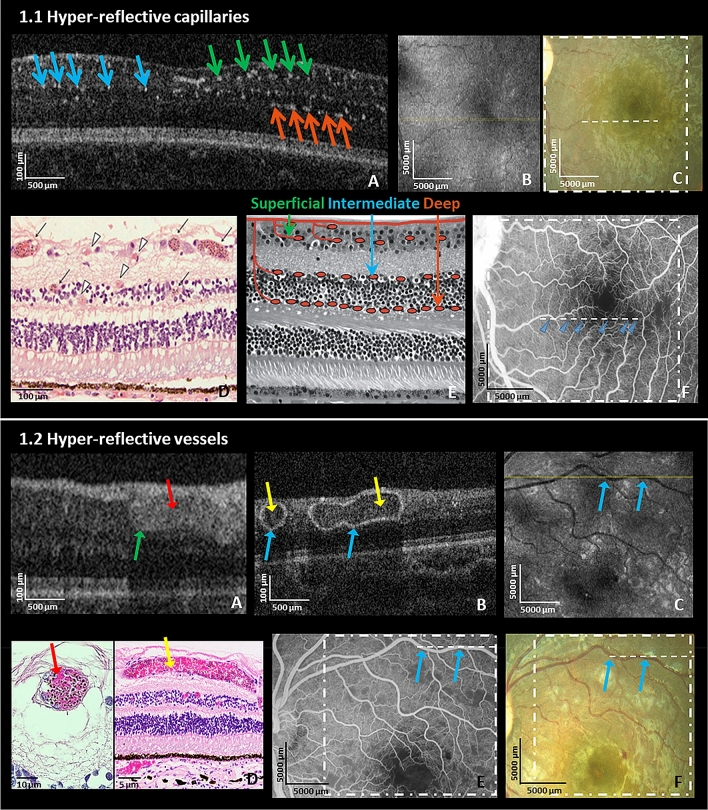
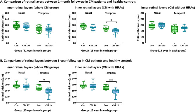
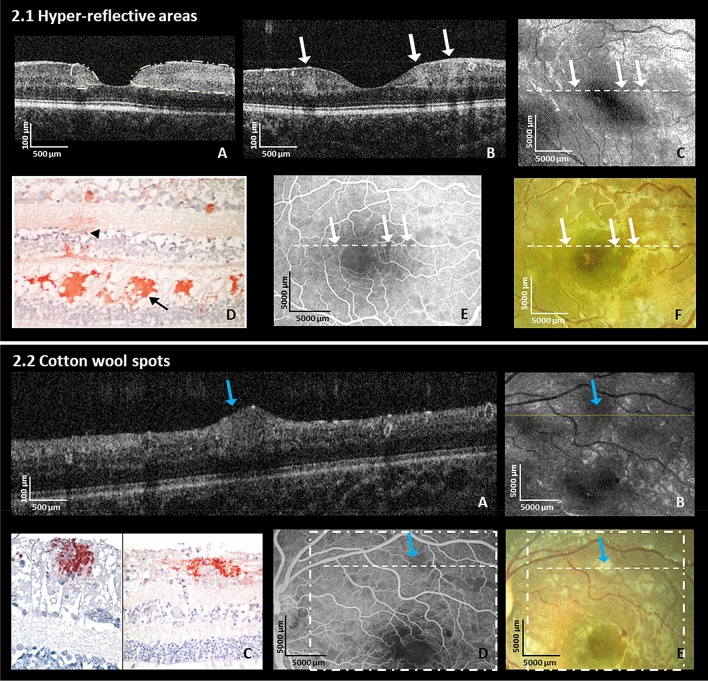
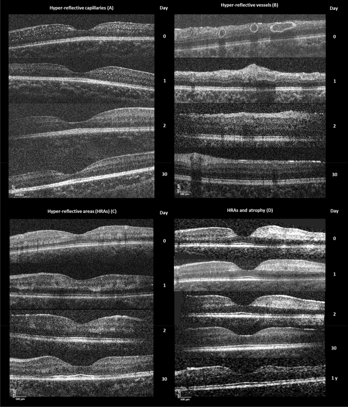
We aimed to investigate structural retinal changes in malarial retinopathy (MR) using hand-held optical coherence tomography (HH-OCT) to assess its diagnostic potential. Children with MR (n = 43) underwent ophthalmoscopy, fluorescein angiography and HH-OCT during admission, 1-month (n = 31) and 1-year (n = 8) post-discharge. Controls were comatose patients without malaria (n = 6) and age/sex-matched healthy children (n = 43). OCT changes and retinal layer thicknesses were compared. On HH-OCT, hyper-reflective areas (HRAs) were seen in the inner retina of 81% of MR patients, corresponding to ischaemic retinal whitening on fundus photography. Cotton wool spots were present in 37% and abnormal hyper-reflective dots, co-localized to capillary plexus, in 93%. Hyper-reflective vessel walls were present in 84%, and intra-retinal cysts in 9%. Vascular changes and cysts resolved within 48 h. HRAs developed into retinal thinning at 1 month (p = 0.027) which was more pronounced after 1 year (p = 0.009). Ischaemic retinal whitening is located within inner retinal layers, distinguishing it from cotton wool spots. Vascular hyper-reflectivity may represent the sequestration of parasitized erythrocytes in vessels, a key CM feature. The mechanisms of post-ischemic retinal atrophy and cerebral atrophy with cognitive impairment may be similar in CM survivors. HH-OCT has the potential for monitoring patients, treatment response and predicting neurological deficits.

摘要

我们旨在利用手持式光学相干断层扫描(HH-OCT)研究疟疾性视网膜病变(MR)的视网膜结构变化,以评估其诊断潜力。患有MR的儿童(n = 43)在入院期间、出院后1个月(n = 31)和1年(n = 8)接受了眼底检查、荧光素血管造影和HH-OCT检查。对照组为无疟疾的昏迷患者(n = 6)和年龄/性别匹配的健康儿童(n = 43)。比较了OCT变化和视网膜层厚度。在HH-OCT上,81%的MR患者视网膜内层可见高反射区(HRA),对应于眼底照片上的缺血性视网膜变白。37%的患者出现棉絮斑,93%的患者在毛细血管丛处出现异常高反射点。84%的患者出现高反射血管壁,9%的患者出现视网膜内囊肿。血管变化和囊肿在48小时内消失。HRA在1个月时发展为视网膜变薄(p = 0.027),1年后更为明显(p = 0.009)。缺血性视网膜变白位于视网膜内层,与棉絮斑不同。血管高反射可能代表寄生虫感染的红细胞在血管中的滞留,这是脑型疟疾的一个关键特征。在脑型疟疾幸存者中,缺血后视网膜萎缩和伴有认知障碍的脑萎缩机制可能相似。HH-OCT有潜力监测患者、治疗反应并预测神经功能缺损。

https://cdn.ncbi.nlm.nih.gov/pmc/blobs/4ac1/8333417/e6d0fe5ab8ac/41598_2021_94495_Fig4_HTML.jpg
https://cdn.ncbi.nlm.nih.gov/pmc/blobs/4ac1/8333417/a7c719b6595b/41598_2021_94495_Fig1_HTML.jpg
https://cdn.ncbi.nlm.nih.gov/pmc/blobs/4ac1/8333417/0f977e234575/41598_2021_94495_Fig2_HTML.jpg
https://cdn.ncbi.nlm.nih.gov/pmc/blobs/4ac1/8333417/108837e6d79c/41598_2021_94495_Fig3_HTML.jpg
https://cdn.ncbi.nlm.nih.gov/pmc/blobs/4ac1/8333417/e6d0fe5ab8ac/41598_2021_94495_Fig4_HTML.jpg

相似文献

1
Cerebral malaria: insight into pathology from optical coherence tomography.脑型疟疾:光学相干断层扫描对病理学的洞察
Sci Rep. 2021 Aug 3;11(1):15722. doi: 10.1038/s41598-021-94495-9.
2
Redefining cerebral malaria by including malaria retinopathy.重新定义脑型疟疾,纳入疟疾视网膜病变。
Future Microbiol. 2011 Mar;6(3):349-55. doi: 10.2217/fmb.11.3.
3
Spectral domain optical coherence tomography findings in eyes with acute ischaemic retinal whitening.急性缺血性视网膜变白患者眼部的频域光学相干断层扫描结果
Br J Ophthalmol. 2015 May;99(5):586-92. doi: 10.1136/bjophthalmol-2014-304900. Epub 2014 Jul 3.
4
In vivo histology of cotton-wool spots using high-resolution optical coherence tomography.使用高分辨率光学相干断层扫描技术对棉絮状斑进行的体内组织学研究。
Am J Ophthalmol. 2006 Apr;141(4):748-50. doi: 10.1016/j.ajo.2005.10.048.
5
Retinopathy in severe malaria in Ghanaian children--overlap between fundus changes in cerebral and non-cerebral malaria.加纳儿童严重疟疾性视网膜病变——脑性疟疾和非脑性疟疾眼底改变的重叠。
Malar J. 2010 Aug 12;9:232. doi: 10.1186/1475-2875-9-232.
6
Optical coherence tomography angiography characterization of acute and late stage Purtscher retinopathy.急性和晚期普尔夏视网膜病变的光学相干断层扫描血管造影特征
Eur J Ophthalmol. 2018 Jul;28(4):NP1-NP6. doi: 10.1177/1120672118769788. Epub 2018 Apr 18.
7
Reversibility of retinal microvascular changes in severe falciparum malaria.重症恶性疟视网膜微血管变化的可逆性
Am J Trop Med Hyg. 2014 Sep;91(3):493-495. doi: 10.4269/ajtmh.14-0116. Epub 2014 Jun 16.
8
Hyperreflective sign in resolved cotton wool spots using high-resolution optical coherence tomography and optical coherence tomography ophthalmoscopy.使用高分辨率光学相干断层扫描和光学相干断层扫描检眼镜观察棉絮斑消退后的高反射征。
Ophthalmology. 2007 Mar;114(3):537-43. doi: 10.1016/j.ophtha.2006.06.054.
9
PARACENTRAL ACUTE MIDDLE MACULOPATHY ASSOCIATED WITH SEVERE PLASMODIUM FALCIPARUM MALARIA.伴有严重恶性疟原虫疟疾的中心旁急性黄斑病变
Retin Cases Brief Rep. 2024 Jan 1;18(1):47-50. doi: 10.1097/ICB.0000000000001330.
10
Photographic and angiographic characterization of the retina of Kenyan children with severe malaria.患有严重疟疾的肯尼亚儿童视网膜的影像学和血管造影特征
Arch Ophthalmol. 1997 Aug;115(8):997-1003. doi: 10.1001/archopht.1997.01100160167005.

引用本文的文献

1
Type II Acute Macular Neuroretinopathy Secondary to Malaria.疟疾继发的II型急性黄斑神经视网膜病变。
Case Rep Ophthalmol Med. 2024 May 29;2024:1577127. doi: 10.1155/2024/1577127. eCollection 2024.
2
Predicting Acute and Post-Recovery Outcomes in Cerebral Malaria and Other Comas by Optical Coherence Tomography (OCT in CM) - A protocol for an observational cohort study of Malawian children.通过光学相干断层扫描预测脑型疟疾和其他昏迷的急性及恢复后结局(脑型疟疾中的光学相干断层扫描)——一项针对马拉维儿童的观察性队列研究方案
Wellcome Open Res. 2023 Apr 14;8:172. doi: 10.12688/wellcomeopenres.19166.1. eCollection 2023.
3
Interplay Between the Immune and Nervous Cognitive Systems in Homeostasis and in Malaria.

本文引用的文献

1
A retinal model of cerebral malaria.脑型疟疾的视网膜模型。
Sci Rep. 2019 Mar 5;9(1):3470. doi: 10.1038/s41598-019-39143-z.
2
Neurodevelopmental Impairments 1 Year After Cerebral Malaria.脑疟疾 1 年后的神经发育损伤。
Pediatrics. 2019 Feb;143(2). doi: 10.1542/peds.2018-1026. Epub 2019 Jan 29.
3
Neurovascular sequestration in paediatric malaria is visible clinically in the retina.神经血管隔离在儿科疟疾中在视网膜上临床可见。
免疫和神经认知系统在稳态和疟疾中的相互作用。
Int J Biol Sci. 2023 Jun 28;19(11):3383-3394. doi: 10.7150/ijbs.82556. eCollection 2023.
4
Retinal imaging technologies in cerebral malaria: a systematic review.脑型疟疾中的视网膜成像技术:系统评价。
Malar J. 2023 Apr 26;22(1):139. doi: 10.1186/s12936-023-04566-7.
5
Hyperreflective spots in cerebral malaria.脑型疟疾中的高反射点。
Eye (Lond). 2023 Oct;37(15):3295. doi: 10.1038/s41433-023-02485-0. Epub 2023 Mar 10.
6
Cerebral malaria-using the retina to study the brain.脑疟疾——从视网膜研究大脑。
Eye (Lond). 2023 Aug;37(12):2379-2384. doi: 10.1038/s41433-023-02432-z. Epub 2023 Feb 14.
7
Identifying effective diagnostic biomarkers for childhood cerebral malaria in Africa integrating coexpression analysis with machine learning algorithm.利用共表达分析与机器学习算法整合,鉴定非洲儿童疟疾的有效诊断生物标志物。
Eur J Med Res. 2023 Feb 13;28(1):76. doi: 10.1186/s40001-022-00980-w.
8
Automated malarial retinopathy detection using transfer learning and multi-camera retinal images.使用迁移学习和多相机视网膜图像的自动疟疾性视网膜病变检测
Biocybern Biomed Eng. 2023 Jan-Mar;43(1):109-123. doi: 10.1016/j.bbe.2022.12.003. Epub 2022 Dec 21.
9
Clinical aspects of malarial retinopathy: a critical review.疟原虫性视网膜病变的临床方面:批判性评价。
Pathog Glob Health. 2023 Jul;117(5):450-461. doi: 10.1080/20477724.2022.2128568. Epub 2022 Oct 19.
10
Handheld optical coherence tomography removes barriers to imaging the eyes of young children.手持式光学相干断层扫描技术消除了对幼儿眼部进行成像的障碍。
Eye (Lond). 2022 May;36(5):907-908. doi: 10.1038/s41433-021-01884-5. Epub 2022 Jan 10.
Elife. 2018 Mar 26;7:e32208. doi: 10.7554/eLife.32208.
4
A review of optical coherence tomography angiography (OCTA).光学相干断层扫描血管造影术(OCTA)综述。
Int J Retina Vitreous. 2015 Apr 15;1:5. doi: 10.1186/s40942-015-0005-8. eCollection 2015.
5
Optical coherence tomography angiography in paracentral acute middle maculopathy secondary to central retinal vein occlusion.光学相干断层扫描血管造影在视网膜中央静脉阻塞继发的黄斑中心凹旁急性中浆病中的应用
Eye (Lond). 2016 Jun;30(6):888-93. doi: 10.1038/eye.2016.57. Epub 2016 Apr 1.
6
Acute macular neuroretinopathy: A comprehensive review of the literature.急性黄斑神经视网膜病变:文献综述
Surv Ophthalmol. 2016 Sep-Oct;61(5):538-65. doi: 10.1016/j.survophthal.2016.03.003. Epub 2016 Mar 10.
7
Grading fluorescein angiograms in malarial retinopathy.疟疾性视网膜病变中荧光素血管造影的分级
Malar J. 2015 Sep 24;14:367. doi: 10.1186/s12936-015-0897-7.
8
Paracentral Acute Middle Maculopathy: What We Knew Then and What We Know Now.中心旁急性黄斑中部病变:我们当时所知与现在所知
Retina. 2015 Oct;35(10):1921-30. doi: 10.1097/IAE.0000000000000785.
9
Brain swelling and death in children with cerebral malaria.脑肿胀和死亡的儿童与脑疟疾。
N Engl J Med. 2015 Mar 19;372(12):1126-37. doi: 10.1056/NEJMoa1400116.
10
Severity of retinopathy parallels the degree of parasite sequestration in the eyes and brains of malawian children with fatal cerebral malaria.视网膜病变的严重程度与患有致命性脑型疟疾的马拉维儿童眼睛和大脑中的寄生虫隔离程度相当。
J Infect Dis. 2015 Jun 15;211(12):1977-86. doi: 10.1093/infdis/jiu592. Epub 2014 Oct 28.