Lin Yi-Jia, Chao Tai-Kuang, Khalil Muhammad-Adil, Lee Yu-Ching, Hong Ding-Zhi, Wu Jia-Jhen, Wang Ching-Wei
Department of Pathology, Tri-Service General Hospital, Taipei 11490, Taiwan.
Institute of Pathology and Parasitology, National Defense Medical Center, Taipei 11490, Taiwan.
Cancers (Basel). 2021 Aug 2;13(15):3891. doi: 10.3390/cancers13153891.
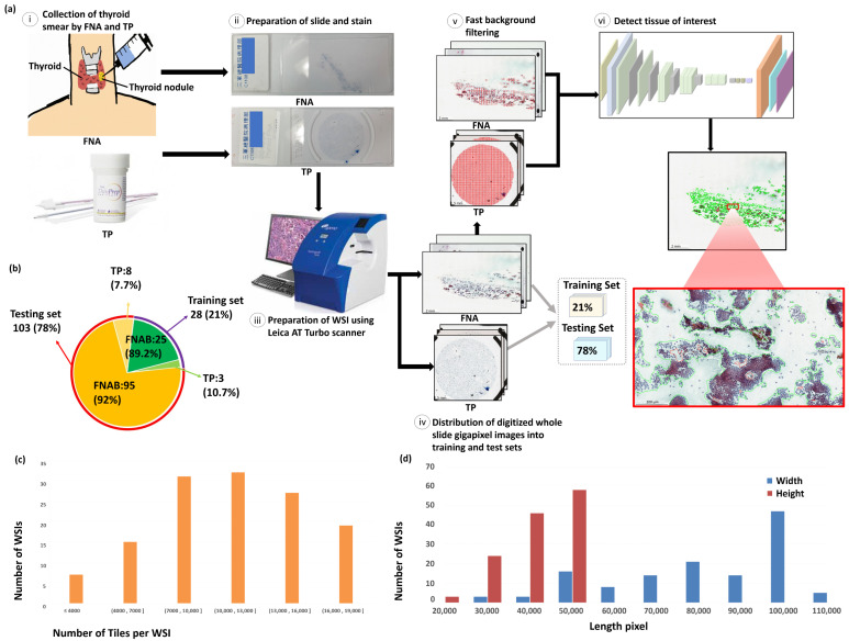
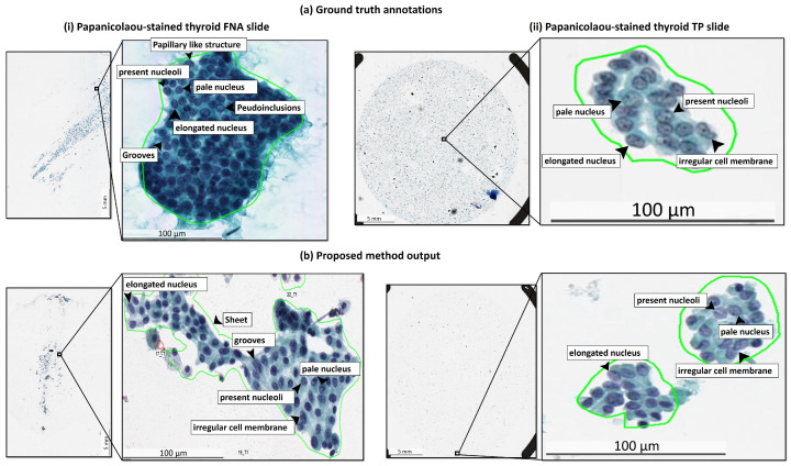
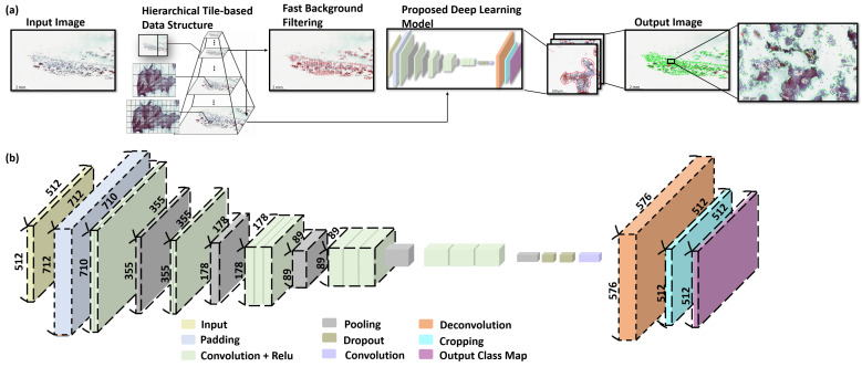
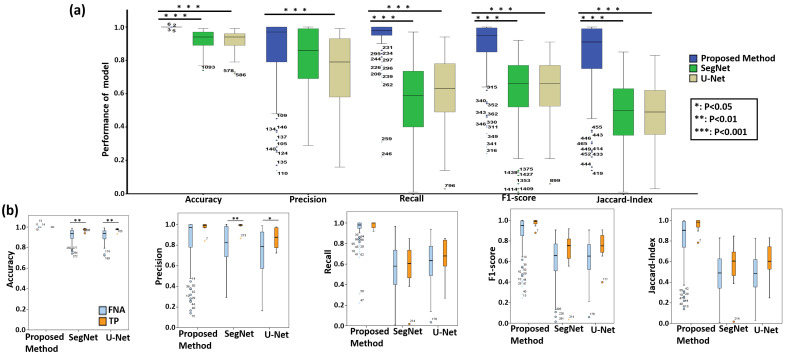
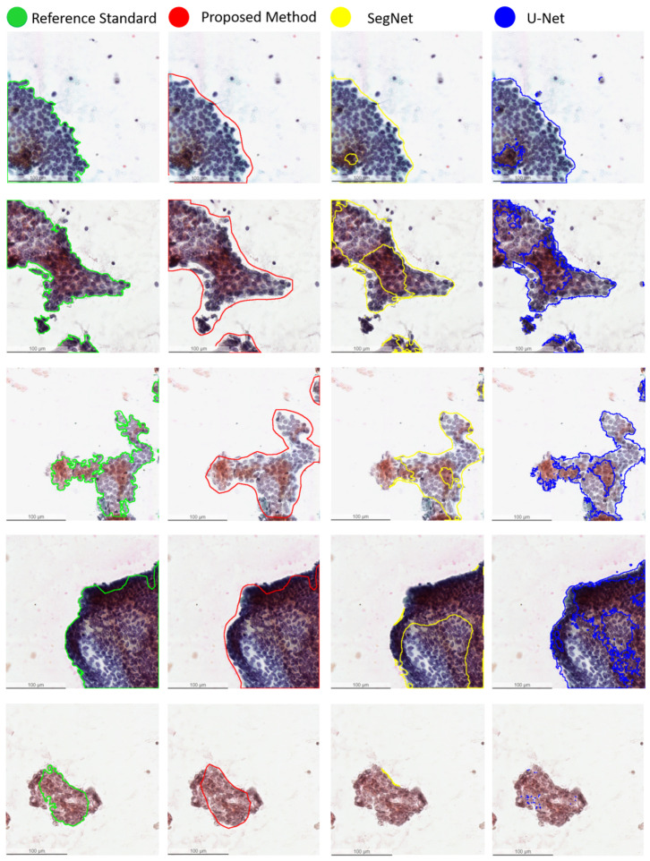
Thyroid cancer is the most common cancer in the endocrine system, and papillary thyroid carcinoma (PTC) is the most prevalent type of thyroid cancer, accounting for 70 to 80% of all thyroid cancer cases. In clinical practice, visual inspection of cytopathological slides is an essential initial method used by the pathologist to diagnose PTC. Manual visual assessment of the whole slide images is difficult, time consuming, and subjective, with a high inter-observer variability, which can sometimes lead to suboptimal patient management due to false-positive and false-negative. In this study, we present a fully automatic, efficient, and fast deep learning framework for fast screening of papanicolaou-stained thyroid fine needle aspiration (FNA) and ThinPrep (TP) cytological slides. To the authors' best of knowledge, this work is the first study to build an automated deep learning framework for identification of PTC from both FNA and TP slides. The proposed deep learning framework is evaluated on a dataset of 131 WSIs, and the results show that the proposed method achieves an accuracy of 99%, precision of 85%, recall of 94% and F1-score of 87% in segmentation of PTC in FNA slides and an accuracy of 99%, precision of 97%, recall of 98%, F1-score of 98%, and Jaccard-Index of 96% in TP slides. In addition, the proposed method significantly outperforms the two state-of-the-art deep learning methods, i.e., U-Net and SegNet, in terms of accuracy, recall, F1-score, and Jaccard-Index (p<0.001). Furthermore, for run-time analysis, the proposed fast screening method takes 0.4 min to process a WSI and is 7.8 times faster than U-Net and 9.1 times faster than SegNet, respectively.
甲状腺癌是内分泌系统中最常见的癌症,而乳头状甲状腺癌(PTC)是甲状腺癌中最常见的类型,占所有甲状腺癌病例的70%至80%。在临床实践中,对细胞病理切片进行目视检查是病理学家诊断PTC的重要初始方法。对整个玻片图像进行人工目视评估既困难又耗时,而且主观,观察者间差异很大,有时会因假阳性和假阴性导致患者管理欠佳。在本研究中,我们提出了一个全自动、高效且快速的深度学习框架,用于快速筛查巴氏染色的甲状腺细针穿刺(FNA)和液基薄层制片(TP)细胞学玻片。据作者所知,这项工作是第一项构建用于从FNA和TP玻片识别PTC的自动化深度学习框架的研究。所提出的深度学习框架在一个包含131个全切片图像(WSI)的数据集上进行了评估,结果表明,所提出的方法在FNA玻片中PTC分割方面的准确率为99%、精确率为85%、召回率为94%、F1分数为87%,在TP玻片中的准确率为99%、精确率为97%、召回率为98%、F1分数为98%、杰卡德指数为96%。此外,在准确率、召回率、F1分数和杰卡德指数方面,所提出的方法显著优于两种最先进的深度学习方法,即U-Net和SegNet(p<0.001)。此外,对于运行时间分析,所提出的快速筛查方法处理一个WSI需要0.4分钟,分别比U-Net快7.8倍,比SegNet快9.1倍。