• 文献检索
  • 文档翻译
  • 深度研究
  • 学术资讯
  • Suppr Zotero 插件Zotero 插件
  • 邀请有礼
  • 套餐&价格
  • 历史记录
应用&插件
Suppr Zotero 插件Zotero 插件浏览器插件Mac 客户端Windows 客户端微信小程序
定价
高级版会员购买积分包购买API积分包
服务
文献检索文档翻译深度研究API 文档MCP 服务
关于我们
关于 Suppr公司介绍联系我们用户协议隐私条款
关注我们

Suppr 超能文献

核心技术专利:CN118964589B侵权必究
粤ICP备2023148730 号-1Suppr @ 2026

文献检索

告别复杂PubMed语法,用中文像聊天一样搜索,搜遍4000万医学文献。AI智能推荐,让科研检索更轻松。

立即免费搜索

文件翻译

保留排版,准确专业,支持PDF/Word/PPT等文件格式,支持 12+语言互译。

免费翻译文档

深度研究

AI帮你快速写综述,25分钟生成高质量综述,智能提取关键信息,辅助科研写作。

立即免费体验

用于乳腺癌诊断的苏木精-伊红全切片图像中转移灶的快速分割

Fast Segmentation of Metastatic Foci in H&E Whole-Slide Images for Breast Cancer Diagnosis.

作者信息

Khalil Muhammad-Adil, Lee Yu-Ching, Lien Huang-Chun, Jeng Yung-Ming, Wang Ching-Wei

机构信息

Graduate Institute of Applied Science and Technology, National Taiwan University of Science and Technology, Taipei 106335, Taiwan.

Department of Pathology, National Taiwan University Hospital, Taipei 100229, Taiwan.

出版信息

Diagnostics (Basel). 2022 Apr 14;12(4):990. doi: 10.3390/diagnostics12040990.

DOI:10.3390/diagnostics12040990
PMID:35454038
原文链接:https://pmc.ncbi.nlm.nih.gov/articles/PMC9030573/
Abstract

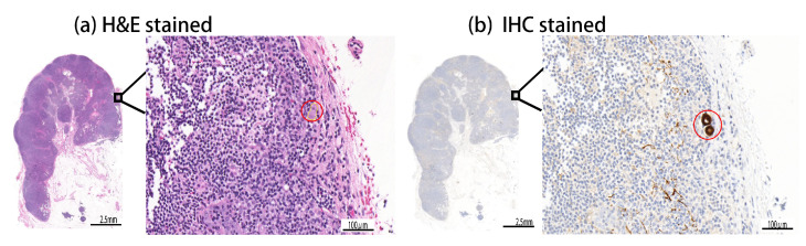
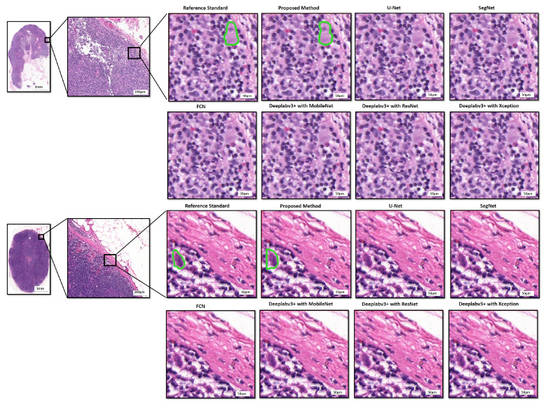
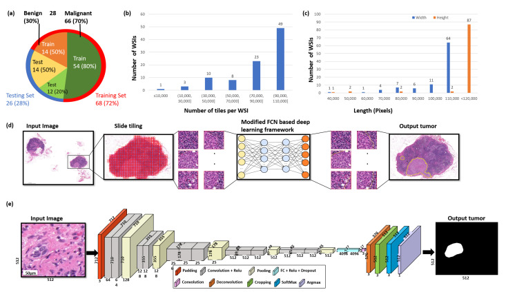
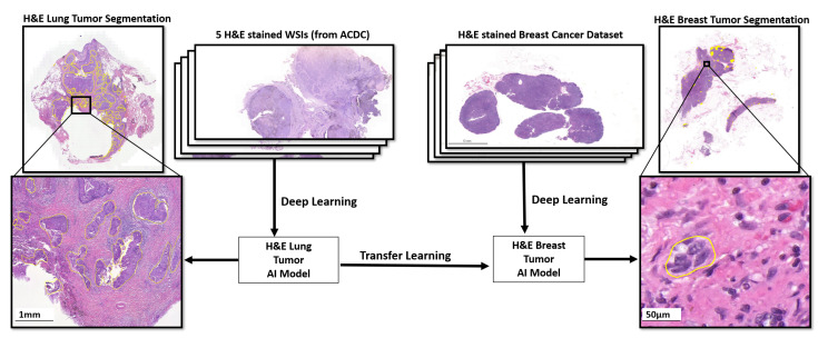
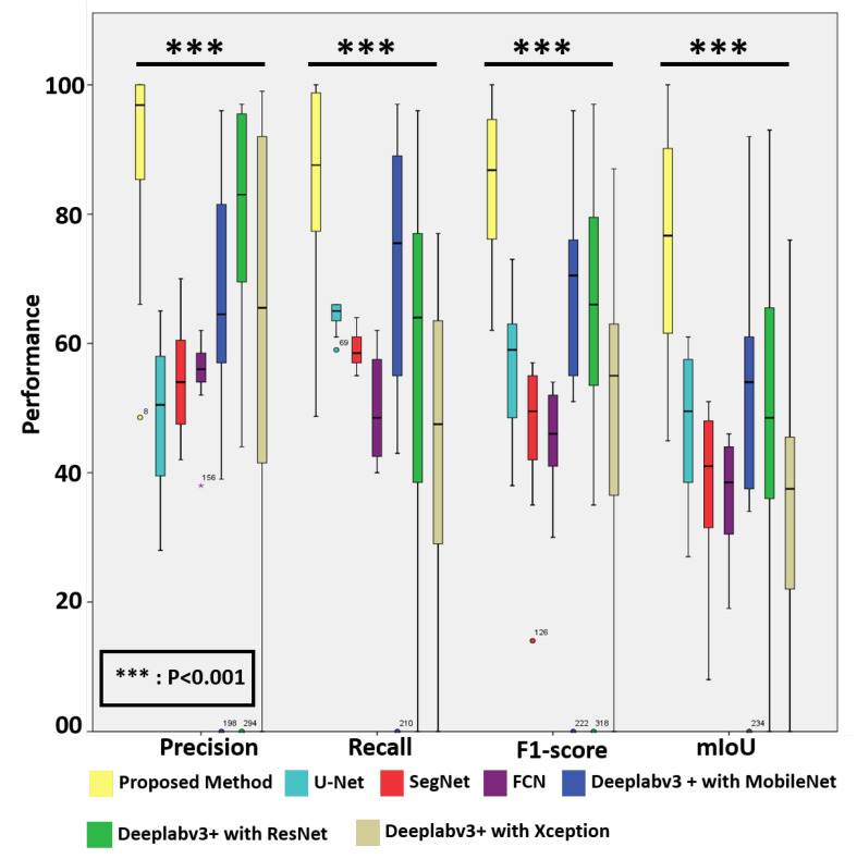
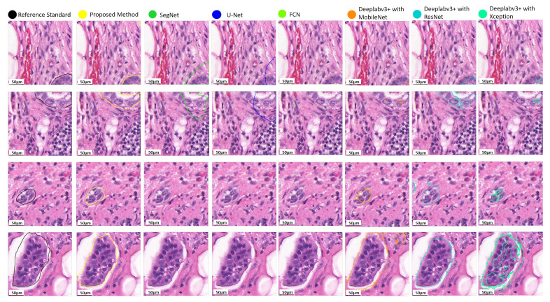
Breast cancer is the leading cause of death for women globally. In clinical practice, pathologists visually scan over enormous amounts of gigapixel microscopic tissue slide images, which is a tedious and challenging task. In breast cancer diagnosis, micro-metastases and especially isolated tumor cells are extremely difficult to detect and are easily neglected because tiny metastatic foci might be missed in visual examinations by medical doctors. However, the literature poorly explores the detection of isolated tumor cells, which could be recognized as a viable marker to determine the prognosis for T1NoMo breast cancer patients. To address these issues, we present a deep learning-based framework for efficient and robust lymph node metastasis segmentation in routinely used histopathological hematoxylin−eosin-stained (H−E) whole-slide images (WSI) in minutes, and a quantitative evaluation is conducted using 188 WSIs, containing 94 pairs of H−E-stained WSIs and immunohistochemical CK(AE1/AE3)-stained WSIs, which are used to produce a reliable and objective reference standard. The quantitative results demonstrate that the proposed method achieves 89.6% precision, 83.8% recall, 84.4% F1-score, and 74.9% mIoU, and that it performs significantly better than eight deep learning approaches, including two recently published models (v3_DCNN and Xception-65), and three variants of Deeplabv3+ with three different backbones, namely, U-Net, SegNet, and FCN, in precision, recall, F1-score, and mIoU (p<0.001). Importantly, the proposed system is shown to be capable of identifying tiny metastatic foci in challenging cases, for which there are high probabilities of misdiagnosis in visual inspection, while the baseline approaches tend to fail in detecting tiny metastatic foci. For computational time comparison, the proposed method takes 2.4 min for processing a WSI utilizing four NVIDIA Geforce GTX 1080Ti GPU cards and 9.6 min using a single NVIDIA Geforce GTX 1080Ti GPU card, and is notably faster than the baseline methods (4-times faster than U-Net and SegNet, 5-times faster than FCN, 2-times faster than the 3 different variants of Deeplabv3+, 1.4-times faster than v3_DCNN, and 41-times faster than Xception-65).

摘要

乳腺癌是全球女性的主要死因。在临床实践中,病理学家需要目视扫描大量数十亿像素的微观组织切片图像,这是一项繁琐且具有挑战性的任务。在乳腺癌诊断中,微转移灶,尤其是孤立肿瘤细胞极难检测,且容易被忽视,因为医生在目视检查时可能会遗漏微小的转移灶。然而,目前文献对孤立肿瘤细胞检测的探讨较少,而孤立肿瘤细胞可被视为确定T1NoMo期乳腺癌患者预后的一个可行标志物。为解决这些问题,我们提出了一个基于深度学习的框架,可在数分钟内对常规使用的苏木精-伊红染色(H-E)全切片图像(WSI)中的淋巴结转移进行高效且稳健的分割,并使用188张WSI进行定量评估,其中包括94对H-E染色的WSI和免疫组化CK(AE1/AE3)染色的WSI,用于生成可靠且客观的参考标准。定量结果表明,所提出的方法实现了89.6%的精度、83.8%的召回率、84.4%的F1分数和74.9%的平均交并比,并且在精度、召回率、F1分数和平均交并比方面,其性能显著优于八种深度学习方法,包括两个最近发表的模型(v3_DCNN和Xception-65),以及带有三种不同骨干网络的Deeplabv3+的三个变体,即U-Net、SegNet和FCN(p<0.001)。重要的是,所提出的系统能够在具有挑战性的病例中识别微小转移灶,而在目视检查中这些病例很可能被误诊,而基线方法往往无法检测到微小转移灶。在计算时间比较方面,所提出的方法使用四张NVIDIA Geforce GTX 1080Ti GPU卡处理一张WSI需要2.4分钟,使用一张NVIDIA Geforce GTX 1080Ti GPU卡需要9.6分钟,并且明显比基线方法更快(比U-Net和SegNet快4倍,比FCN快5倍,比Deeplabv3+的三种不同变体快2倍,比v3_DCNN快1.4倍,比Xception-65快41倍)。

https://cdn.ncbi.nlm.nih.gov/pmc/blobs/9704/9030573/2a659cab7615/diagnostics-12-00990-g007.jpg
https://cdn.ncbi.nlm.nih.gov/pmc/blobs/9704/9030573/7483b32ff5cd/diagnostics-12-00990-g001.jpg
https://cdn.ncbi.nlm.nih.gov/pmc/blobs/9704/9030573/1a248713d0a7/diagnostics-12-00990-g002.jpg
https://cdn.ncbi.nlm.nih.gov/pmc/blobs/9704/9030573/76765e8c33e3/diagnostics-12-00990-g003.jpg
https://cdn.ncbi.nlm.nih.gov/pmc/blobs/9704/9030573/1c4d0ce83898/diagnostics-12-00990-g004.jpg
https://cdn.ncbi.nlm.nih.gov/pmc/blobs/9704/9030573/9981ac8ebbf0/diagnostics-12-00990-g005.jpg
https://cdn.ncbi.nlm.nih.gov/pmc/blobs/9704/9030573/5224771b97bd/diagnostics-12-00990-g006.jpg
https://cdn.ncbi.nlm.nih.gov/pmc/blobs/9704/9030573/2a659cab7615/diagnostics-12-00990-g007.jpg

相似文献

1
Fast Segmentation of Metastatic Foci in H&E Whole-Slide Images for Breast Cancer Diagnosis.用于乳腺癌诊断的苏木精-伊红全切片图像中转移灶的快速分割
Diagnostics (Basel). 2022 Apr 14;12(4):990. doi: 10.3390/diagnostics12040990.
2
A Soft Label Deep Learning to Assist Breast Cancer Target Therapy and Thyroid Cancer Diagnosis.一种用于辅助乳腺癌靶向治疗和甲状腺癌诊断的软标签深度学习
Cancers (Basel). 2022 Oct 28;14(21):5312. doi: 10.3390/cancers14215312.
3
Deep Learning Using Endobronchial-Ultrasound-Guided Transbronchial Needle Aspiration Image to Improve the Overall Diagnostic Yield of Sampling Mediastinal Lymphadenopathy.使用支气管内超声引导下经支气管针吸活检图像的深度学习来提高纵隔淋巴结病变采样的总体诊断率。
Diagnostics (Basel). 2022 Sep 16;12(9):2234. doi: 10.3390/diagnostics12092234.
4
Fast cross-staining alignment of gigapixel whole slide images with application to prostate cancer and breast cancer analysis.千兆像素全玻片图像的快速交叉染色对齐及其在前列腺癌和乳腺癌分析中的应用。
Sci Rep. 2022 Jul 8;12(1):11623. doi: 10.1038/s41598-022-15962-5.
5
Deep Learning Fast Screening Approach on Cytological Whole Slides for Thyroid Cancer Diagnosis.用于甲状腺癌诊断的细胞学全切片深度学习快速筛查方法
Cancers (Basel). 2021 Aug 2;13(15):3891. doi: 10.3390/cancers13153891.
6
Automatic Multi-Stain Registration of Whole Slide Images in Histopathology.自动多染色切片图像在组织病理学中的配准。
Annu Int Conf IEEE Eng Med Biol Soc. 2021 Nov;2021:3622-3625. doi: 10.1109/EMBC46164.2021.9629970.
7
Diagnostic Assessment of Deep Learning Algorithms for Detection of Lymph Node Metastases in Women With Breast Cancer.用于检测乳腺癌女性患者淋巴结转移的深度学习算法的诊断评估
JAMA. 2017 Dec 12;318(22):2199-2210. doi: 10.1001/jama.2017.14585.
8
Prototypical multiple instance learning for predicting lymph node metastasis of breast cancer from whole-slide pathological images.用于从全切片病理图像预测乳腺癌淋巴结转移的典型多实例学习
Med Image Anal. 2023 Apr;85:102748. doi: 10.1016/j.media.2023.102748. Epub 2023 Jan 13.
9
A Fast and Refined Cancer Regions Segmentation Framework in Whole-slide Breast Pathological Images.一种用于全玻片乳腺病理图像的快速精细化癌症区域分割框架。
Sci Rep. 2019 Jan 29;9(1):882. doi: 10.1038/s41598-018-37492-9.
10
Segmentation of Breast Tubules in H&E Images Based on a DKS-DoubleU-Net Model.基于 DKS-DoubleU-Net 模型的 H&E 图像乳腺小管分割。
Biomed Res Int. 2022 Sep 30;2022:2961610. doi: 10.1155/2022/2961610. eCollection 2022.

引用本文的文献

1
Mask-Guided and Fidelity-Constrained Deep Learning Model for Accurate Translation of Brain CT Images to Diffusion MRI Images in Acute Stroke Patients.用于急性中风患者脑CT图像准确转换为扩散磁共振成像图像的掩码引导和保真度约束深度学习模型
J Imaging Inform Med. 2025 Sep 2. doi: 10.1007/s10278-025-01649-6.
2
Automatic segmentation of clear cell renal cell carcinoma based on deep learning and a preliminary exploration of the tumor microenvironment.基于深度学习的透明细胞肾细胞癌自动分割及肿瘤微环境的初步探索
Transl Androl Urol. 2025 Jul 30;14(7):2059-2074. doi: 10.21037/tau-2025-400. Epub 2025 Jul 25.
3
Out-of-distribution generalization for segmentation of lymph node metastasis in breast cancer.

本文引用的文献

1
Novel Transfer Learning Approach for Medical Imaging with Limited Labeled Data.用于有限标注数据的医学成像的新型迁移学习方法。
Cancers (Basel). 2021 Mar 30;13(7):1590. doi: 10.3390/cancers13071590.
2
Deep Learning Methods for Lung Cancer Segmentation in Whole-Slide Histopathology Images-The ACDC@LungHP Challenge 2019.深度学习方法在全切片组织病理学图像肺癌分割中的应用——2019 年 ACDC@LungHP 挑战赛。
IEEE J Biomed Health Inform. 2021 Feb;25(2):429-440. doi: 10.1109/JBHI.2020.3039741. Epub 2021 Feb 5.
3
Cancer statistics, 2020.癌症统计数据,2020 年。
用于乳腺癌淋巴结转移分割的分布外泛化
Sci Rep. 2025 Jan 7;15(1):1127. doi: 10.1038/s41598-024-80495-y.
4
HI-Net: A novel histopathologic image segmentation model for metastatic breast cancer via lightweight dataset construction.HI-Net:一种通过轻量级数据集构建用于转移性乳腺癌的新型组织病理学图像分割模型。
Heliyon. 2024 Sep 27;10(19):e38410. doi: 10.1016/j.heliyon.2024.e38410. eCollection 2024 Oct 15.
5
Deep learning to assess microsatellite instability directly from histopathological whole slide images in endometrial cancer.深度学习直接从子宫内膜癌的组织病理学全切片图像评估微卫星不稳定性。
NPJ Digit Med. 2024 May 29;7(1):143. doi: 10.1038/s41746-024-01131-7.
6
Artificial intelligence in digital pathology: a systematic review and meta-analysis of diagnostic test accuracy.数字病理学中的人工智能:诊断测试准确性的系统评价与荟萃分析
NPJ Digit Med. 2024 May 4;7(1):114. doi: 10.1038/s41746-024-01106-8.
7
Computational methods for metastasis detection in lymph nodes and characterization of the metastasis-free lymph node microarchitecture: A systematic-narrative hybrid review.淋巴结转移检测及无转移淋巴结微结构特征分析的计算方法:一项系统叙述性混合综述
J Pathol Inform. 2024 Feb 4;15:100367. doi: 10.1016/j.jpi.2024.100367. eCollection 2024 Dec.
8
Efficient Convolution Network to Assist Breast Cancer Diagnosis and Target Therapy.用于辅助乳腺癌诊断和靶向治疗的高效卷积网络。
Cancers (Basel). 2023 Aug 6;15(15):3991. doi: 10.3390/cancers15153991.
9
Value of Artificial Intelligence in Evaluating Lymph Node Metastases.人工智能在评估淋巴结转移中的价值
Cancers (Basel). 2023 Apr 26;15(9):2491. doi: 10.3390/cancers15092491.
10
Computational Pathology for Breast Cancer and Gynecologic Cancer.乳腺癌和妇科癌症的计算病理学
Cancers (Basel). 2023 Feb 2;15(3):942. doi: 10.3390/cancers15030942.
CA Cancer J Clin. 2020 Jan;70(1):7-30. doi: 10.3322/caac.21590. Epub 2020 Jan 8.
4
Prediction of Lymph Node Metastasis in Breast Cancer by Gene Expression and Clinicopathological Models: Development and Validation within a Population-Based Cohort.基于基因表达和临床病理模型预测乳腺癌的淋巴结转移:基于人群队列的开发和验证。
Clin Cancer Res. 2019 Nov 1;25(21):6368-6381. doi: 10.1158/1078-0432.CCR-19-0075. Epub 2019 Jul 24.
5
Fast and accurate tumor segmentation of histology images using persistent homology and deep convolutional features.利用持久同调与深度卷积特征实现快速准确的组织学图像肿瘤分割。
Med Image Anal. 2019 Jul;55:1-14. doi: 10.1016/j.media.2019.03.014. Epub 2019 Apr 4.
6
Artificial intelligence in neuropathology: deep learning-based assessment of tauopathy.神经病理学中的人工智能:基于深度学习的 tau 病评估。
Lab Invest. 2019 Jul;99(7):1019-1029. doi: 10.1038/s41374-019-0202-4. Epub 2019 Feb 15.
7
From Detection of Individual Metastases to Classification of Lymph Node Status at the Patient Level: The CAMELYON17 Challenge.从检测单个转移灶到对患者水平的淋巴结状态进行分类:CAMELYON17 挑战赛。
IEEE Trans Med Imaging. 2019 Feb;38(2):550-560. doi: 10.1109/TMI.2018.2867350.
8
Segmentation of Nuclei in Histopathology Images by Deep Regression of the Distance Map.基于距离图深度回归的组织病理学图像细胞核分割。
IEEE Trans Med Imaging. 2019 Feb;38(2):448-459. doi: 10.1109/TMI.2018.2865709.
9
A Fast and Refined Cancer Regions Segmentation Framework in Whole-slide Breast Pathological Images.一种用于全玻片乳腺病理图像的快速精细化癌症区域分割框架。
Sci Rep. 2019 Jan 29;9(1):882. doi: 10.1038/s41598-018-37492-9.
10
Detection and classification of cancer in whole slide breast histopathology images using deep convolutional networks.使用深度卷积网络对全切片乳腺组织病理学图像中的癌症进行检测和分类。
Pattern Recognit. 2018 Dec;84:345-356. doi: 10.1016/j.patcog.2018.07.022. Epub 2018 Jul 20.