Dai Manna, Xiao Gao, Fiondella Lance, Shao Ming, Zhang Yu Shrike
Division of Engineering in Medicine, Department of Medicine, Brigham and Women's Hospital, Harvard Medical School, Cambridge, MA 02139, USA.
John A. Paulson School of Engineering and Applied Sciences, Harvard University, Cambridge, Massachusetts 02138, USA.
Sens Actuators A Phys. 2021 Nov 1;331. doi: 10.1016/j.sna.2021.112928. Epub 2021 Jun 18.
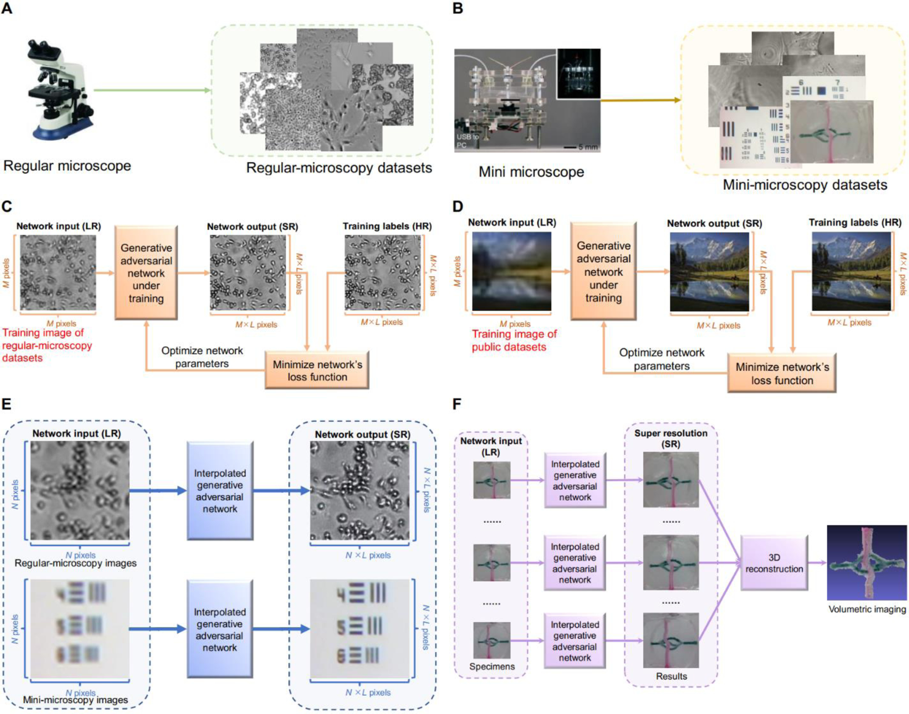
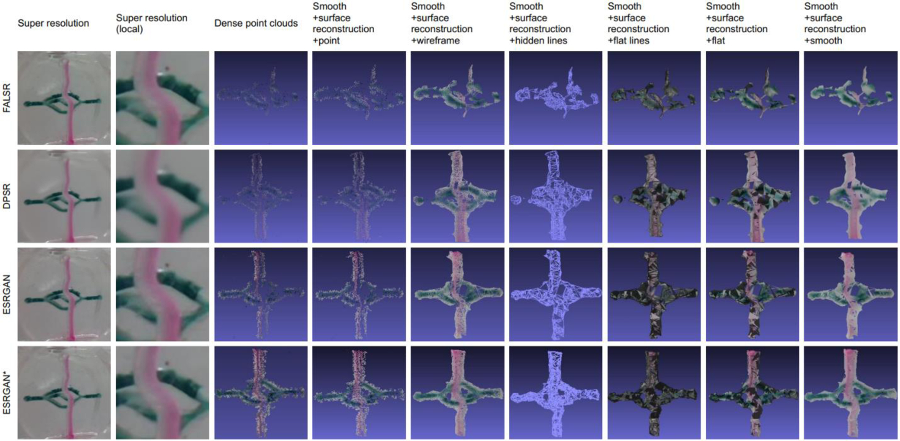
Artificial intelligence algorithms that aid mini-microscope imaging are attractive for numerous applications. In this paper, we optimize artificial intelligence techniques to provide clear, and natural biomedical imaging. We demonstrate that a deep learning-enabled super-resolution method can significantly enhance the spatial resolution of mini-microscopy and regular-microscopy. This data-driven approach trains a generative adversarial network to transform low-resolution images into super-resolved ones. Mini-microscopic images and regular-microscopic images acquired with different optical microscopes under various magnifications are collected as our experimental benchmark datasets. The only input to this generative-adversarial-network-based method are images from the datasets down-sampled by the Bicubic interpolation. We use independent test set to evaluate this deep learning approach with other deep learning-based algorithms through qualitative and quantitative comparisons. To clearly present the improvements achieved by this generative-adversarial-network-based method, we zoom into the local features to explore and highlight the qualitative differences. We also employ the peak signal-to-noise ratio and the structural similarity, to quantitatively compare alternative super-resolution methods. The quantitative results illustrate that super-resolution images obtained from our approach with interpolation parameter =0.25 more closely match those of the original high-resolution images than to those obtained by any of the alternative state-of-the-art method. These results are significant for fields that use microscopy tools, such as biomedical imaging of engineered living systems. We also utilize this generative adversarial network-based algorithm to optimize the resolution of biomedical specimen images and then generate three-dimensional reconstruction, so as to enhance the ability of three-dimensional imaging throughout the entire volumes for spatial-temporal analyses of specimen structures.
辅助微型显微镜成像的人工智能算法在众多应用中颇具吸引力。在本文中,我们优化人工智能技术以提供清晰、自然的生物医学成像。我们证明,一种基于深度学习的超分辨率方法能够显著提高微型显微镜和常规显微镜的空间分辨率。这种数据驱动的方法训练一个生成对抗网络,将低分辨率图像转换为超分辨率图像。我们收集了在不同放大倍数下用不同光学显微镜获取的微型显微镜图像和常规显微镜图像,作为我们的实验基准数据集。这种基于生成对抗网络的方法的唯一输入是通过双立方插值下采样的数据集图像。我们使用独立测试集,通过定性和定量比较,将这种深度学习方法与其他基于深度学习的算法进行评估。为了清晰展示这种基于生成对抗网络的方法所取得的改进,我们放大局部特征以探索和突出定性差异。我们还采用峰值信噪比和结构相似性,对替代超分辨率方法进行定量比较。定量结果表明,与任何替代的现有最先进方法相比,我们的方法在插值参数 = 0.25 时获得的超分辨率图像与原始高分辨率图像的匹配度更高。这些结果对于使用显微镜工具的领域具有重要意义,例如工程化生命系统的生物医学成像。我们还利用这种基于生成对抗网络的算法来优化生物医学标本图像的分辨率,然后生成三维重建,以增强在整个体积上进行标本结构时空分析的三维成像能力。