Translational Imaging Center, University of Southern California, Los Angeles, CA, 90089, USA.
Molecular and Computational Biology Section, University of Southern California, Los Angeles, CA, 90089, USA.
Commun Biol. 2020 Feb 14;3(1):74. doi: 10.1038/s42003-020-0787-6.
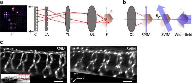
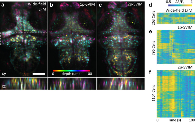
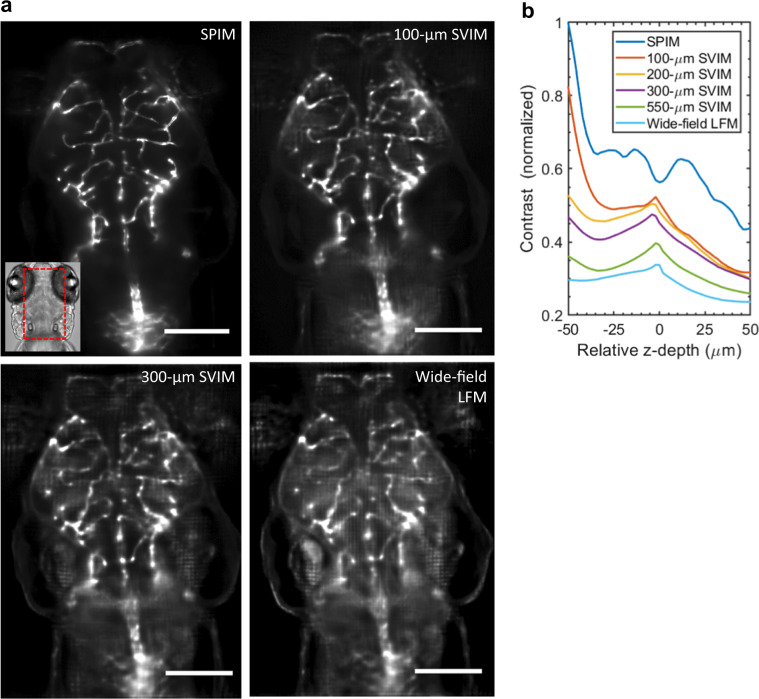
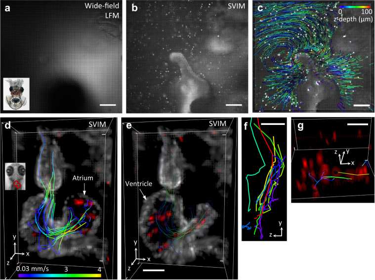
Light-field fluorescence microscopy uniquely provides fast, synchronous volumetric imaging by capturing an extended volume in one snapshot, but often suffers from low contrast due to the background signal generated by its wide-field illumination strategy. We implemented light-field-based selective volume illumination microscopy (SVIM), where illumination is confined to only the volume of interest, removing the background generated from the extraneous sample volume, and dramatically enhancing the image contrast. We demonstrate the capabilities of SVIM by capturing cellular-resolution 3D movies of flowing bacteria in seawater as they colonize their squid symbiotic partner, as well as of the beating heart and brain-wide neural activity in larval zebrafish. These applications demonstrate the breadth of imaging applications that we envision SVIM will enable, in capturing tissue-scale 3D dynamic biological systems at single-cell resolution, fast volumetric rates, and high contrast to reveal the underlying biology.
光场荧光显微镜通过在一次拍摄中捕获扩展体积,独特地提供快速、同步的体积成像,但由于其宽场照明策略产生的背景信号,通常对比度较低。我们实现了基于光场的选择性体积照明显微镜 (SVIM),其中照明仅局限于感兴趣的体积,去除了来自多余样品体积产生的背景,极大地提高了图像对比度。我们通过拍摄在海水中流动的细菌殖民其鱿鱼共生伙伴时的细胞分辨率 3D 电影,以及在幼虫斑马鱼的跳动心脏和大脑广泛的神经活动,展示了 SVIM 的功能。这些应用展示了我们设想 SVIM 将能够实现的成像应用的广泛性,以单细胞分辨率、快速体积速率和高对比度捕获组织尺度的 3D 动态生物系统,以揭示潜在的生物学。