Di Tella Sonia, Blasi Valeria, Cabinio Monia, Bergsland Niels, Buccino Giovanni, Baglio Francesca
Istituto di Ricovero e Cura a Carattere Scientifico (IRCCS) Fondazione Don Carlo Gnocchi ONLUS, Milan, Italy.
Department of Psychology, Università Cattolica del Sacro Cuore, Milan, Italy.
Front Aging Neurosci. 2021 Jul 29;13:694676. doi: 10.3389/fnagi.2021.694676. eCollection 2021.
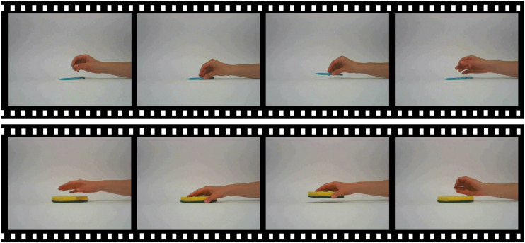
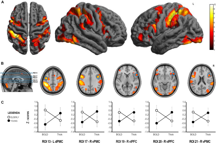
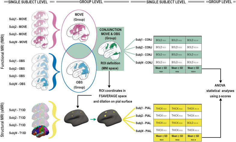
Aging is the major risk factor for chronic age-related neurological diseases such as neurodegenerative disorders and neurovascular injuries. Exploiting the multimodal nature of the Mirror Neuron System (MNS), rehabilitative interventions have been proposed based on motor-resonance mechanisms in recent years. Despite the considerable evidence of the MNS' functionality in young adults, further investigation of the action-observation matching system is required in aging, where well-known structural and functional brain changes occur. Twenty-one healthy young adults (mean age 26.66y) and 19 healthy elderly participants (mean age 71.47y) underwent a single MRI evaluation including a T1-3D high-resolution and functional MRI (fMRI) with mirror task. Morphological and functional BOLD data were derived from MRI images to highlight cortical activations associated with the task; to detect differences between the two groups (Young, Elderly) in the two MRI indexes (BOLD and thickness z-scores) using mixed factorial ANOVA (GroupIndex analyses); and to investigate the presence of different cortical lateralization of the BOLD signal in the two groups. In the entire sample, the activation of a bilateral MNS fronto-parietal network was highlighted. The mixed ANOVA (pFDR-corr < 0.05) revealed significant interactions between BOLD signal and cortical thickness in left dorsal premotor cortex, right ventral premotor and prefrontal cortices. A different cortical lateralization of the BOLD signal in frontal lobe activity between groups was also found. Data herein reported suggest that age-related cortical thinning of the MNS is coupled with increased interhemispheric symmetry along with premotor and prefrontal cortex recruitment. These physiological changes of MNS resemble the aging of the motor and cognitive neural systems, suggesting specific but also common aging and compensatory mechanisms.
衰老是慢性年龄相关性神经疾病(如神经退行性疾病和神经血管损伤)的主要风险因素。近年来,基于镜像神经元系统(MNS)的多模态特性,人们提出了基于运动共振机制的康复干预措施。尽管有大量证据表明MNS在年轻人中具有功能,但在发生了众所周知的大脑结构和功能变化的老年人中,仍需要对动作观察匹配系统进行进一步研究。21名健康的年轻成年人(平均年龄26.66岁)和19名健康的老年参与者(平均年龄71.47岁)接受了单次MRI评估,包括T1-3D高分辨率和功能性MRI(fMRI)镜像任务。从MRI图像中获取形态学和功能性BOLD数据,以突出与任务相关的皮质激活;使用混合因子方差分析(组×指标分析)检测两组(年轻人、老年人)在两个MRI指标(BOLD和厚度z分数)上的差异;并研究两组中BOLD信号不同的皮质侧化情况。在整个样本中,突出显示了双侧MNS额顶叶网络激活。混合方差分析(pFDR校正<0.05)显示,左侧背侧运动前皮质、右侧腹侧运动前皮质和前额叶皮质的BOLD信号与皮质厚度之间存在显著交互作用。还发现两组之间额叶活动中BOLD信号的皮质侧化不同。本文报道的数据表明,与年龄相关的MNS皮质变薄与半球间对称性增加以及运动前皮质和前额叶皮质的募集有关。MNS的这些生理变化类似于运动和认知神经系统的衰老,提示了特定但也常见的衰老和代偿机制。