Department of Radiology, Weill Cornell Medicine, New York, NY, USA.
Department of Pathology, University of Colorado School of Medicine, Aurora, CO, USA.
Korean J Radiol. 2021 Oct;22(10):1650-1657. doi: 10.3348/kjr.2020.1391. Epub 2021 Jul 26.
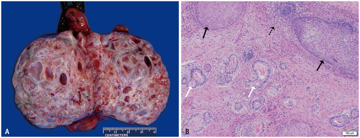
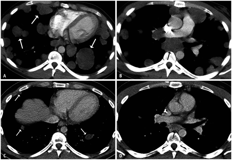
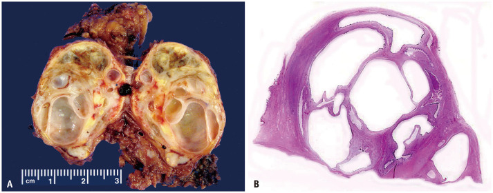
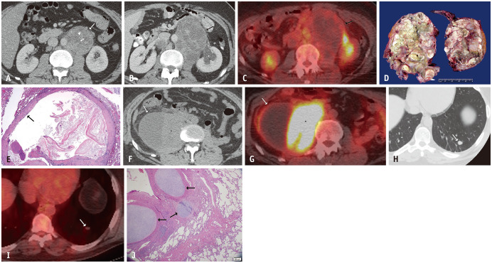
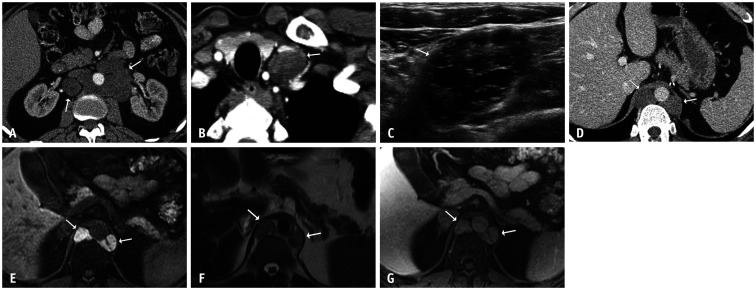
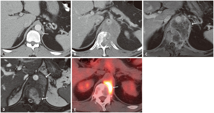
Metastatic mature teratoma is a common radiologic and histopathologic finding after chemotherapy for metastatic non-seminomatous germ cell tumors. The leading theory for these residual tumors is the selective chemotherapy resistance of teratomas versus the high chemotherapy sensitivity of the embryonal components. Growing teratoma syndrome is a relatively rare phenomenon defined as an enlarging residual mass histologically proven to be a mature teratoma in the setting of normal serum tumor markers. Metastatic mature teratomas should be resected because of their malignant potential and occasional progression to growing teratoma syndrome with the invasion of the surrounding structures. CT is the preferred imaging modality for post-chemotherapy surveillance and should cover all sites of potential metastatic disease. This article reviews the clinical, pathologic, and multimodality imaging features of metastatic mature teratomas in patients with primary testicular non-seminomatous germ cell tumors.
化疗治疗转移性非精原细胞瘤生殖细胞肿瘤后,成熟性畸胎瘤转移是一种常见的放射影像学和组织病理学表现。对于这些残留肿瘤,主要理论是畸胎瘤对化疗的选择性耐药性与胚胎成分的高化疗敏感性。生长性畸胎瘤综合征是一种相对罕见的现象,定义为在血清肿瘤标志物正常的情况下,残留肿块增大,组织学证实为成熟性畸胎瘤。由于其恶性潜能以及偶尔进展为生长性畸胎瘤综合征并侵犯周围结构,转移性成熟性畸胎瘤应行切除术。CT 是化疗后监测的首选影像学方法,应涵盖所有潜在转移疾病的部位。本文综述了原发性睾丸非精原细胞瘤生殖细胞肿瘤患者中转移性成熟性畸胎瘤的临床、病理和多模态影像学特征。