Bocchetta Martina, Malpetti Maura, Todd Emily G, Rowe James B, Rohrer Jonathan D
Dementia Research Centre, Department of Neurodegenerative Disease, UCL Queen Square Institute of Neurology, University College London, London, UK.
Department of Clinical Neurosciences and Cambridge University Hospitals NHS Trust, University of Cambridge, Cambridge, UK.
Brain Commun. 2021 Jul 16;3(3):fcab158. doi: 10.1093/braincomms/fcab158. eCollection 2021.
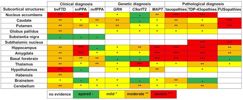
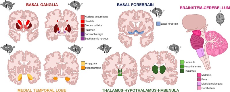
Whilst initial anatomical studies of frontotemporal dementia focussed on cortical involvement, the relevance of subcortical structures to the pathophysiology of frontotemporal dementia has been increasingly recognized over recent years. Key structures affected include the caudate, putamen, nucleus accumbens, and globus pallidus within the basal ganglia, the hippocampus and amygdala within the medial temporal lobe, the basal forebrain, and the diencephalon structures of the thalamus, hypothalamus and habenula. At the most posterior aspect of the brain, focal involvement of brainstem and cerebellum has recently also been shown in certain subtypes of frontotemporal dementia. Many of the neuroimaging studies on subcortical structures in frontotemporal dementia have been performed in clinically defined sporadic cases. However, investigations of genetically- and pathologically-confirmed forms of frontotemporal dementia are increasingly common and provide molecular specificity to the changes observed. Furthermore, detailed analyses of sub-nuclei and subregions within each subcortical structure are being added to the literature, allowing refinement of the patterns of subcortical involvement. This review focuses on the existing literature on structural imaging and neuropathological studies of subcortical anatomy across the spectrum of frontotemporal dementia, along with investigations of brain-behaviour correlates that examine the cognitive sequelae of specific subcortical involvement: it aims to 'look beneath the surface' and summarize the patterns of subcortical involvement have been described in frontotemporal dementia.
虽然额颞叶痴呆最初的解剖学研究集中在皮质受累方面,但近年来,皮质下结构与额颞叶痴呆病理生理学的相关性越来越受到认可。受影响的关键结构包括基底神经节内的尾状核、壳核、伏隔核和苍白球,内侧颞叶内的海马体和杏仁核,基底前脑,以及丘脑、下丘脑和缰核等间脑结构。在脑的最后部,脑干和小脑的局灶性受累最近也在某些额颞叶痴呆亚型中被发现。许多关于额颞叶痴呆皮质下结构的神经影像学研究是在临床定义的散发性病例中进行的。然而,对基因和病理证实的额颞叶痴呆形式的研究越来越普遍,并为观察到的变化提供了分子特异性。此外,对每个皮质下结构内的亚核和亚区域的详细分析也不断被纳入文献,从而使皮质下受累模式更加精确。本综述聚焦于现有关于额颞叶痴呆全谱中皮质下解剖结构的结构成像和神经病理学研究的文献,以及对脑-行为相关性的研究,这些研究考察了特定皮质下受累的认知后遗症:其目的是“透过表面深入观察”,总结额颞叶痴呆中已描述的皮质下受累模式。