Lee Bo Mi, Park Sang Jun, Noh Insup, Kim Chun-Ho
Laboratory of Tissue Engineering, Korea Institute of Radiological and Medical Sciences, 01812, Seoul, Korea.
Department of Convergence program of Biomedical Engineering & Biomaterials, The Graduate School, Seoul National University of Science and Technology, Seoul, Korea.
Biomater Res. 2021 Aug 30;25(1):27. doi: 10.1186/s40824-021-00228-4.
The molecular weight of hyaluronic acid (HyA) depends on the type of organ in the body. When HyA of the desired molecular weight is implanted into the human body for regeneration of damaged tissue, it is degraded by hyaluronidase in associated with an inflammatory response. This study sought to evaluate the effects of HyA molecular weight and concentration on pro- and anti-inflammatory responses in murine macrophages.
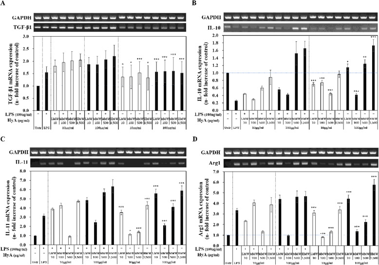
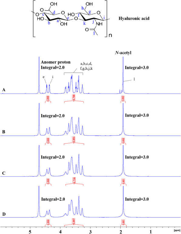
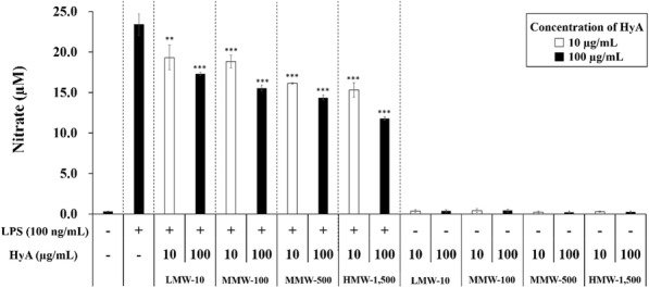
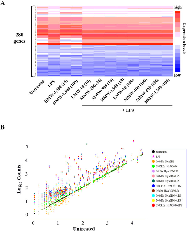
The structures and molecular weights of HyAs (LMW-10, MMW-100, MMW-500, and HMW-1,500) were confirmed by H NMR and gel permeation chromatography (GPC), respectively. After treatment of murine macrophages with a low (10 µg/mL) or high (100 µg/mL) concentration of each molecular weight HyA, cells were stimulated with lipopolysaccharide (LPS) and changes in immune response in both LPS-stimulated and untreated macrophages were evaluated by assessing nitric oxide (NO) production, and analyzing expression of pro- and anti-inflammatory genes including by RT-PCR.
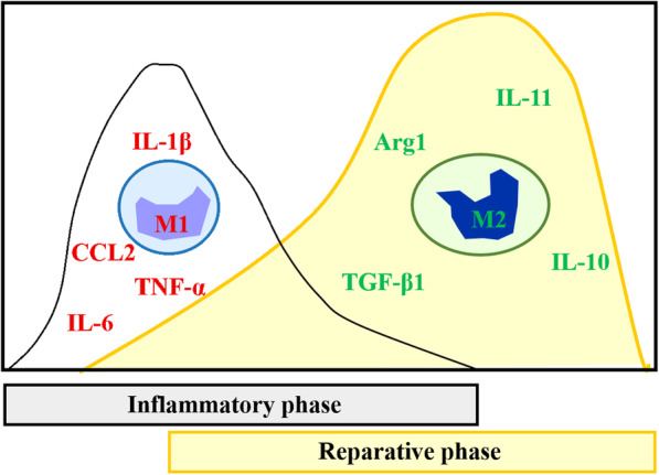
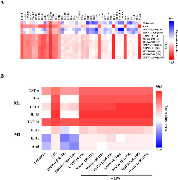
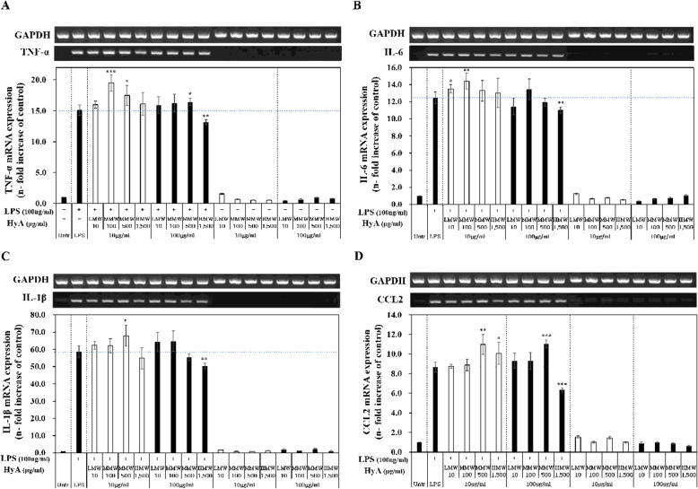
Molecular weights of LMW-10, MMW-100, MMW-500, and HMW-1,500 were 13,241 ± 161, 96,531 ± 1,167, 512,657 ± 8,545, and 1,249,500 ± 37,477 Da, respectively. NO production by LPS-stimulated macrophages was decreased by increasing concentrations and molecular weights of HyA. At a high concentration of 100 µg/mL, HMW-1,500 reduced NO production in LPS-stimulated macrophages to about 45 %. Using NanoString technology, we also found that the immune-related genes TNF-α, IL-6, IL-1β, TGF-β1, IL-10, IL-11, CCL2, and Arg1 were specifically over-expressed in LPS-stimulated macrophages treated with various molecular weights of HyA. An RT-PCR analysis of gene expression showed that HMW-1,500 decreased expression of classically activated (M1) macrophage genes, such as TNF-α, IL-6, CCL2, and IL-1β, in LPS-stimulated macrophages, whereas medium molecular-weight HyA (MMW-100 and MMW-500) instead increased expression levels of these genes. HMW-1,500 at a high concentration (100 µg/mL) significantly decreased expression of pro-inflammatory genes in LPS-stimulated macrophages. Expression of genes associated with anti-inflammatory responses (M2 phenotype), such as TGF-β1, IL-10, IL-11, and Arg1, were increased by high concentrations of MMW-500 and HMW-1,500 in LPS-stimulated macrophages.
High molecular-weight HyA (i.e., > 1,250 kDa) inhibits pro-inflammatory responses in LPS-stimulated macrophages and induces anti-inflammatory responses in a concentration dependent manner.
透明质酸(HyA)的分子量取决于其在体内的器官类型。当将具有所需分子量的HyA植入人体以促进受损组织再生时,它会在炎症反应中被透明质酸酶降解。本研究旨在评估HyA分子量和浓度对小鼠巨噬细胞促炎和抗炎反应的影响。
分别通过核磁共振氢谱(¹H NMR)和凝胶渗透色谱法(GPC)确认HyA(低分子量-10,中分子量-100,中分子量-500和高分子量-1500)的结构和分子量。用每种分子量的低浓度(10μg/mL)或高浓度(100μg/mL)HyA处理小鼠巨噬细胞后,用脂多糖(LPS)刺激细胞,并通过评估一氧化氮(NO)产生以及分析包括通过逆转录聚合酶链反应(RT-PCR)检测的促炎和抗炎基因的表达,来评估LPS刺激和未处理的巨噬细胞中免疫反应的变化。
低分子量-10、中分子量-100、中分子量-500和高分子量-1500的分子量分别为13,241±161、96,531±1,167、512,657±8,545和1,249,500±37,477 Da。LPS刺激的巨噬细胞产生的NO随着HyA浓度和分子量的增加而减少。在100μg/mL的高浓度下,高分子量-1500将LPS刺激的巨噬细胞中的NO产生降低至约45%。使用NanoString技术,我们还发现免疫相关基因肿瘤坏死因子-α(TNF-α)、白细胞介素-6(IL-6)、白细胞介素-1β(IL-1β)、转化生长因子-β1(TGF-β1)、白细胞介素-10(IL-10)、白细胞介素-11(IL-11)、趋化因子配体2(CCL2)和精氨酸酶1(Arg1)在经不同分子量HyA处理的LPS刺激的巨噬细胞中特异性过表达。基因表达的RT-PCR分析表明,高分子量-1500降低了LPS刺激的巨噬细胞中经典活化(M1)巨噬细胞基因如TNF-α、IL-6、CCL2和IL-1β的表达,而中分子量HyA(中分子量-100和中分子量-500)反而增加了这些基因的表达水平。高浓度(100μg/mL)的高分子量-1500显著降低了LPS刺激的巨噬细胞中促炎基因的表达。在LPS刺激的巨噬细胞中,高浓度的中分子量-500和高分子量-1500增加了与抗炎反应(M2表型)相关的基因如TGF-β1、IL-10、IL-11和Arg1的表达。
高分子量HyA(即>1250 kDa)抑制LPS刺激的巨噬细胞中的促炎反应,并以浓度依赖性方式诱导抗炎反应。