Department of Orthopaedic Surgery, Inje University Haeundae Paik Hospital, Busan, Korea.
Department of Occupational and Environmental Medicine, Inje University Haeundae Paik Hospital, Busan, Korea.
Clin Orthop Surg. 2021 Sep;13(3):315-319. doi: 10.4055/cios20253. Epub 2021 Jul 16.
Modified tension band wiring is one of the most preferred surgical methods for transverse patellar fractures. However, the optimal depth or sagittal position of a Kirschner wire (K-wire) in modified tension band wiring has yet to be determined. The purpose of this study was to evaluate whether the depth of a K-wire affects the biomechanical characteristics of modified tension band wiring using the finite-element method.
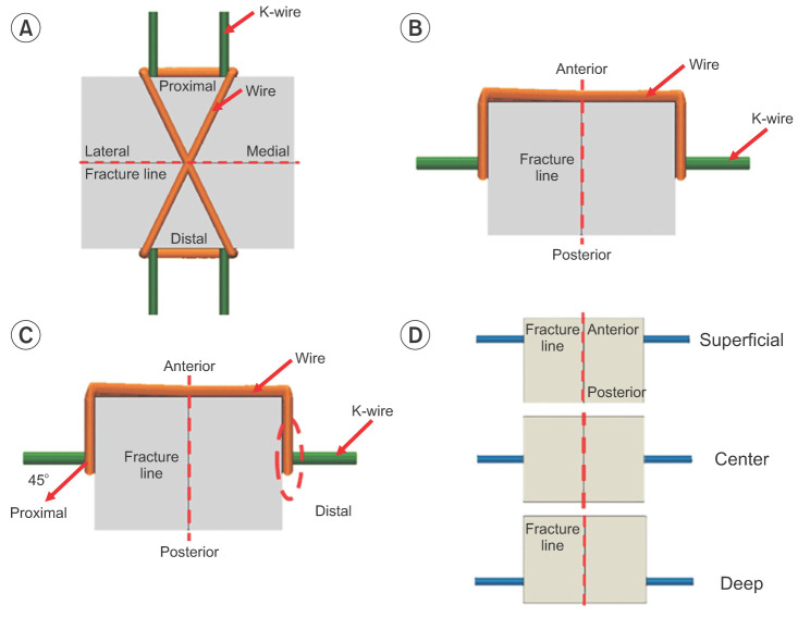
A patella model was designed with a cuboid shape (length, 34.3 mm; width, 44.8 mm; and thickness, 22.4 mm) and divided into the cortical and cancellous bone parts. A transverse fracture line was formed on the midline of the cuboid shape model. The cuboidal model was applied to modified tension band wiring. The depth or sagittal position of the K-wire was divided into superficial, center, and deep. With the Abaqus v2017 program (Dassault System Inc.), the distal part of the model was fixed, and a tensile load of 850 N was applied to the proximal part of the model at an angle of 45°. The maximum pressures of the cortical and cancellous bones at the fracture plane were measured. The largest von Mises values of the K-wire and stainless steel wire were also measured. The fracture gap on the distracted or anterior side was measured.
In deep K-wire placement, the highest peak von Mises values of the cortical and cancellous bones were observed. The K-wire and stainless steel wire showed the highest von Mises values in deep K-wire placement. The fracture gap was also largest in deep K-wire placement.
The depth of the K-wire affects the biomechanical characteristics of modified tension band wiring. Deep placement of the K-wire will be more favorable for bone union than the empirically known 5-mm anterior or center placement of the K-wire.
改良张力带钢丝固定是治疗横断髌骨骨折最常用的手术方法之一。然而,改良张力带钢丝固定中克氏针(K 线)的最佳深度或矢状位置尚未确定。本研究旨在通过有限元法评估 K 线的深度是否会影响改良张力带钢丝固定的生物力学特性。
设计了一个具有长方体形状(长 34.3mm,宽 44.8mm,厚 22.4mm)的髌骨模型,并分为皮质骨和松质骨部分。在长方体模型的中线上形成横断骨折线。将长方体模型应用于改良张力带钢丝固定。K 线的深度或矢状位置分为浅层、中心和深层。使用 Abaqus v2017 程序(达索系统公司),固定模型的远端,在 45°角向模型的近端施加 850N 的拉伸载荷。测量骨折平面皮质骨和松质骨的最大压力。还测量了 K 线和不锈钢丝的最大 von Mises 值。测量分散或前侧的骨折间隙。
在深部 K 线放置时,皮质骨和松质骨的最高峰值 von Mises 值。在深部 K 线放置时,K 线和不锈钢丝显示出最高的 von Mises 值。深部 K 线放置时骨折间隙也最大。
K 线的深度会影响改良张力带钢丝固定的生物力学特性。与经验所知的 K 线前或中心 5mm 放置相比,深部 K 线放置更有利于骨愈合。