• 文献检索
  • 文档翻译
  • 深度研究
  • 学术资讯
  • Suppr Zotero 插件Zotero 插件
  • 邀请有礼
  • 套餐&价格
  • 历史记录
应用&插件
Suppr Zotero 插件Zotero 插件浏览器插件Mac 客户端Windows 客户端微信小程序
定价
高级版会员购买积分包购买API积分包
服务
文献检索文档翻译深度研究API 文档MCP 服务
关于我们
关于 Suppr公司介绍联系我们用户协议隐私条款
关注我们

Suppr 超能文献

核心技术专利:CN118964589B侵权必究
粤ICP备2023148730 号-1Suppr @ 2026

文献检索

告别复杂PubMed语法,用中文像聊天一样搜索,搜遍4000万医学文献。AI智能推荐,让科研检索更轻松。

立即免费搜索

文件翻译

保留排版,准确专业,支持PDF/Word/PPT等文件格式,支持 12+语言互译。

免费翻译文档

深度研究

AI帮你快速写综述,25分钟生成高质量综述,智能提取关键信息,辅助科研写作。

立即免费体验

一种用于评估乳腺癌放疗患者心脏结构所接受剂量的自动分割方法的应用。

Application of an automatic segmentation method for evaluating cardiac structure doses received by breast radiotherapy patients.

作者信息

Jung Jae Won, Mille Matthew M, Ky Bonnie, Kenworthy Walter, Lee Choonik, Yeom Yeon Soo, Kwag Aaron, Bosch Walter, MacDonald Shannon, Cahlon Oren, Bekelman Justin E, Lee Choonsik

机构信息

Department of Physics, East Carolina University, Greenville, NC 27858, United States.

Division of Cancer Epidemiology and Genetics, National Cancer Institute, National Institutes of Health, Rockville, MD 20850, United States.

出版信息

Phys Imaging Radiat Oncol. 2021 Aug 23;19:138-144. doi: 10.1016/j.phro.2021.08.005. eCollection 2021 Jul.

DOI:10.1016/j.phro.2021.08.005
PMID:34485719
原文链接:https://pmc.ncbi.nlm.nih.gov/articles/PMC8397890/
Abstract

BACKGROUND AND PURPOSE

Quantifying radiation dose to cardiac substructures is important for research on the etiology and prevention of complications following radiotherapy; however, segmentation of substructures is challenging. In this study we demonstrate the application of our atlas-based automatic segmentation method to breast cancer radiotherapy plans for generating radiation doses in support of late effects research.

MATERIAL AND METHODS

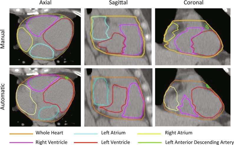
We applied our segmentation method to contour heart substructures on the computed tomography (CT) images of 70 breast cancer patients who received external photon radiotherapy. Two cardiologists provided manual segmentation of the whole heart (WH), left/right atria, left/right ventricles, and left anterior descending artery (LAD). The automatically contours were compared with manual delineations to evaluate similarity in terms of geometry and dose.

RESULTS

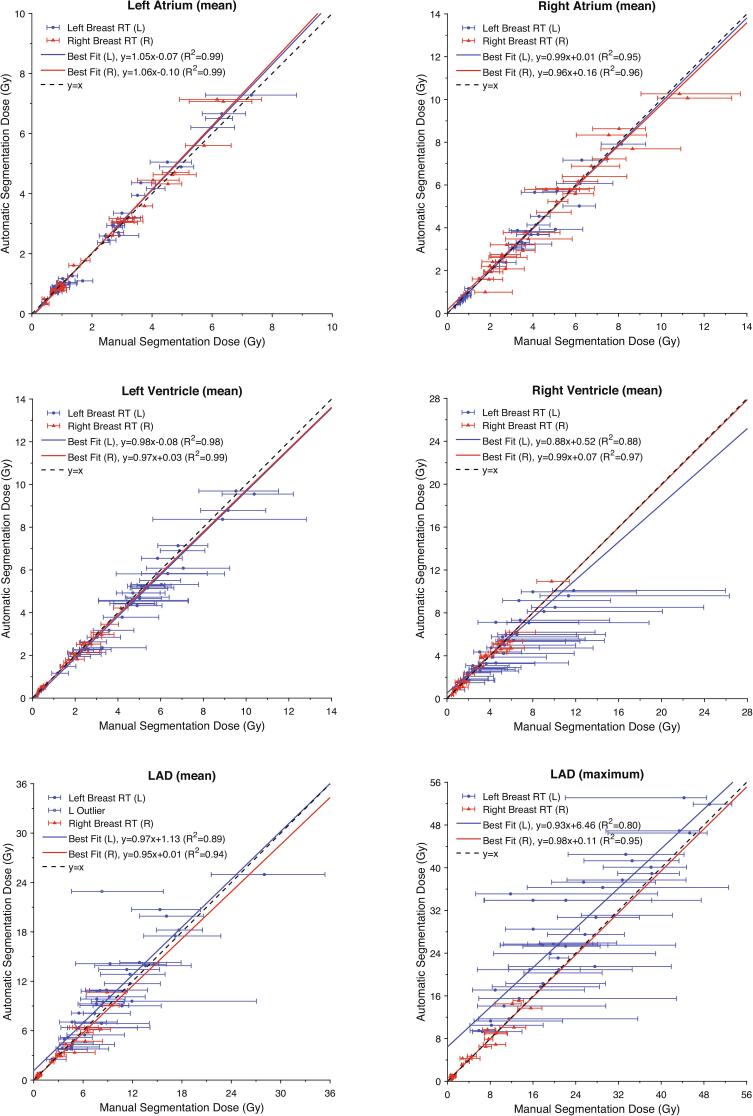
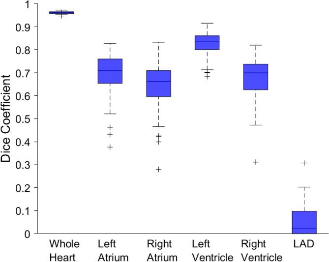
The mean Dice similarity coefficient between manual and automatic segmentations was 0.96 for the WH, 0.65 to 0.82 for the atria and ventricles, and 0.06 for the LAD. The mean average surface distance was 1.2 mm for the WH, 3.4 to 4.1 mm for the atria and ventricles, and 6.4 mm for the LAD. We found the dose to the cardiac substructures based on our automatic segmentation agrees with manual segmentation within expected observer variability. For left breast patients, the mean absolute difference in mean dose was 0.1 Gy for the WH, 0.2 to 0.7 Gy for the atria and ventricles, and 1.8 Gy for the LAD. For right breast patients, these values were 0.0 Gy, 0.1 to 0.4 Gy, and 0.4 Gy, respectively.

CONCLUSION

Our automatic segmentation method will facilitate the development of radiotherapy prescriptive criteria for mitigating cardiovascular complications.

摘要

背景与目的

量化心脏亚结构的辐射剂量对于放射治疗后并发症的病因学及预防研究具有重要意义;然而,亚结构的分割具有挑战性。在本研究中,我们展示了基于图谱的自动分割方法在乳腺癌放射治疗计划中的应用,以生成辐射剂量,支持晚期效应研究。

材料与方法

我们将分割方法应用于70例接受外照射光子放疗的乳腺癌患者的计算机断层扫描(CT)图像上,以勾勒心脏亚结构。两名心脏病专家对整个心脏(WH)、左/右心房、左/右心室以及左前降支动脉(LAD)进行手动分割。将自动勾勒结果与手动描绘结果进行比较,以评估几何形状和剂量方面的相似性。

结果

手动分割与自动分割之间的平均骰子相似系数,对于WH为0.96,对于心房和心室为0.65至0.82,对于LAD为0.06。平均表面距离,对于WH为1.2毫米,对于心房和心室为3.4至4.1毫米,对于LAD为6.4毫米。我们发现基于自动分割得出的心脏亚结构剂量与手动分割结果在预期的观察者变异性范围内一致。对于左乳患者,平均剂量的平均绝对差异,对于WH为0.1 Gy,对于心房和心室为0.2至0.7 Gy,对于LAD为1.8 Gy。对于右乳患者,这些值分别为0.0 Gy、0.1至0.4 Gy和0.4 Gy。

结论

我们的自动分割方法将有助于制定减轻心血管并发症的放射治疗处方标准。

https://cdn.ncbi.nlm.nih.gov/pmc/blobs/c09f/8397890/e23f48f2aa50/gr3.jpg
https://cdn.ncbi.nlm.nih.gov/pmc/blobs/c09f/8397890/2396790e5b4e/gr1.jpg
https://cdn.ncbi.nlm.nih.gov/pmc/blobs/c09f/8397890/ffbccb1b86e9/gr2.jpg
https://cdn.ncbi.nlm.nih.gov/pmc/blobs/c09f/8397890/e23f48f2aa50/gr3.jpg
https://cdn.ncbi.nlm.nih.gov/pmc/blobs/c09f/8397890/2396790e5b4e/gr1.jpg
https://cdn.ncbi.nlm.nih.gov/pmc/blobs/c09f/8397890/ffbccb1b86e9/gr2.jpg
https://cdn.ncbi.nlm.nih.gov/pmc/blobs/c09f/8397890/e23f48f2aa50/gr3.jpg

相似文献

1
Application of an automatic segmentation method for evaluating cardiac structure doses received by breast radiotherapy patients.一种用于评估乳腺癌放疗患者心脏结构所接受剂量的自动分割方法的应用。
Phys Imaging Radiat Oncol. 2021 Aug 23;19:138-144. doi: 10.1016/j.phro.2021.08.005. eCollection 2021 Jul.
2
Automatic segmentation of cardiac structures for breast cancer radiotherapy.用于乳腺癌放疗的心脏结构自动分割
Phys Imaging Radiat Oncol. 2019 Dec 5;12:44-48. doi: 10.1016/j.phro.2019.11.007. eCollection 2019 Oct.
3
Atlas-based auto-segmentation for delineating the heart and cardiac substructures in breast cancer radiation therapy.基于图谱的自动分割在乳腺癌放射治疗中心脏和心脏亚结构勾画中的应用。
Acta Oncol. 2022 Feb;61(2):247-254. doi: 10.1080/0284186X.2021.1967445. Epub 2021 Aug 24.
4
A deep learning segmentation method to assess dose to organs at risk during breast radiotherapy.一种用于评估乳腺癌放疗期间危及器官剂量的深度学习分割方法。
Phys Imaging Radiat Oncol. 2023 Nov 21;28:100520. doi: 10.1016/j.phro.2023.100520. eCollection 2023 Oct.
5
Cardiac Substructure Segmentation and Dosimetry Using a Novel Hybrid Magnetic Resonance and Computed Tomography Cardiac Atlas.使用新型磁共振与计算机断层扫描心脏图谱进行心脏亚结构分割和剂量学评估。
Int J Radiat Oncol Biol Phys. 2019 Mar 15;103(4):985-993. doi: 10.1016/j.ijrobp.2018.11.025. Epub 2018 Nov 22.
6
Is mean heart dose a relevant surrogate parameter of left ventricle and coronary arteries exposure during breast cancer radiotherapy: a dosimetric evaluation based on individually-determined radiation dose (BACCARAT study).平均心脏剂量是否是乳腺癌放疗过程中左心室和冠状动脉暴露的相关替代参数:基于个体确定的辐射剂量的剂量学评估(BACCARAT 研究)。
Radiat Oncol. 2019 Feb 7;14(1):29. doi: 10.1186/s13014-019-1234-z.
7
Validation of a Fully Automated Hybrid Deep Learning Cardiac Substructure Segmentation Tool for Contouring and Dose Evaluation in Lung Cancer Radiotherapy.全自动混合深度学习心脏亚结构分割工具在肺癌放疗中的轮廓勾画和剂量评估的验证。
Clin Oncol (R Coll Radiol). 2023 Jun;35(6):370-381. doi: 10.1016/j.clon.2023.03.005. Epub 2023 Mar 11.
8
Feasibility of multi-atlas cardiac segmentation from thoracic planning CT in a probabilistic framework.基于概率框架的胸部计划 CT 多图谱心脏分割的可行性。
Phys Med Biol. 2019 Apr 8;64(8):085006. doi: 10.1088/1361-6560/ab0ea6.
9
Automatic segmentation of cardiac substructures from noncontrast CT images: accurate enough for dosimetric analysis?从非对比 CT 图像自动分割心脏亚结构:是否足以进行剂量分析?
Acta Oncol. 2019 Jan;58(1):81-87. doi: 10.1080/0284186X.2018.1521985. Epub 2018 Oct 11.
10
Validation of separate multi-atlases for auto segmentation of cardiac substructures in CT-scans acquired in deep inspiration breath hold and free breathing.在深吸气屏气和自由呼吸采集的 CT 扫描中,用于自动分割心脏亚结构的独立多图谱的验证。
Radiother Oncol. 2021 Oct;163:46-54. doi: 10.1016/j.radonc.2021.07.025. Epub 2021 Jul 31.

引用本文的文献

1
Narrative Review: Cardiotoxicities and Cardiac-Sparing Techniques in Radiotherapy.叙述性综述:放射治疗中的心脏毒性和心脏保护技术
Technol Cancer Res Treat. 2024 Jan-Dec;23:15330338241301211. doi: 10.1177/15330338241301211.
2
Cardiac substructure delineation in radiation therapy - A state-of-the-art review.放射治疗中心脏亚结构的描绘——一项最新综述。
J Med Imaging Radiat Oncol. 2024 Dec;68(8):914-949. doi: 10.1111/1754-9485.13668. Epub 2024 May 17.
3
A deep learning segmentation method to assess dose to organs at risk during breast radiotherapy.

本文引用的文献

1
Automatic segmentation of cardiac structures for breast cancer radiotherapy.用于乳腺癌放疗的心脏结构自动分割
Phys Imaging Radiat Oncol. 2019 Dec 5;12:44-48. doi: 10.1016/j.phro.2019.11.007. eCollection 2019 Oct.
2
Cardiotoxicity of breast cancer radiotherapy - overview of current results.乳腺癌放疗的心脏毒性——当前结果概述
Rep Pract Oncol Radiother. 2020 Mar-Apr;25(2):182-186. doi: 10.1016/j.rpor.2019.12.008. Epub 2019 Dec 9.
3
Cardiac substructure segmentation with deep learning for improved cardiac sparing.
一种用于评估乳腺癌放疗期间危及器官剂量的深度学习分割方法。
Phys Imaging Radiat Oncol. 2023 Nov 21;28:100520. doi: 10.1016/j.phro.2023.100520. eCollection 2023 Oct.
4
Automated contouring and statistical process control for plan quality in a breast clinical trial.一项乳腺癌临床试验中计划质量的自动轮廓描绘与统计过程控制
Phys Imaging Radiat Oncol. 2023 Aug 23;28:100486. doi: 10.1016/j.phro.2023.100486. eCollection 2023 Oct.
5
Interpretable Machine Learning for Choosing Radiation Dose-volume Constraints on Cardio-pulmonary Substructures Associated with Overall Survival in NRG Oncology RTOG 0617.用于选择与 NRG 肿瘤学 RTOG 0617 总生存相关的心肺亚结构的辐射剂量-体积约束的可解释机器学习。
Int J Radiat Oncol Biol Phys. 2023 Dec 1;117(5):1270-1286. doi: 10.1016/j.ijrobp.2023.06.009. Epub 2023 Jun 19.
6
Advances in automatic delineation of target volume and cardiac substructure in breast cancer radiotherapy (Review).乳腺癌放疗中靶区体积及心脏亚结构自动勾画的进展(综述)
Oncol Lett. 2023 Feb 2;25(3):110. doi: 10.3892/ol.2023.13697. eCollection 2023 Mar.
7
Association of cardiac calcium burden with overall survival after radiotherapy for non-small cell lung cancer.非小细胞肺癌放疗后心脏钙负荷与总生存期的关联
Phys Imaging Radiat Oncol. 2023 Jan 5;25:100410. doi: 10.1016/j.phro.2023.01.001. eCollection 2023 Jan.
8
Synthetic contrast-enhanced computed tomography generation using a deep convolutional neural network for cardiac substructure delineation in breast cancer radiation therapy: a feasibility study.基于深度卷积神经网络的合成对比增强计算机断层扫描生成技术在乳腺癌放疗中心脏亚结构勾画中的可行性研究。
Radiat Oncol. 2022 Apr 22;17(1):83. doi: 10.1186/s13014-022-02051-0.
深度学习进行心脏亚结构分割,以实现更好的心脏保护。
Med Phys. 2020 Feb;47(2):576-586. doi: 10.1002/mp.13940. Epub 2019 Dec 29.
4
Pragmatic randomised clinical trial of proton versus photon therapy for patients with non-metastatic breast cancer: the Radiotherapy Comparative Effectiveness (RadComp) Consortium trial protocol.质子与光子放疗治疗非转移性乳腺癌患者的实用随机临床试验:放射治疗比较效果(RadComp)联盟试验方案。
BMJ Open. 2019 Oct 15;9(10):e025556. doi: 10.1136/bmjopen-2018-025556.
5
Coronary Artery Dose-Volume Parameters Predict Risk of Calcification After Radiation Therapy.冠状动脉剂量体积参数可预测放疗后钙化风险。
J Cardiovasc Imaging. 2019 Oct;27(4):268-279. doi: 10.4250/jcvi.2019.27.e38.
6
Evaluation of algorithms for Multi-Modality Whole Heart Segmentation: An open-access grand challenge.多模态全心脏分割算法评估:一个开放获取的大型挑战赛。
Med Image Anal. 2019 Dec;58:101537. doi: 10.1016/j.media.2019.101537. Epub 2019 Aug 1.
7
Feasibility of multi-atlas cardiac segmentation from thoracic planning CT in a probabilistic framework.基于概率框架的胸部计划 CT 多图谱心脏分割的可行性。
Phys Med Biol. 2019 Apr 8;64(8):085006. doi: 10.1088/1361-6560/ab0ea6.
8
Cardiac Substructure Segmentation and Dosimetry Using a Novel Hybrid Magnetic Resonance and Computed Tomography Cardiac Atlas.使用新型磁共振与计算机断层扫描心脏图谱进行心脏亚结构分割和剂量学评估。
Int J Radiat Oncol Biol Phys. 2019 Mar 15;103(4):985-993. doi: 10.1016/j.ijrobp.2018.11.025. Epub 2018 Nov 22.
9
Automatic segmentation of cardiac substructures from noncontrast CT images: accurate enough for dosimetric analysis?从非对比 CT 图像自动分割心脏亚结构:是否足以进行剂量分析?
Acta Oncol. 2019 Jan;58(1):81-87. doi: 10.1080/0284186X.2018.1521985. Epub 2018 Oct 11.
10
Geometric and dosimetric evaluation of atlas based auto-segmentation of cardiac structures in breast cancer patients.基于图谱的乳腺癌患者心脏结构自动分割的几何和剂量学评估。
Radiother Oncol. 2019 Feb;131:215-220. doi: 10.1016/j.radonc.2018.07.013. Epub 2018 Aug 11.