文献检索文档翻译深度研究
Suppr Zotero 插件Zotero 插件
邀请有礼套餐&价格历史记录

新学期,新优惠

限时优惠:9月1日-9月22日

30天高级会员仅需29元

1天体验卡首发特惠仅需5.99元

了解详情
不再提醒
插件&应用
Suppr Zotero 插件Zotero 插件浏览器插件Mac 客户端Windows 客户端微信小程序
高级版
套餐订阅购买积分包
AI 工具
文献检索文档翻译深度研究
关于我们
关于 Suppr公司介绍联系我们用户协议隐私条款
关注我们

Suppr 超能文献

核心技术专利:CN118964589B侵权必究
粤ICP备2023148730 号-1Suppr @ 2025

空间转录组和单细胞转录组数据的整合揭示了小鼠器官发生。

Integration of spatial and single-cell transcriptomic data elucidates mouse organogenesis.

机构信息

Wellcome-Medical Research Council Cambridge Stem Cell Institute, University of Cambridge, Cambridge, UK.

Department of Physiology, Development and Neuroscience, University of Cambridge, Cambridge, UK.

出版信息

Nat Biotechnol. 2022 Jan;40(1):74-85. doi: 10.1038/s41587-021-01006-2. Epub 2021 Sep 6.


DOI:10.1038/s41587-021-01006-2
PMID:34489600
原文链接:https://pmc.ncbi.nlm.nih.gov/articles/PMC8763645/
Abstract

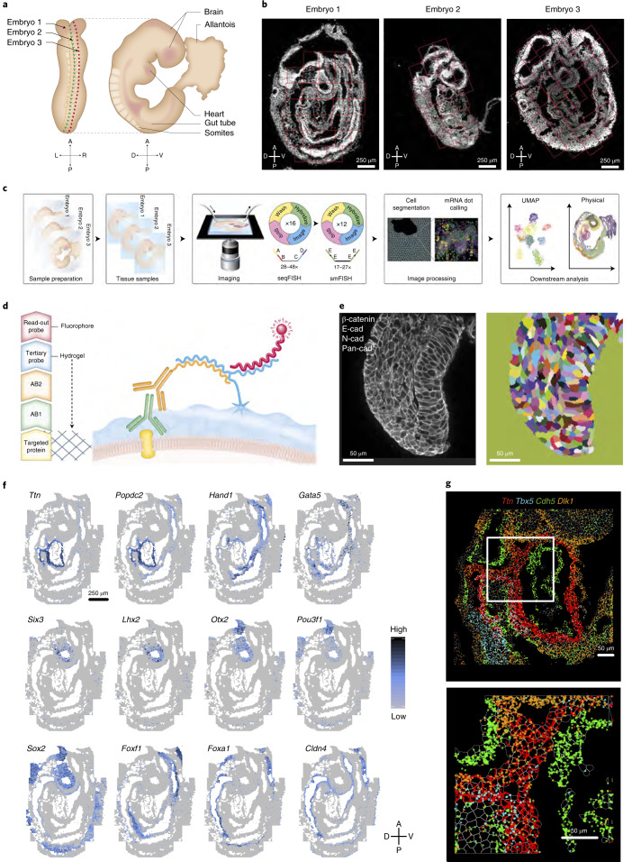
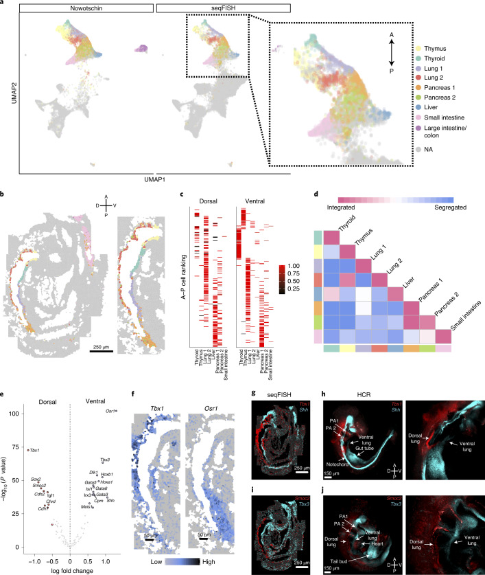
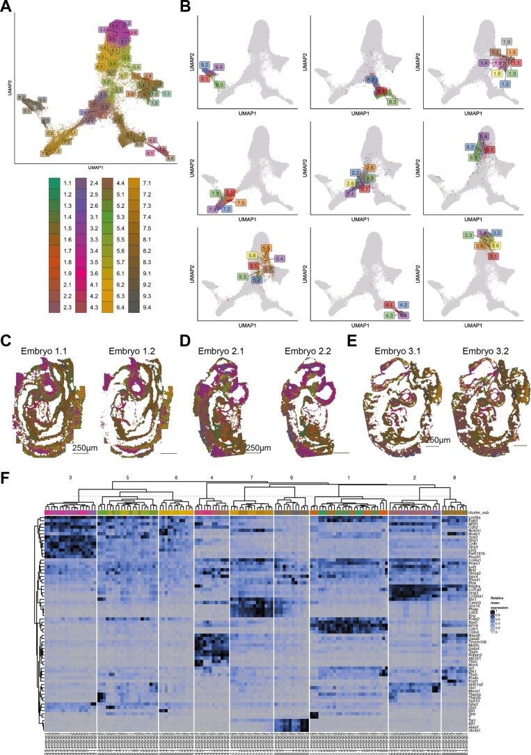
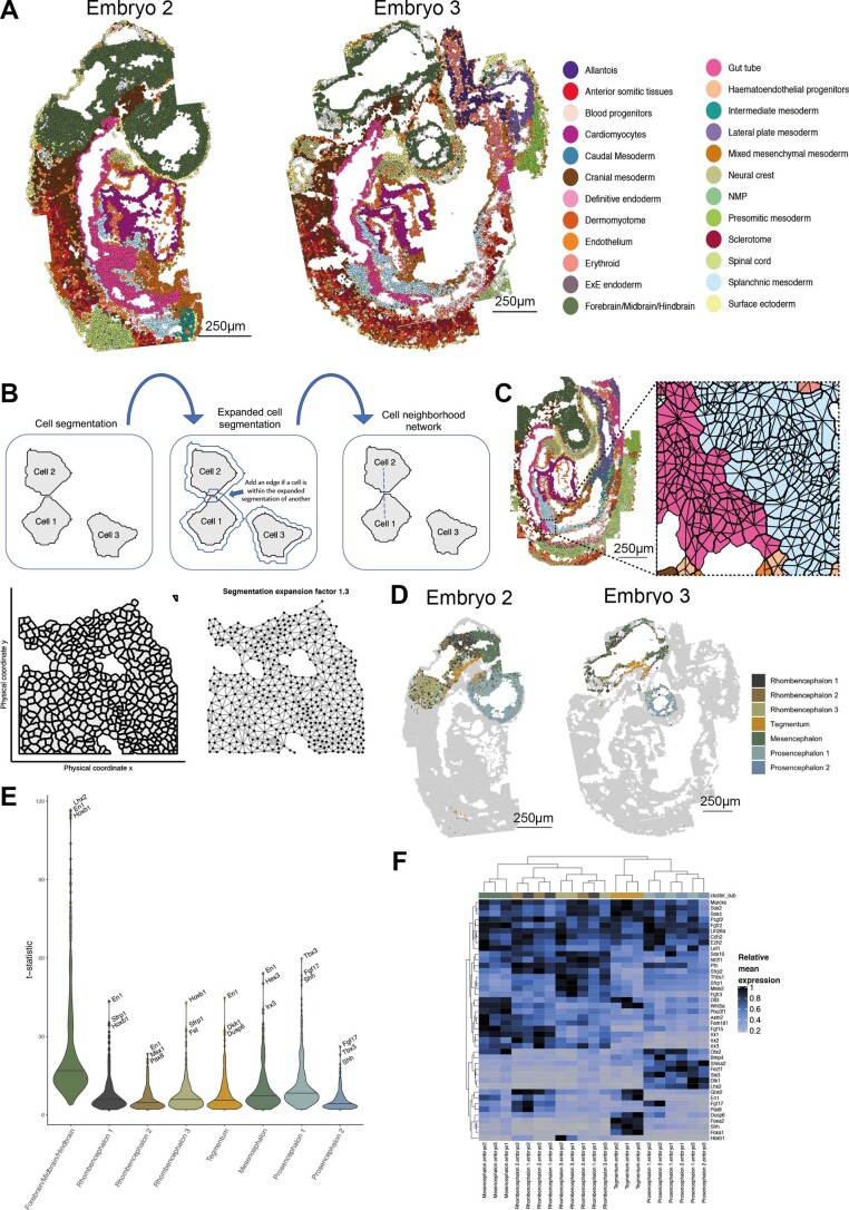
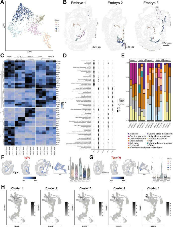
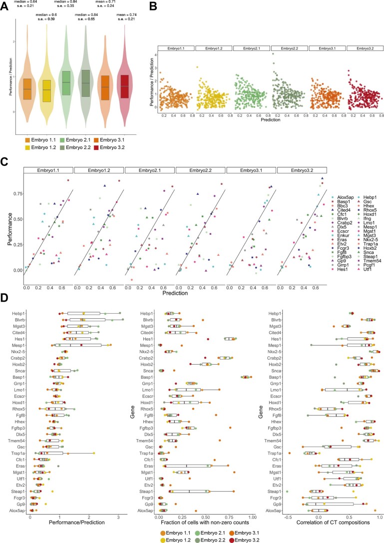
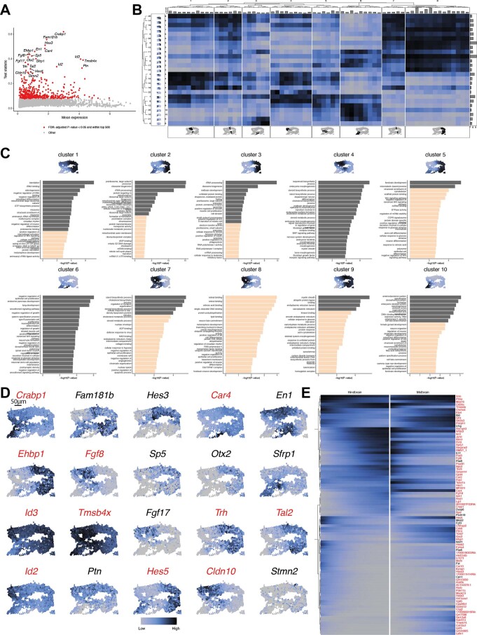
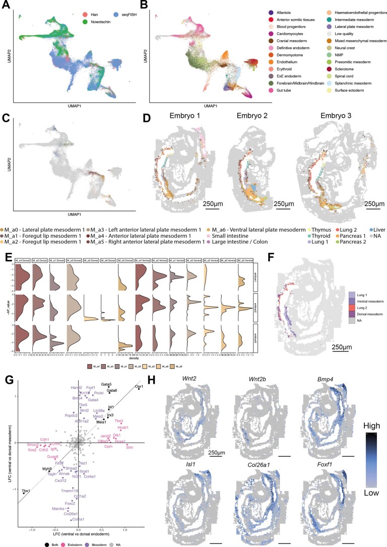
Molecular profiling of single cells has advanced our knowledge of the molecular basis of development. However, current approaches mostly rely on dissociating cells from tissues, thereby losing the crucial spatial context of regulatory processes. Here, we apply an image-based single-cell transcriptomics method, sequential fluorescence in situ hybridization (seqFISH), to detect mRNAs for 387 target genes in tissue sections of mouse embryos at the 8-12 somite stage. By integrating spatial context and multiplexed transcriptional measurements with two single-cell transcriptome atlases, we characterize cell types across the embryo and demonstrate that spatially resolved expression of genes not profiled by seqFISH can be imputed. We use this high-resolution spatial map to characterize fundamental steps in the patterning of the midbrain-hindbrain boundary (MHB) and the developing gut tube. We uncover axes of cell differentiation that are not apparent from single-cell RNA-sequencing (scRNA-seq) data, such as early dorsal-ventral separation of esophageal and tracheal progenitor populations in the gut tube. Our method provides an approach for studying cell fate decisions in complex tissues and development.

摘要

单细胞的分子谱分析提高了我们对发育分子基础的认识。然而,目前的方法大多依赖于将细胞从组织中分离出来,从而失去了调控过程的关键空间背景。在这里,我们应用基于图像的单细胞转录组学方法——顺序荧光原位杂交(seqFISH),在 8-12 体节阶段的小鼠胚胎组织切片中检测 387 个靶基因的 mRNA。通过整合空间背景和带有两个单细胞转录组图谱的多路转录测量,我们对胚胎中的细胞类型进行了描述,并证明了可以对 seqFISH 未分析的基因的空间表达进行推断。我们使用这个高分辨率的空间图谱来描述中脑-后脑边界(MHB)和发育中的肠道管的图案形成的基本步骤。我们揭示了一些从单细胞 RNA 测序(scRNA-seq)数据中不明显的细胞分化轴,例如肠道管中食管和气管祖细胞群体的早期背腹分离。我们的方法为研究复杂组织和发育中的细胞命运决定提供了一种方法。

https://cdn.ncbi.nlm.nih.gov/pmc/blobs/5ea7/8763645/2b56a659c421/41587_2021_1006_Fig14_ESM.jpg
https://cdn.ncbi.nlm.nih.gov/pmc/blobs/5ea7/8763645/d1b27e5ab891/41587_2021_1006_Fig1_HTML.jpg
https://cdn.ncbi.nlm.nih.gov/pmc/blobs/5ea7/8763645/3fc12290daa3/41587_2021_1006_Fig2_HTML.jpg
https://cdn.ncbi.nlm.nih.gov/pmc/blobs/5ea7/8763645/6c6d6d2db42e/41587_2021_1006_Fig3_HTML.jpg
https://cdn.ncbi.nlm.nih.gov/pmc/blobs/5ea7/8763645/90f88c022a90/41587_2021_1006_Fig4_HTML.jpg
https://cdn.ncbi.nlm.nih.gov/pmc/blobs/5ea7/8763645/5796e31e7c7a/41587_2021_1006_Fig5_ESM.jpg
https://cdn.ncbi.nlm.nih.gov/pmc/blobs/5ea7/8763645/ad9e08bd4f39/41587_2021_1006_Fig6_ESM.jpg
https://cdn.ncbi.nlm.nih.gov/pmc/blobs/5ea7/8763645/9d32e847623a/41587_2021_1006_Fig7_ESM.jpg
https://cdn.ncbi.nlm.nih.gov/pmc/blobs/5ea7/8763645/6d53fd5b8452/41587_2021_1006_Fig8_ESM.jpg
https://cdn.ncbi.nlm.nih.gov/pmc/blobs/5ea7/8763645/86f7b9332350/41587_2021_1006_Fig9_ESM.jpg
https://cdn.ncbi.nlm.nih.gov/pmc/blobs/5ea7/8763645/82d64fc83170/41587_2021_1006_Fig10_ESM.jpg
https://cdn.ncbi.nlm.nih.gov/pmc/blobs/5ea7/8763645/a9df0628b4b3/41587_2021_1006_Fig11_ESM.jpg
https://cdn.ncbi.nlm.nih.gov/pmc/blobs/5ea7/8763645/d07d8603ae14/41587_2021_1006_Fig12_ESM.jpg
https://cdn.ncbi.nlm.nih.gov/pmc/blobs/5ea7/8763645/92f240381559/41587_2021_1006_Fig13_ESM.jpg
https://cdn.ncbi.nlm.nih.gov/pmc/blobs/5ea7/8763645/2b56a659c421/41587_2021_1006_Fig14_ESM.jpg

相似文献

[1]
Integration of spatial and single-cell transcriptomic data elucidates mouse organogenesis.

Nat Biotechnol. 2022-1

[2]
Transcriptome-scale super-resolved imaging in tissues by RNA seqFISH.

Nature. 2019-3-25

[3]
Spatiotemporal transcriptomic atlas of mouse organogenesis using DNA nanoball-patterned arrays.

Cell. 2022-5-12

[4]
Computational solutions for spatial transcriptomics.

Comput Struct Biotechnol J. 2022-9-1

[5]
Concordance of MERFISH spatial transcriptomics with bulk and single-cell RNA sequencing.

Life Sci Alliance. 2023-1

[6]
Spatial transcriptome profiling by MERFISH reveals subcellular RNA compartmentalization and cell cycle-dependent gene expression.

Proc Natl Acad Sci U S A. 2019-9-9

[7]
Spatial reconstruction of single-cell gene expression data.

Nat Biotechnol. 2015-5

[8]
Characterizing spatial gene expression heterogeneity in spatially resolved single-cell transcriptomic data with nonuniform cellular densities.

Genome Res. 2021-10

[9]
Dynamics and Spatial Genomics of the Nascent Transcriptome by Intron seqFISH.

Cell. 2018-6-7

[10]
A single-cell atlas of spatial and temporal gene expression in the mouse cranial neural plate.

Elife. 2025-4-7

引用本文的文献

[1]
Spatial Transcriptomics Decodes Breast Cancer Microenvironment Heterogeneity: From Multidimensional Dynamic Profiling to Precision Therapy Blueprint Construction.

Biomolecules. 2025-7-24

[2]
Multi-scale and multi-context interpretable mapping of cell states across heterogeneous spatial samples.

Nat Commun. 2025-8-21

[3]
Finding spatially variable ligand-receptor interactions with functional support from downstream genes.

Nat Commun. 2025-8-21

[4]
LSGI: interpretable spatial gradient analysis for spatial transcriptomics data.

Genome Biol. 2025-8-8

[5]
SpaSEG: unsupervised deep learning for multi-task analysis of spatially resolved transcriptomics.

Genome Biol. 2025-7-29

[6]
Advanced omics approaches in liver transplant settings: current applications and future prospectives.

Front Immunol. 2025-7-11

[7]
GeneSurfer enables transcriptome-wide exploration and annotation of gene co-expression modules in 3D spatial transcriptomics data.

iScience. 2025-6-6

[8]
High-resolution mapping of single cells in spatial context.

Nat Commun. 2025-7-15

[9]
Refinement strategies for Tangram for reliable single-cell to spatial mapping.

Bioinformatics. 2025-7-1

[10]
spRefine Denoises and Imputes Spatial Transcriptomics with a Reference-Free Framework Powered by Genomic Language Model.

bioRxiv. 2025-7-7

本文引用的文献

[1]
Imaging cell lineage with a synthetic digital recording system.

Science. 2021-4-9

[2]
Diverse Routes toward Early Somites in the Mouse Embryo.

Dev Cell. 2021-1-11

[3]
maintains levels critical for normal somite segmentation clock function.

Elife. 2020-11-19

[4]
Single cell transcriptomics identifies a signaling network coordinating endoderm and mesoderm diversification during foregut organogenesis.

Nat Commun. 2020-8-27

[5]
Epigenetic regulator function through mouse gastrulation.

Nature. 2020-7-29

[6]
Investigating higher-order interactions in single-cell data with scHOT.

Nat Methods. 2020-7-13

[7]
The lateral plate mesoderm.

Development. 2020-6-19

[8]
Delineating the early transcriptional specification of the mammalian trachea and esophagus.

Elife. 2020-6-9

[9]
Cell-fate plasticity, adhesion and cell sorting complementarily establish a sharp midbrain-hindbrain boundary.

Development. 2020-6-8

[10]
Single-cell and spatial transcriptomics reveal somitogenesis in gastruloids.

Nature. 2020-2-19

文献AI研究员

20分钟写一篇综述,助力文献阅读效率提升50倍

立即体验

用中文搜PubMed

大模型驱动的PubMed中文搜索引擎

马上搜索

推荐工具

医学文档翻译智能文献检索