Unit on Neural Circuits and Adaptive Behaviors, Clinical and Translational Neuroscience Branch, National Institute of Mental Health, Bethesda, MD, USA.
Inscopix Inc., Palo Alto, CA, USA.
J Neurosci Methods. 2017 Nov 1;291:238-248. doi: 10.1016/j.jneumeth.2017.08.016. Epub 2017 Aug 19.
In vivo optical imaging of neural activity provides important insights into brain functions at the single-cell level. Cranial windows and virally delivered calcium indicators are commonly used for imaging cortical activity through two-photon microscopes in head-fixed animals. Recently, head-mounted one-photon microscopes have been developed for freely behaving animals. However, minimizing tissue damage from the virus injection procedure and maintaining window clarity for imaging can be technically challenging.
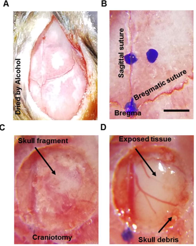
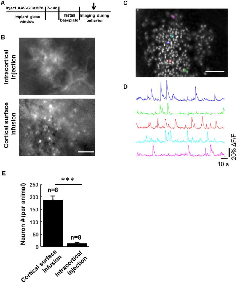
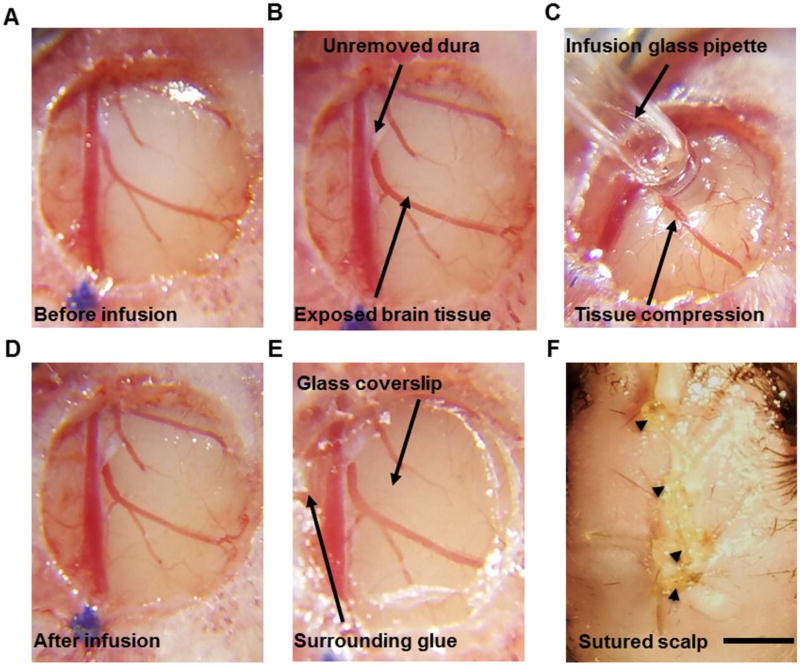
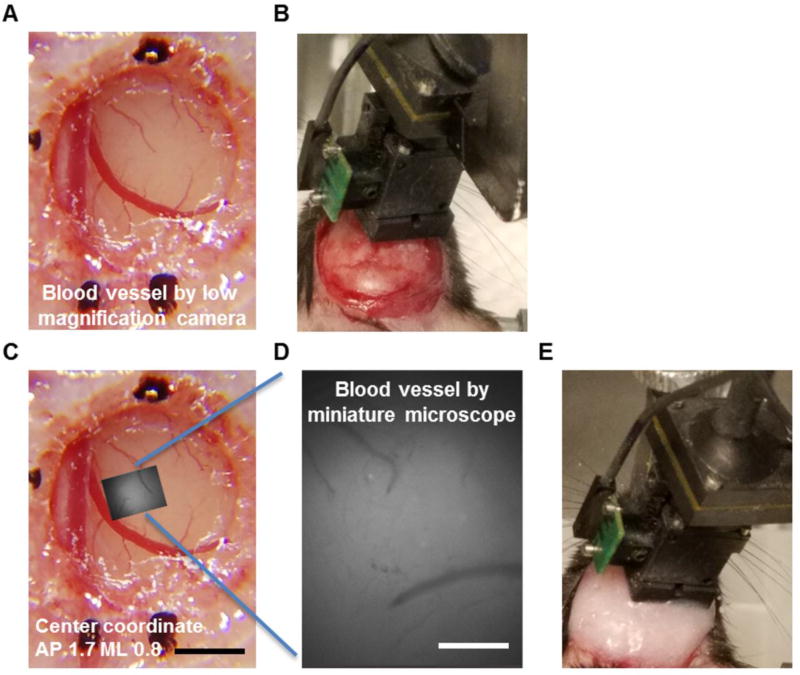
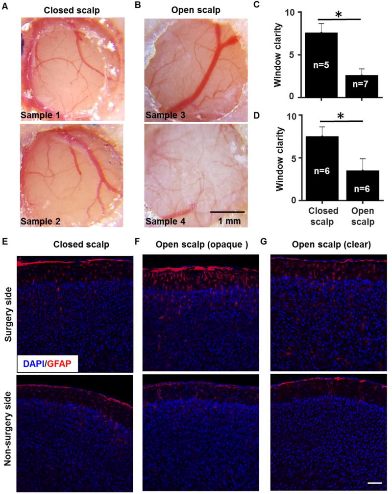
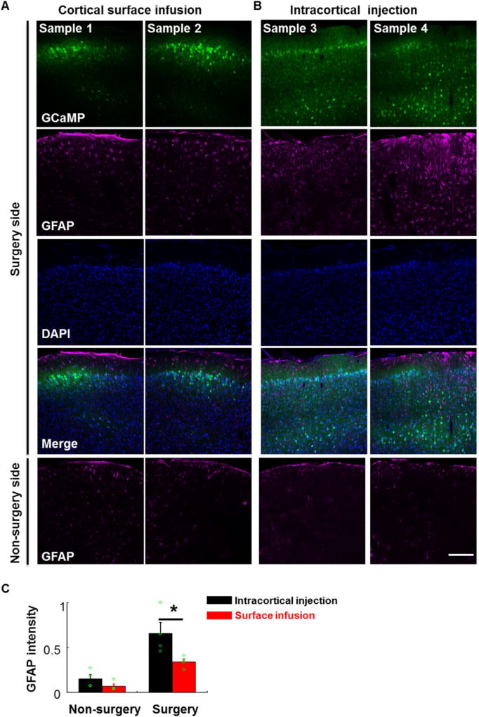
We used a wide-diameter glass pipette at the cortical surface for infusing the viral calcium reporter AAV-GCaMP6 into the cortex. After infusion, the scalp skin over the implanted optical window was sutured to facilitate postoperative recovery. The sutured scalp was removed approximately two weeks later and a miniature microscope was attached above the window to image neuronal activity in freely moving mice.
We found that cortical surface virus infusion efficiently labeled neurons in superficial layers, and scalp skin suturing helped to maintain the long-term clarity of optical windows. As a result, several hundred neurons could be recorded in freely moving animals.
Compared to intracortical virus injection and open-scalp postoperative recovery, our methods minimized tissue damage and dura overgrowth underneath the optical window, and significantly increased the experimental success rate and the yield of identified neurons.
Our improved cranial surgery technique allows for high-yield calcium imaging of cortical neurons with head-mounted microscopes in freely behaving animals. This technique may be beneficial for other optical applications such as two-photon microscopy, multi-site imaging, and optogenetic modulation.
在体神经活动光学成像技术为单细胞水平的大脑功能研究提供了重要的见解。颅窗和病毒传递的钙指示剂常用于通过固定在头部的动物的双光子显微镜对皮质活动进行成像。最近,为自由活动的动物开发了头戴式单光子显微镜。然而,病毒注射过程中最小化组织损伤并保持成像窗口的清晰度在技术上具有挑战性。
我们使用皮质表面的宽直径玻璃吸管将病毒钙报告基因 AAV-GCaMP6 注入皮质。注入后,将头皮皮瓣缝合在植入的光学窗口上,以促进术后恢复。大约两周后,去除缝合的头皮,并在窗口上方安装微型显微镜以对自由移动的小鼠进行神经元活动成像。
我们发现皮质表面病毒输注可有效地标记浅层神经元,头皮皮瓣缝合有助于保持光学窗口的长期清晰度。结果,在自由移动的动物中可以记录到数百个神经元。
与皮质内病毒注射和开颅术后恢复相比,我们的方法最大限度地减少了组织损伤和光学窗口下方的硬脑膜过度生长,并显著提高了实验成功率和鉴定神经元的产量。
我们改进的颅手术技术允许在自由活动的动物中使用头戴式显微镜进行高产量皮质神经元钙成像。该技术可能有益于其他光学应用,如双光子显微镜、多部位成像和光遗传学调节。