Department of Biological Physics, ELTE Eötvös Loránd University, Budapest, Hungary.
CellSorter Scientific Company for Innovations, Erdőalja út 174, 1037, Budapest, Hungary.
Sci Rep. 2021 Sep 16;11(1):18500. doi: 10.1038/s41598-021-97734-1.
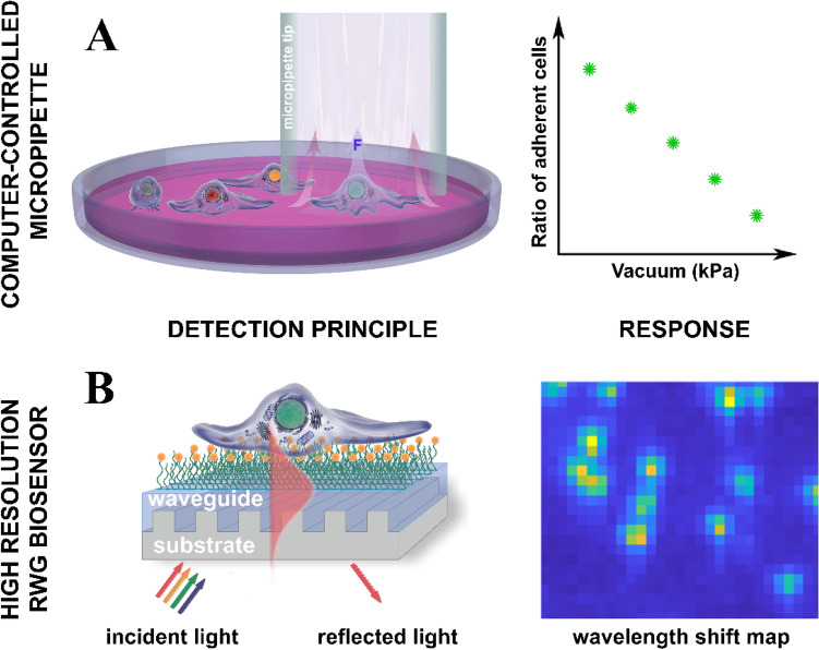
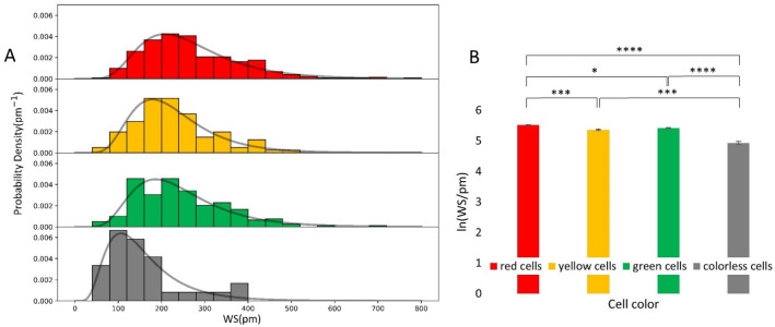
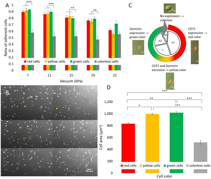
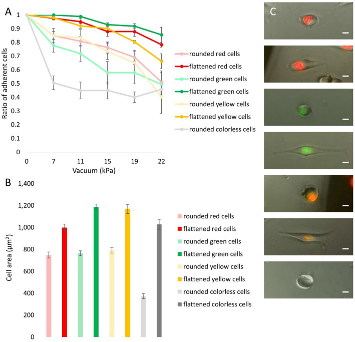
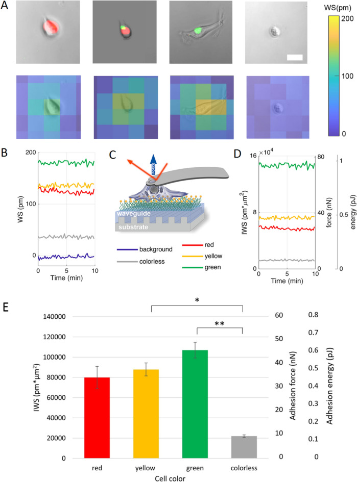
The high throughput, cost effective and sensitive quantification of cell adhesion strength at the single-cell level is still a challenging task. The adhesion force between tissue cells and their environment is crucial in all multicellular organisms. Integrins transmit force between the intracellular cytoskeleton and the extracellular matrix. This force is not only a mechanical interaction but a way of signal transduction as well. For instance, adhesion-dependent cells switch to an apoptotic mode in the lack of adhesion forces. Adhesion of tumor cells is a potential therapeutic target, as it is actively modulated during tissue invasion and cell release to the bloodstream resulting in metastasis. We investigated the integrin-mediated adhesion between cancer cells and their RGD (Arg-Gly-Asp) motif displaying biomimetic substratum using the HeLa cell line transfected by the Fucci fluorescent cell cycle reporter construct. We employed a computer-controlled micropipette and a high spatial resolution label-free resonant waveguide grating-based optical sensor calibrated to adhesion force and energy at the single-cell level. We found that the overall adhesion strength of single cancer cells is approximately constant in all phases except the mitotic (M) phase with a significantly lower adhesion. Single-cell evanescent field based biosensor measurements revealed that at the mitotic phase the cell material mass per unit area inside the cell-substratum contact zone is significantly less, too. Importantly, the weaker mitotic adhesion is not simply a direct consequence of the measured smaller contact area. Our results highlight these differences in the mitotic reticular adhesions and confirm that cell adhesion is a promising target of selective cancer drugs as the vast majority of normal, differentiated tissue cells do not enter the M phase and do not divide.
高通量、经济高效且灵敏地定量单细胞水平的细胞黏附强度仍然是一项具有挑战性的任务。组织细胞与其环境之间的黏附力在所有多细胞生物中都至关重要。整合素在细胞内细胞骨架和细胞外基质之间传递力。这种力不仅是一种机械相互作用,也是一种信号转导方式。例如,在缺乏黏附力的情况下,依赖黏附的细胞会转换为凋亡模式。肿瘤细胞的黏附是一个潜在的治疗靶点,因为它在组织侵袭和细胞释放到血液中导致转移时会被积极调节。我们使用转染了 Fucci 荧光细胞周期报告构建体的 HeLa 细胞系,研究了癌细胞与展示 RGD(精氨酸-甘氨酸-天冬氨酸)基序的仿生基质之间的整合素介导的黏附。我们采用了计算机控制的微管移液器和高空间分辨率的无标记共振波导光栅光学传感器,该传感器可校准至单细胞水平的黏附力和能量。我们发现,除了有丝分裂(M)期外,单个癌细胞的整体黏附强度在所有相中基本保持不变,而在有丝分裂期的黏附力明显降低。基于单细胞渐逝场的生物传感器测量结果表明,在有丝分裂期,细胞-基质接触区域内单位面积的细胞物质质量明显减少。重要的是,较弱的有丝分裂黏附力并非简单地直接导致测量到的较小接触面积的结果。我们的研究结果突出了有丝分裂网状黏附的这些差异,并证实了细胞黏附是选择性癌症药物的有前途的靶点,因为绝大多数正常分化的组织细胞不会进入 M 期,也不会分裂。