• 文献检索
  • 文档翻译
  • 深度研究
  • 学术资讯
  • Suppr Zotero 插件Zotero 插件
  • 邀请有礼
  • 套餐&价格
  • 历史记录
应用&插件
Suppr Zotero 插件Zotero 插件浏览器插件Mac 客户端Windows 客户端微信小程序
定价
高级版会员购买积分包购买API积分包
服务
文献检索文档翻译深度研究API 文档MCP 服务
关于我们
关于 Suppr公司介绍联系我们用户协议隐私条款
关注我们

Suppr 超能文献

核心技术专利:CN118964589B侵权必究
粤ICP备2023148730 号-1Suppr @ 2026

文献检索

告别复杂PubMed语法,用中文像聊天一样搜索,搜遍4000万医学文献。AI智能推荐,让科研检索更轻松。

立即免费搜索

文件翻译

保留排版,准确专业,支持PDF/Word/PPT等文件格式,支持 12+语言互译。

免费翻译文档

深度研究

AI帮你快速写综述,25分钟生成高质量综述,智能提取关键信息,辅助科研写作。

立即免费体验

高通量机器人流体力显微镜测量细胞周期中单细胞粘附参数的种群分布。

Population distributions of single-cell adhesion parameters during the cell cycle from high-throughput robotic fluidic force microscopy.

机构信息

Nanobiosensorics Laboratory, Institute of Technical Physics and Materials Science, Centre for Energy Research, Budapest, Hungary.

Department of Electronics Technology, Faculty of Electrical Engineering and Informatics, Budapest University of Technology and Economics, Budapest, Hungary.

出版信息

Sci Rep. 2022 May 11;12(1):7747. doi: 10.1038/s41598-022-11770-z.

DOI:10.1038/s41598-022-11770-z
PMID:35546603
原文链接:https://pmc.ncbi.nlm.nih.gov/articles/PMC9095720/
Abstract

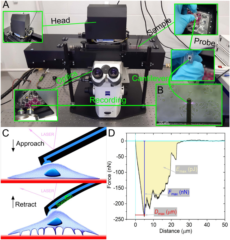
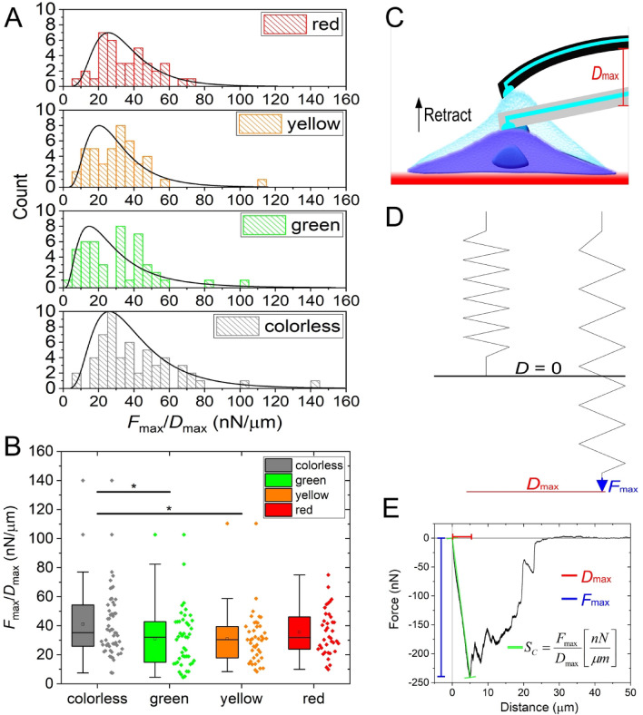
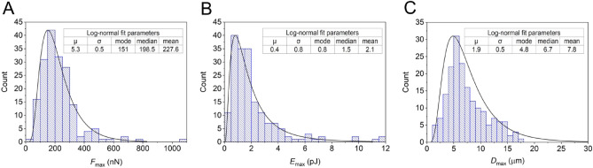
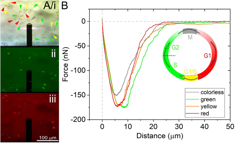
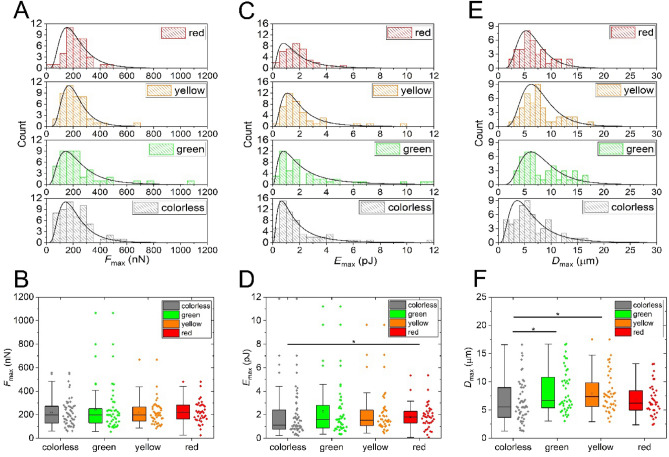
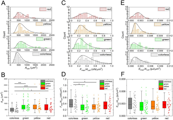
Single-cell adhesion plays an essential role in biological and biomedical sciences, but its precise measurement for a large number of cells is still a challenging task. At present, typical force measuring techniques usually offer low throughput, a few cells per day, and therefore are unable to uncover phenomena emerging at the population level. In this work, robotic fluidic force microscopy (FluidFM) was utilized to measure the adhesion parameters of cells in a high-throughput manner to study their population distributions in-depth. The investigated cell type was the genetically engineered HeLa Fucci construct with cell cycle-dependent expression of fluorescent proteins. This feature, combined with the high-throughput measurement made it possible for the first time to characterize the single-cell adhesion distributions at various stages of the cell cycle. It was found that parameters such as single-cell adhesion force and energy follow a lognormal population distribution. Therefore, conclusions based on adhesion data of a low number of cells or treating the population as normally distributed can be misleading. Moreover, we found that the cell area was significantly the smallest, and the area normalized maximal adhesion force was significantly the largest for the colorless cells (the mitotic (M) and early G1 phases). Notably, the parameter characterizing the elongation of the cells until the maximum level of force between the cell and its substratum was also dependent on the cell cycle, which quantity was the smallest for the colorless cells. A novel parameter, named the spring coefficient of the cell, was introduced as the fraction of maximal adhesion force and maximal cell elongation during the mechanical detachment, which was found to be significantly the largest for the colorless cells. Cells in the M phase adhere in atypical way, with so-called reticular adhesions, which are different from canonical focal adhesions. We first revealed that reticular adhesion can exert a higher force per unit area than canonical focal adhesions, and cells in this phase are significantly stiffer. The possible biological consequences of these findings were also discussed, together with the practical relevance of the observed population-level adhesion phenomena.

摘要

单细胞黏附在生物和生物医学科学中起着至关重要的作用,但对大量细胞进行精确测量仍然是一项具有挑战性的任务。目前,典型的力测量技术通常提供低通量,每天只有几个细胞,因此无法揭示在群体水平上出现的现象。在这项工作中,我们利用机器人流体力学显微镜(FluidFM)以高通量的方式测量细胞的黏附参数,深入研究它们的群体分布。所研究的细胞类型是具有荧光蛋白细胞周期依赖性表达的基因工程 HeLa Fucci 构建体。这一特征,再加上高通量测量,使得首次有可能对细胞周期各阶段的单细胞黏附分布进行特征描述。结果发现,单细胞黏附力和能量等参数呈对数正态分布。因此,基于少量细胞的黏附数据得出的结论或假设群体呈正态分布可能会产生误导。此外,我们发现无色细胞(有丝分裂(M)和早期 G1 期)的细胞面积最小,归一化的最大黏附力最大。值得注意的是,细胞在达到与基质之间的最大力水平之前的伸长参数也依赖于细胞周期,无色细胞的这个参数最小。引入了一个新的参数,称为细胞的弹簧系数,它是在机械分离过程中最大黏附力和最大细胞伸长的分数,无色细胞的这个参数最大。M 期的细胞以非典型的方式黏附,形成所谓的网状黏附,与典型的黏附斑不同。我们首次揭示了网状黏附可以比典型的黏附斑施加更高的单位面积力,并且这个阶段的细胞明显更硬。还讨论了这些发现的可能生物学后果,以及观察到的群体水平黏附现象的实际意义。

https://cdn.ncbi.nlm.nih.gov/pmc/blobs/8b80/9095720/b7f6d8d63df4/41598_2022_11770_Fig6_HTML.jpg
https://cdn.ncbi.nlm.nih.gov/pmc/blobs/8b80/9095720/400d181a9180/41598_2022_11770_Fig1_HTML.jpg
https://cdn.ncbi.nlm.nih.gov/pmc/blobs/8b80/9095720/e51f70d35e6b/41598_2022_11770_Fig2_HTML.jpg
https://cdn.ncbi.nlm.nih.gov/pmc/blobs/8b80/9095720/f380809bdcdc/41598_2022_11770_Fig3_HTML.jpg
https://cdn.ncbi.nlm.nih.gov/pmc/blobs/8b80/9095720/e3dd0a78c971/41598_2022_11770_Fig4_HTML.jpg
https://cdn.ncbi.nlm.nih.gov/pmc/blobs/8b80/9095720/3e26a4ab30d8/41598_2022_11770_Fig5_HTML.jpg
https://cdn.ncbi.nlm.nih.gov/pmc/blobs/8b80/9095720/b7f6d8d63df4/41598_2022_11770_Fig6_HTML.jpg

相似文献

1
Population distributions of single-cell adhesion parameters during the cell cycle from high-throughput robotic fluidic force microscopy.高通量机器人流体力显微镜测量细胞周期中单细胞粘附参数的种群分布。
Sci Rep. 2022 May 11;12(1):7747. doi: 10.1038/s41598-022-11770-z.
2
Single-cell adhesion force kinetics of cell populations from combined label-free optical biosensor and robotic fluidic force microscopy.单细胞黏附力动力学分析:基于无标记光学生物传感器和机器人流体力学显微镜的细胞群体。
Sci Rep. 2020 Jan 9;10(1):61. doi: 10.1038/s41598-019-56898-7.
3
Cell-substratum and cell-cell adhesion forces and single-cell mechanical properties in mono- and multilayer assemblies from robotic fluidic force microscopy.通过机器人流体力显微镜研究单层和多层组件中的细胞-基质及细胞-细胞粘附力和单细胞力学特性。
Eur J Cell Biol. 2022 Sep-Nov;101(4):151273. doi: 10.1016/j.ejcb.2022.151273. Epub 2022 Aug 31.
4
4D Force Detection of Cell Adhesion and Contractility.细胞黏附与收缩力的 4D 力检测
Nano Lett. 2023 Apr 12;23(7):2467-2475. doi: 10.1021/acs.nanolett.2c03733. Epub 2023 Mar 28.
5
Single-cell adhesion strength and contact density drops in the M phase of cancer cells.癌细胞 M 期的单细胞黏附强度和接触密度下降。
Sci Rep. 2021 Sep 16;11(1):18500. doi: 10.1038/s41598-021-97734-1.
6
Variation in cell-substratum adhesion in relation to cell cycle phases.细胞与基质粘附的变化与细胞周期阶段的关系。
Exp Cell Res. 2004 Feb 1;293(1):58-67. doi: 10.1016/j.yexcr.2003.10.005.
7
Extending applications of AFM to fluidic AFM in single living cell studies.将原子力显微镜的应用扩展到用于单细胞研究的流体原子力显微镜中。
J Cell Physiol. 2022 Aug;237(8):3222-3238. doi: 10.1002/jcp.30809. Epub 2022 Jun 13.
8
Single-cell force spectroscopy of fluid flow-tuned cell adhesion for dissecting hemodynamics in tumor metastasis.单细胞力谱分析流体调节细胞黏附在肿瘤转移中剖析血流动力学的作用
Nanoscale. 2023 Dec 21;16(1):360-372. doi: 10.1039/d3nr04439d.
9
Cell-Cycle-Dependent Regulation of Cell Adhesions: Adhering to the Schedule: Three papers reveal unexpected properties of adhesion structures as cells progress through the cell cycle.细胞周期依赖性细胞黏附调控:遵循时间表:三篇论文揭示了细胞在细胞周期中进行时黏附结构的意外性质。
Bioessays. 2019 Jan;41(1):e1800165. doi: 10.1002/bies.201800165. Epub 2018 Nov 28.
10
Mechanical cues direct focal adhesion dynamics.机械信号引导粘着斑动力学。
Prog Mol Biol Transl Sci. 2014;126:103-34. doi: 10.1016/B978-0-12-394624-9.00005-1.

引用本文的文献

1
Determination of the Minimum Cell-to-Cell Adhesion Time Using Optical Tweezers in Leukemia and Lymphoma Research.在白血病和淋巴瘤研究中使用光镊测定细胞间最小黏附时间
Methods Protoc. 2025 Jun 4;8(3):59. doi: 10.3390/mps8030059.
2
Single-Cell Fluidic Force Spectroscopy Reveals Dynamic Mechanical Fingerprints of Malignancy in Breast Cancer.单细胞流体力谱揭示乳腺癌恶性转化的动态力学指纹图谱。
ACS Appl Mater Interfaces. 2024 Sep 25;16(38):50147-50159. doi: 10.1021/acsami.4c06335. Epub 2024 Aug 6.
3
Continuous distribution of cancer cells in the cell cycle unveiled by AI-segmented imaging of 37,000 HeLa FUCCI cells.

本文引用的文献

1
Quantification of the Adhesion Strength of to Tooth Enamel.对……与牙釉质附着力的定量分析。 你提供的原文中“of”后面似乎缺少内容,请你核对一下完整文本,以便我能更准确地翻译。
Microorganisms. 2021 Oct 25;9(11):2213. doi: 10.3390/microorganisms9112213.
2
Cell cycle control in cancer.癌症中的细胞周期调控。
Nat Rev Mol Cell Biol. 2022 Jan;23(1):74-88. doi: 10.1038/s41580-021-00404-3. Epub 2021 Sep 10.
3
Actin cytoskeleton deregulation confers midostaurin resistance in FLT3-mutant acute myeloid leukemia.肌动蛋白细胞骨架失调赋予 FLT3 突变型急性髓系白血病对米哚妥林的耐药性。
通过对37000个HeLa FUCCI细胞进行人工智能分割成像揭示细胞周期中癌细胞的连续分布。
Heliyon. 2024 Apr 23;10(9):e30239. doi: 10.1016/j.heliyon.2024.e30239. eCollection 2024 May 15.
4
Label-free biomolecular and cellular methods in small molecule epigallocatechin-gallate research.小分子表没食子儿茶素没食子酸酯研究中的无标记生物分子和细胞方法。
Heliyon. 2024 Feb 5;10(3):e25603. doi: 10.1016/j.heliyon.2024.e25603. eCollection 2024 Feb 15.
5
Hydrodynamic function and spring constant calibration of FluidFM micropipette cantilevers.FluidFM微吸管悬臂的流体动力学功能和弹簧常数校准
Microsyst Nanoeng. 2024 Feb 18;10:26. doi: 10.1038/s41378-023-00629-6. eCollection 2024.
6
Recent advances in single-cell subcellular sampling.单细胞亚细胞采样的最新进展。
Chem Commun (Camb). 2023 May 2;59(36):5312-5328. doi: 10.1039/d3cc00573a.
7
4D Force Detection of Cell Adhesion and Contractility.细胞黏附与收缩力的 4D 力检测
Nano Lett. 2023 Apr 12;23(7):2467-2475. doi: 10.1021/acs.nanolett.2c03733. Epub 2023 Mar 28.
8
Single-cell temporal transcriptomics from tiny cytoplasmic biopsies.从微小的细胞质活检中进行单细胞时间转录组学研究。
Cell Rep Methods. 2022 Oct 17;2(10):100319. doi: 10.1016/j.crmeth.2022.100319. eCollection 2022 Oct 24.
Commun Biol. 2021 Jun 25;4(1):799. doi: 10.1038/s42003-021-02215-w.
4
Staphylococcus aureus vWF-binding protein triggers a strong interaction between clumping factor A and host vWF.金黄色葡萄球菌 vWF 结合蛋白引发聚集因子 A 与宿主 vWF 之间的强烈相互作用。
Commun Biol. 2021 Apr 12;4(1):453. doi: 10.1038/s42003-021-01986-6.
5
A Review of Single-Cell Adhesion Force Kinetics and Applications.单细胞黏附力动力学及其应用综述。
Cells. 2021 Mar 5;10(3):577. doi: 10.3390/cells10030577.
6
Spatiotemporal dissection of the cell cycle with single-cell proteogenomics.单细胞蛋白质基因组学解析细胞周期的时空动态。
Nature. 2021 Feb;590(7847):649-654. doi: 10.1038/s41586-021-03232-9. Epub 2021 Feb 24.
7
Simultaneous time-varying viscosity, elasticity, and mass measurements of single adherent cancer cells across cell cycle.在细胞周期内对单个贴壁癌细胞的同时时变粘度、弹性和质量进行测量。
Sci Rep. 2020 Jul 30;10(1):12803. doi: 10.1038/s41598-020-69638-z.
8
Single-cell adhesion force kinetics of cell populations from combined label-free optical biosensor and robotic fluidic force microscopy.单细胞黏附力动力学分析:基于无标记光学生物传感器和机器人流体力学显微镜的细胞群体。
Sci Rep. 2020 Jan 9;10(1):61. doi: 10.1038/s41598-019-56898-7.
9
The production of L- and D-phenylalanines using engineered phenylalanine ammonia lyases from Petroselinum crispum.利用来自皱叶欧芹的工程化苯丙氨酸解氨酶生产 L-和 D-苯丙氨酸。
Sci Rep. 2019 Dec 27;9(1):20123. doi: 10.1038/s41598-019-56554-0.
10
Adhesion force measurements on functionalized microbeads: An in-depth comparison of computer controlled micropipette and fluidic force microscopy.功能化微球的粘附力测量:计算机控制微移液器和流体力显微镜的深入比较。
J Colloid Interface Sci. 2019 Nov 1;555:245-253. doi: 10.1016/j.jcis.2019.07.102. Epub 2019 Jul 31.