Tang Shuiying, Xu Bihong, Li Jincheng, Zhong Meifeng, Hong Ziyang, Zhao Wei, Zeng Tao, He Xiaofeng
Division of Vascular and Interventional Radiology, Department of General Surgery, Nanfang Hospital, Southern Medical University, Guangzhou, China.
Interventional Radiology and Pathology, Nanfang Hospital, Department of Pathology, Southern Medical University, Guangzhou, China.
Ann Transl Med. 2021 Aug;9(15):1257. doi: 10.21037/atm-21-3233.
Oxidative stress is an important factor in the modulation of both tumorigenesis and anticancer responses. Ozone (O) is a strong oxidant that causes redox reactions and exerts anticancer effects in various types of cancer cells. However, the pathways involved in O-induced cell death are not well understood.
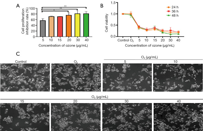
human hepatocellular carcinoma (HCC) BEL7402 cells were treated with various O concentrations to evaluate O cytotoxicity by Cell Counting Kit-8 (CCK-8) assay and flow cytometry. The regulatory mechanisms were analyzed by western blot analysis. , an HCC model was established to evaluate the inhibition of HCC with O treatment.
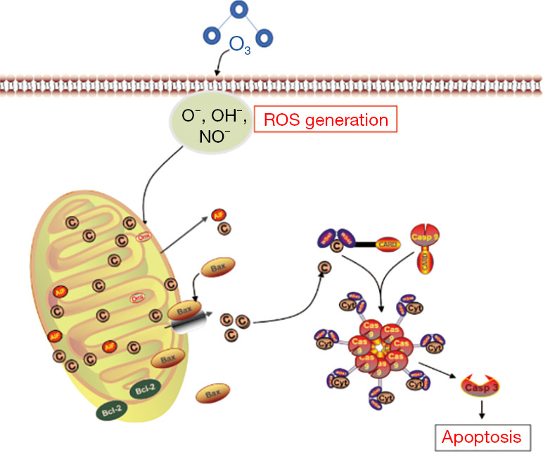
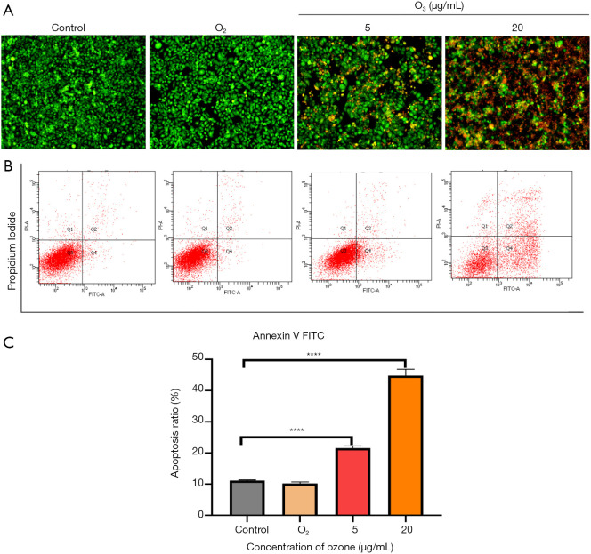
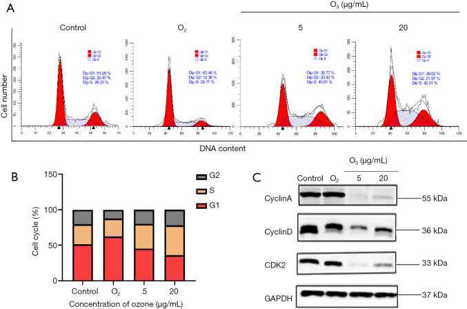
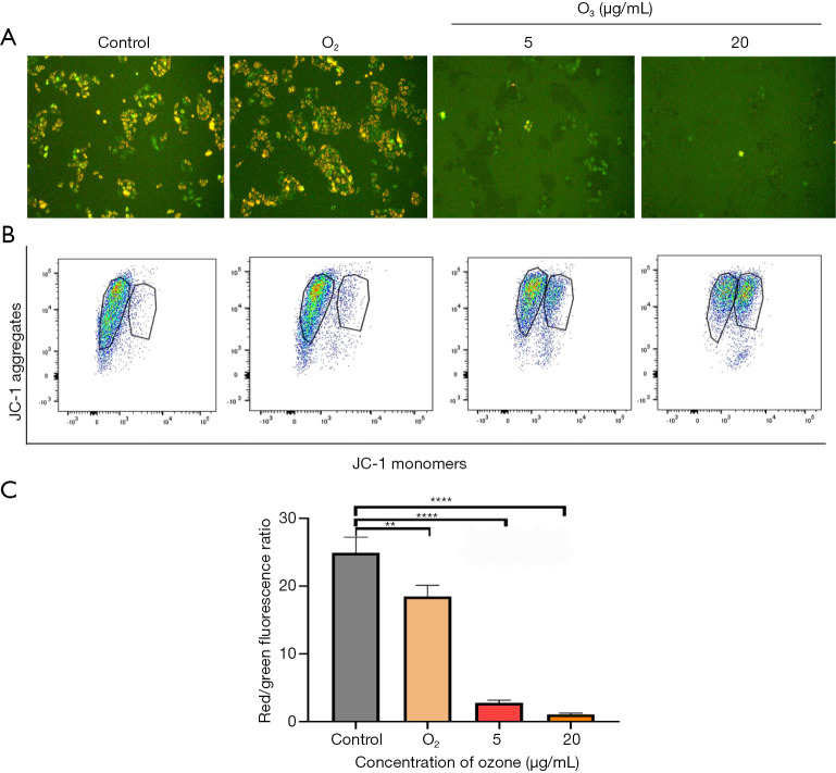
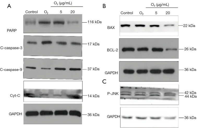
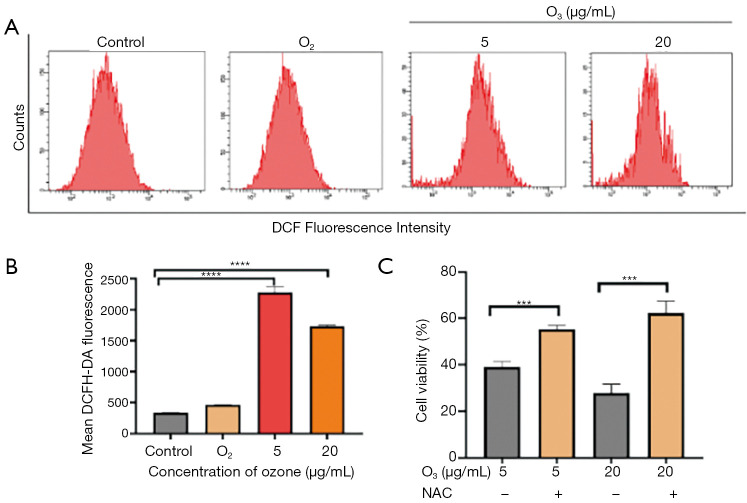
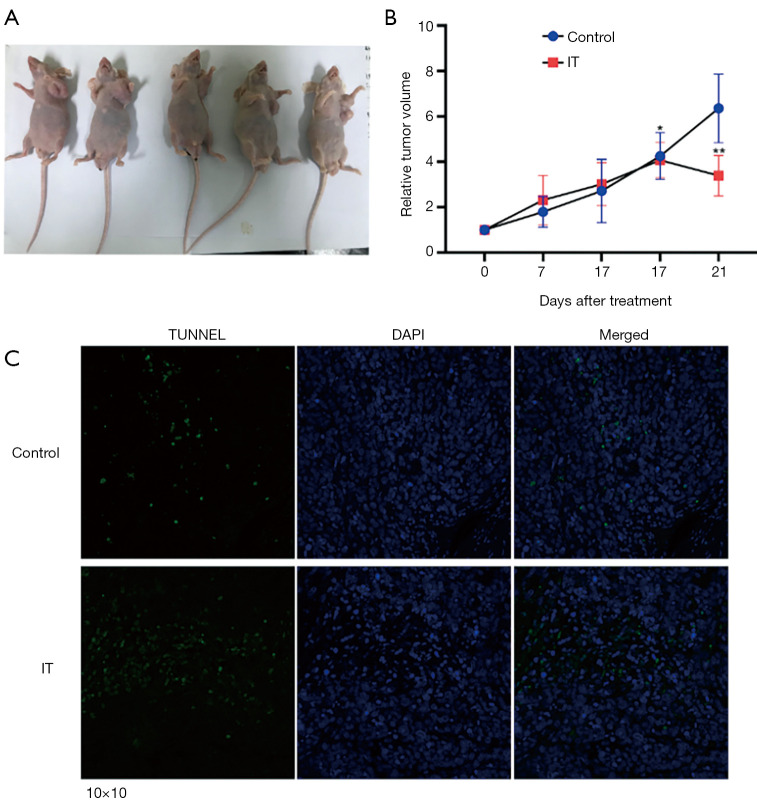
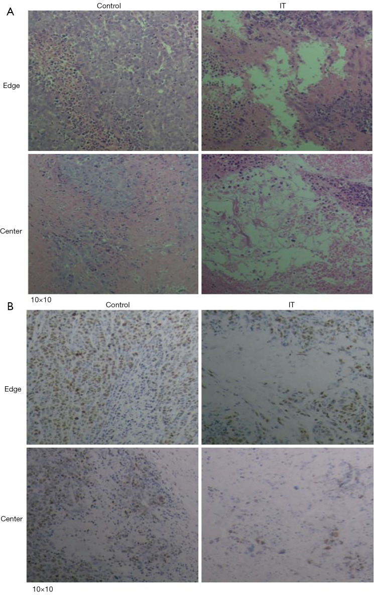
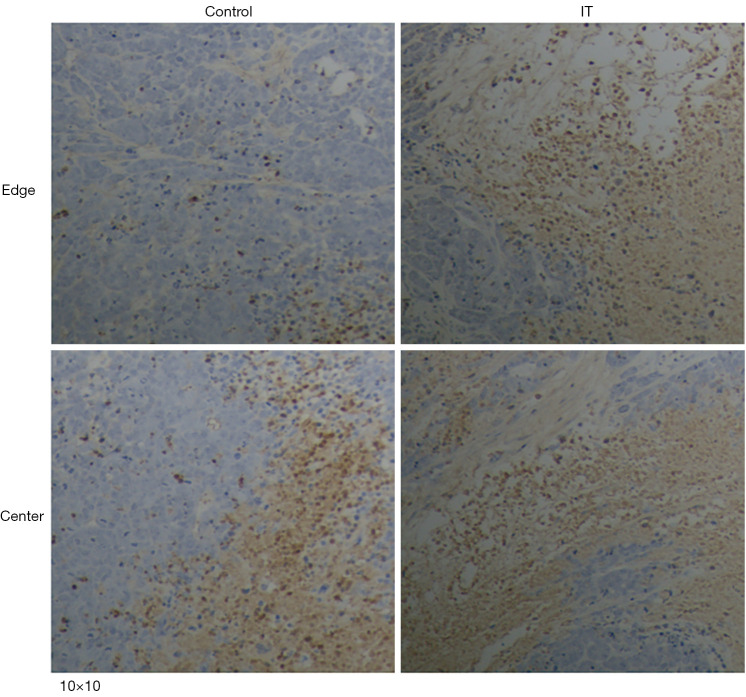
cells treated with O exhibited a round and small morphology with nuclear shrinkage and fragmentation. The CCK-8 assay confirmed the potent cytotoxic activity of O against BEL7402 cells (IC value of 5 µg/mL). Acridine orange/ethidium bromide (AO/EB) staining revealed apoptosis of BEL7402 cells after O treatment. Flow cytometry analysis showed that S phase cell cycle arrest and apoptosis increased with O exposure. In addition, O3 exposure reduced the mitochondrial membrane potential (ΔΨm) and induced reactive oxygen species (ROS) accumulation. Western blot analysis showed that O exposure reduced B-cell lymphoma 2 (BCL-2) expression and increased cleaved poly ADP-ribose polymerase (PARP), cytochrome C (Cyt-C), caspase-3, caspase-9, and p-JNK expression. , treatment with intratumor injection O (20 µg/mL) inhibited HCC growth.
Overall, our findings showed that O induces BEL7402 cell apoptosis via the intrinsic mitochondria-dependent pathway. Therefore, O has therapeutic potential for HCC.
氧化应激是肿瘤发生和抗癌反应调节中的一个重要因素。臭氧(O₃)是一种强氧化剂,可引起氧化还原反应并在各种类型的癌细胞中发挥抗癌作用。然而,臭氧诱导细胞死亡所涉及的途径尚不完全清楚。
用不同浓度的臭氧处理人肝癌(HCC)BEL7402细胞,通过细胞计数试剂盒-8(CCK-8)检测和流式细胞术评估臭氧的细胞毒性。通过蛋白质免疫印迹分析来分析其调控机制。此外,建立肝癌模型以评估臭氧治疗对肝癌的抑制作用。
经臭氧处理的细胞呈现圆形且体积较小的形态,伴有核固缩和核碎裂。CCK-8检测证实了臭氧对BEL7402细胞具有强大的细胞毒性活性(IC₅₀值为5μg/mL)。吖啶橙/溴化乙锭(AO/EB)染色显示臭氧处理后BEL7402细胞发生凋亡。流式细胞术分析表明,随着臭氧暴露,S期细胞周期停滞和凋亡增加。此外,臭氧暴露降低了线粒体膜电位(ΔΨm)并诱导了活性氧(ROS)积累。蛋白质免疫印迹分析表明,臭氧暴露降低了B细胞淋巴瘤2(BCL-2)的表达,并增加了裂解的聚ADP核糖聚合酶(PARP)、细胞色素C(Cyt-C)、半胱天冬酶-3、半胱天冬酶-9和磷酸化JNK的表达。此外,瘤内注射臭氧(20μg/mL)治疗可抑制肝癌生长。
总体而言,我们的研究结果表明,臭氧通过内在的线粒体依赖性途径诱导BEL7402细胞凋亡。因此,臭氧对肝癌具有治疗潜力。