• 文献检索
  • 文档翻译
  • 深度研究
  • 学术资讯
  • Suppr Zotero 插件Zotero 插件
  • 邀请有礼
  • 套餐&价格
  • 历史记录
应用&插件
Suppr Zotero 插件Zotero 插件浏览器插件Mac 客户端Windows 客户端微信小程序
定价
高级版会员购买积分包购买API积分包
服务
文献检索文档翻译深度研究API 文档MCP 服务
关于我们
关于 Suppr公司介绍联系我们用户协议隐私条款
关注我们

Suppr 超能文献

核心技术专利:CN118964589B侵权必究
粤ICP备2023148730 号-1Suppr @ 2026

文献检索

告别复杂PubMed语法,用中文像聊天一样搜索,搜遍4000万医学文献。AI智能推荐,让科研检索更轻松。

立即免费搜索

文件翻译

保留排版,准确专业,支持PDF/Word/PPT等文件格式,支持 12+语言互译。

免费翻译文档

深度研究

AI帮你快速写综述,25分钟生成高质量综述,智能提取关键信息,辅助科研写作。

立即免费体验

当前用于放射性坏死和假性进展诊断的新兴 MRI 工具。

Current emerging MRI tools for radionecrosis and pseudoprogression diagnosis.

机构信息

Department of Neuroradiology, Sorbonne Université, Assistance Publique-Hôpitaux de Paris, Groupe Hospitalier Pitié-Salpêtrière-Charles Foix.

Sorbonne Université, INSERM, CNRS, Assistance Publique-Hôpitaux de Paris, Institut du Cerveau et de la Moelle épinière, boulevard de l'Hôpital, Paris.

出版信息

Curr Opin Oncol. 2021 Nov 1;33(6):597-607. doi: 10.1097/CCO.0000000000000793.

DOI:10.1097/CCO.0000000000000793
PMID:34534142
原文链接:https://pmc.ncbi.nlm.nih.gov/articles/PMC8528135/
Abstract

PURPOSE OF REVIEW

This review aims to cover current MRI techniques for assessing treatment response in brain tumors, with a focus on radio-induced lesions.

RECENT FINDINGS

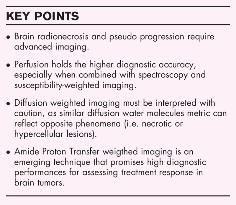
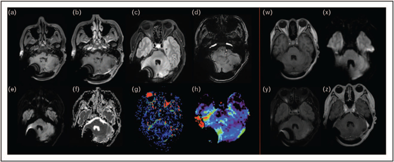
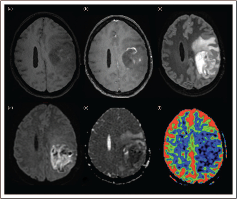
Pseudoprogression and radionecrosis are common radiological entities after brain tumor irradiation and are difficult to distinguish from real progression, with major consequences on daily patient care. To date, shortcomings of conventional MRI have been largely recognized but morphological sequences are still used in official response assessment criteria. Several complementary advanced techniques have been proposed but none of them have been validated, hampering their clinical use. Among advanced MRI, brain perfusion measures increase diagnostic accuracy, especially when added with spectroscopy and susceptibility-weighted imaging. However, lack of reproducibility, because of several hard-to-control variables, is still a major limitation for their standardization in routine protocols. Amide Proton Transfer is an emerging molecular imaging technique that promises to offer new metrics by indirectly quantifying intracellular mobile proteins and peptide concentration. Preliminary studies suggest that this noncontrast sequence may add key biomarkers in tumor evaluation, especially in posttherapeutic settings.

SUMMARY

Benefits and pitfalls of conventional and advanced imaging on posttreatment assessment are discussed and the potential added value of APT in this clinicoradiological evolving scenario is introduced.

摘要

目的综述

本综述旨在介绍目前用于评估脑肿瘤治疗反应的 MRI 技术,重点是放射性损伤。

最近的发现

脑肿瘤放疗后常见的放射性病变包括假性进展和放射性坏死,难以与真正的进展相区分,这对患者的日常治疗有重大影响。迄今为止,常规 MRI 的局限性已得到广泛认可,但形态学序列仍用于官方的反应评估标准。已经提出了几种补充的高级技术,但都没有得到验证,这阻碍了它们的临床应用。在高级 MRI 中,脑灌注测量可提高诊断准确性,尤其是与波谱和磁敏感加权成像相结合时。然而,由于难以控制的多种变量,缺乏可重复性仍然是将其标准化纳入常规方案的主要限制。酰胺质子转移是一种新兴的分子成像技术,有望通过间接定量细胞内可移动蛋白和肽浓度提供新的指标。初步研究表明,这种无对比序列可能在肿瘤评估中增加关键的生物标志物,特别是在治疗后。

总结

讨论了常规和高级影像学在治疗后评估中的优缺点,并介绍了 APT 在这种临床影像学不断发展的情况下的潜在附加价值。

https://cdn.ncbi.nlm.nih.gov/pmc/blobs/2610/8528135/8efc0991cff2/coonc-33-597-g004.jpg
https://cdn.ncbi.nlm.nih.gov/pmc/blobs/2610/8528135/60fe3d5f3f2c/coonc-33-597-g001.jpg
https://cdn.ncbi.nlm.nih.gov/pmc/blobs/2610/8528135/86a992a5262f/coonc-33-597-g002.jpg
https://cdn.ncbi.nlm.nih.gov/pmc/blobs/2610/8528135/c8799f0117e3/coonc-33-597-g003.jpg
https://cdn.ncbi.nlm.nih.gov/pmc/blobs/2610/8528135/8efc0991cff2/coonc-33-597-g004.jpg

相似文献

1
Current emerging MRI tools for radionecrosis and pseudoprogression diagnosis.当前用于放射性坏死和假性进展诊断的新兴 MRI 工具。
Curr Opin Oncol. 2021 Nov 1;33(6):597-607. doi: 10.1097/CCO.0000000000000793.
2
Treatment-related changes in glioblastoma: a review on the controversies in response assessment criteria and the concepts of true progression, pseudoprogression, pseudoresponse and radionecrosis.治疗相关的胶质母细胞瘤变化:对反应评估标准的争议和真正进展、假性进展、假性缓解和放射性坏死概念的综述。
Clin Transl Oncol. 2018 Aug;20(8):939-953. doi: 10.1007/s12094-017-1816-x. Epub 2017 Dec 7.
3
Imaging Glioblastoma Posttreatment: Progression, Pseudoprogression, Pseudoresponse, Radiation Necrosis.胶质母细胞瘤治疗后的影像学表现:进展、假性进展、假性反应、放射性坏死
Radiol Clin North Am. 2019 Nov;57(6):1199-1216. doi: 10.1016/j.rcl.2019.07.003. Epub 2019 Aug 16.
4
What effective technique to differentiate radiation brain necrosis from a tumor progression in patients treated with radiation: A monocentric retrospective study combining the MRI TRAMs technique and the (F)-dopa PET/CT.有哪些有效的技术可以区分接受放疗的患者的放射性脑坏死与肿瘤进展:一项结合 MRI TRAMs 技术和(F)-多巴 PET/CT 的单中心回顾性研究。
Cancer Radiother. 2023 Jun;27(4):273-280. doi: 10.1016/j.canrad.2022.12.002. Epub 2023 Apr 19.
5
Pseudoprogression, radionecrosis, inflammation or true tumor progression? challenges associated with glioblastoma response assessment in an evolving therapeutic landscape.假性进展、放射性坏死、炎症还是真正的肿瘤进展?在不断发展的治疗领域中,与胶质母细胞瘤反应评估相关的挑战。
J Neurooncol. 2017 Sep;134(3):495-504. doi: 10.1007/s11060-017-2375-2. Epub 2017 Apr 5.
6
Umbrella review and network meta-analysis of diagnostic imaging test accuracy studies in Differentiating between brain tumor progression versus pseudoprogression and radionecrosis.针对脑肿瘤进展与假性进展和放射性坏死的鉴别诊断,对诊断成像试验准确性研究的伞状评价和网络荟萃分析。
J Neurooncol. 2024 Jan;166(1):1-15. doi: 10.1007/s11060-023-04528-8. Epub 2024 Jan 11.
7
Dynamic susceptibility contrast (DSC) perfusion MRI in differential diagnosis between radionecrosis and neoangiogenesis in cerebral metastases using rCBV, rCBF and K2.动态对比磁共振灌注成像(DSC)在脑转移瘤放射性坏死与新生血管形成鉴别诊断中的应用:rCBV、rCBF 和 K2 的价值。
Radiol Med. 2018 Jul;123(7):545-552. doi: 10.1007/s11547-018-0866-7. Epub 2018 Mar 5.
8
Applying amide proton transfer-weighted MRI to distinguish pseudoprogression from true progression in malignant gliomas.应用酰胺质子转移加权磁共振成像鉴别恶性胶质瘤的假性进展与真性进展。
J Magn Reson Imaging. 2016 Aug;44(2):456-62. doi: 10.1002/jmri.25159. Epub 2016 Jan 20.
9
Differentiating radiation necrosis from tumor progression in brain metastases treated with stereotactic radiotherapy: utility of intravoxel incoherent motion perfusion MRI and correlation with histopathology.在立体定向放疗治疗脑转移瘤中,从肿瘤进展中鉴别放射性坏死:应用体素内不相干运动灌注 MRI 的效用及其与组织病理学的相关性。
J Neurooncol. 2017 Sep;134(2):433-441. doi: 10.1007/s11060-017-2545-2. Epub 2017 Jul 3.
10
Shape Features of the Lesion Habitat to Differentiate Brain Tumor Progression from Pseudoprogression on Routine Multiparametric MRI: A Multisite Study.病灶形态特征有助于区分常规多参数 MRI 上脑肿瘤进展与假性进展:一项多中心研究。
AJNR Am J Neuroradiol. 2018 Dec;39(12):2187-2193. doi: 10.3174/ajnr.A5858. Epub 2018 Nov 1.

引用本文的文献

1
Imaging cancer metabolism using magnetic resonance.利用磁共振成像技术研究癌症代谢
Npj Imaging. 2024 Jan 11;2(1):1. doi: 10.1038/s44303-023-00004-0.
2
Understanding the Radiobiology of Central Nervous System Diseases in the Golden Age of Radiosurgery-Does It Matter?在放射外科的黄金时代理解中枢神经系统疾病的放射生物学——这重要吗?
Brain Sci. 2025 Jun 17;15(6):649. doi: 10.3390/brainsci15060649.
3
Brain MRI and regional vulnerabilities to radiation necrosis: investigating the impact of stereotactic radiotherapy in brain metastases treatment.

本文引用的文献

1
Fully automated analysis combining [F]-FET-PET and multiparametric MRI including DSC perfusion and APTw imaging: a promising tool for objective evaluation of glioma progression.结合 [F]-FET-PET 和多参数 MRI(包括 DSC 灌注和 APTw 成像)的全自动分析:一种用于客观评估胶质瘤进展的有前途的工具。
Eur J Nucl Med Mol Imaging. 2021 Dec;48(13):4445-4455. doi: 10.1007/s00259-021-05427-8. Epub 2021 Jun 25.
2
MRI biomarkers in neuro-oncology.神经肿瘤学中的 MRI 生物标志物。
Nat Rev Neurol. 2021 Aug;17(8):486-500. doi: 10.1038/s41582-021-00510-y. Epub 2021 Jun 20.
3
Radionecrosis (RN) in patients with brain metastases treated with stereotactic radiosurgery (SRS) and immunotherapy.
脑磁共振成像与放射性坏死的区域易损性:探究立体定向放射治疗在脑转移瘤治疗中的影响
Front Radiol. 2025 Apr 9;5:1554017. doi: 10.3389/fradi.2025.1554017. eCollection 2025.
4
Fluid-Suppressed Amide Proton Transfer-Weighted Imaging Outperforms Leakage-Corrected Dynamic Susceptibility Contrast Perfusion in Distinguishing Progression from Radionecrosis in Brain Metastases.在鉴别脑转移瘤放射性坏死进展方面,液体抑制酰胺质子转移加权成像优于渗漏校正动态磁敏感对比灌注成像。
Cancers (Basel). 2025 Mar 31;17(7):1175. doi: 10.3390/cancers17071175.
5
Case Report: Clinical awareness about the effect of laser interstitial thermal therapy on pediatric high-grade brain tumors after radiotherapy.病例报告:关于激光间质热疗对放疗后小儿高级别脑肿瘤影响的临床认识。
Front Surg. 2025 Jan 17;11:1462074. doi: 10.3389/fsurg.2024.1462074. eCollection 2024.
6
APT imaging of hepatocellular carcinoma signals an effective therapeutic response in advance of tumor shrinkage.肝细胞癌的APT成像在肿瘤缩小之前就预示着有效的治疗反应。
Hepat Oncol. 2024 Dec 31;11(1):2389031. doi: 10.1080/20450923.2024.2389031. Epub 2024 Sep 12.
7
Amide proton transfer weighted MRI measurements yield consistent and repeatable results in patients with gliomas: a prospective test-retest study.酰胺质子转移加权磁共振成像测量在胶质瘤患者中产生一致且可重复的结果:一项前瞻性重测研究。
Eur Radiol. 2025 Jun;35(6):3367-3379. doi: 10.1007/s00330-024-11197-2. Epub 2024 Dec 18.
8
A study method using early dynamic acquisition of [F]fluorodopa positron emission tomography for the differential diagnosis between progression and radionecrosis of brain metastases after radiotherapy.一种利用早期动态采集[F]氟多巴正电子发射断层扫描进行放疗后脑转移瘤进展与放射性坏死鉴别诊断的研究方法。
EJNMMI Res. 2024 Oct 9;14(1):93. doi: 10.1186/s13550-024-01158-7.
9
Are Dual-Phase F-Fluorodeoxyglucose PET-mpMRI Diagnostic Performances to Distinguish Brain Tumour Radionecrosis/Recurrence after Cranial Radiotherapy Usable in Routine?双相F-氟脱氧葡萄糖PET-mpMRI用于区分颅脑放疗后脑肿瘤放射性坏死/复发的诊断性能能否用于常规?
Cancers (Basel). 2024 Sep 21;16(18):3216. doi: 10.3390/cancers16183216.
10
Clinical and radiological effects of Bevacizumab for the treatment of radionecrosis after stereotactic brain radiotherapy.贝伐单抗治疗立体定向脑放疗后放射性坏死的临床和影像学效果。
BMC Cancer. 2024 Jul 30;24(1):918. doi: 10.1186/s12885-024-12643-6.
接受立体定向放射外科治疗(SRS)和免疫治疗的脑转移瘤患者的放射性坏死(RN)
Int J Neurosci. 2023 Feb;133(2):186-193. doi: 10.1080/00207454.2021.1900843. Epub 2021 Apr 19.
4
Bevacizumab for radiation necrosis following radiotherapy of brain metastatic disease: a systematic review & meta-analysis.贝伐单抗治疗脑转移瘤放疗后放射性坏死:系统评价与荟萃分析。
BMC Cancer. 2021 Feb 16;21(1):167. doi: 10.1186/s12885-021-07889-3.
5
Clinical routine acquisition protocol for 3D relaxation-compensated APT and rNOE CEST-MRI of the human brain at 3T.临床常规获取协议 3T 下人类大脑的 3D 弛豫补偿 APT 和 rNOE CEST-MRI
Magn Reson Med. 2021 Jul;86(1):393-404. doi: 10.1002/mrm.28699. Epub 2021 Feb 14.
6
Imaging Glioblastoma Posttreatment: Progression, Pseudoprogression, Pseudoresponse, Radiation Necrosis.脑胶质瘤治疗后影像学表现:进展、假性进展、假性缓解、放射性坏死。
Neuroimaging Clin N Am. 2021 Feb;31(1):103-120. doi: 10.1016/j.nic.2020.09.010.
7
Recognizing Radiation-induced Changes in the Central Nervous System: Where to Look and What to Look For.识别中枢神经系统的放射性改变:观察什么及如何观察。
Radiographics. 2021 Jan-Feb;41(1):224-248. doi: 10.1148/rg.2021200064. Epub 2020 Nov 20.
8
Magnetic resonance spectroscopy of isocitrate dehydrogenase mutated gliomas: current knowledge on the neurochemical profile.异柠檬酸脱氢酶突变型脑胶质瘤的磁共振波谱分析:神经化学特征的现有知识。
Curr Opin Neurol. 2020 Aug;33(4):413-421. doi: 10.1097/WCO.0000000000000833.
9
Abscopal effect in radioimmunotherapy.放射免疫治疗中的远隔效应。
Int Immunopharmacol. 2020 Aug;85:106663. doi: 10.1016/j.intimp.2020.106663. Epub 2020 Jun 7.
10
Brain metastasis formation and irradiation by stereotactic radiation therapy combined with immunotherapy: A systematic review.脑转移瘤形成及立体定向放疗联合免疫治疗的放射效应:系统综述。
Crit Rev Oncol Hematol. 2020 May;149:102923. doi: 10.1016/j.critrevonc.2020.102923. Epub 2020 Mar 3.