• 文献检索
  • 文档翻译
  • 深度研究
  • 学术资讯
  • Suppr Zotero 插件Zotero 插件
  • 邀请有礼
  • 套餐&价格
  • 历史记录
应用&插件
Suppr Zotero 插件Zotero 插件浏览器插件Mac 客户端Windows 客户端微信小程序
定价
高级版会员购买积分包购买API积分包
服务
文献检索文档翻译深度研究API 文档MCP 服务
关于我们
关于 Suppr公司介绍联系我们用户协议隐私条款
关注我们

Suppr 超能文献

核心技术专利:CN118964589B侵权必究
粤ICP备2023148730 号-1Suppr @ 2026

文献检索

告别复杂PubMed语法,用中文像聊天一样搜索,搜遍4000万医学文献。AI智能推荐,让科研检索更轻松。

立即免费搜索

文件翻译

保留排版,准确专业,支持PDF/Word/PPT等文件格式,支持 12+语言互译。

免费翻译文档

深度研究

AI帮你快速写综述,25分钟生成高质量综述,智能提取关键信息,辅助科研写作。

立即免费体验

肺癌 H&E 染色数字切片中三级淋巴结构(TLS)的鉴定和密度评估。

Tertiary lymphoid structures (TLS) identification and density assessment on H&E-stained digital slides of lung cancer.

机构信息

Department of Pathology, UCL Cancer Institute, University College London, London, United Kingdom.

Centre for Medical Image Computing, University College London, London, United Kingdom.

出版信息

PLoS One. 2021 Sep 23;16(9):e0256907. doi: 10.1371/journal.pone.0256907. eCollection 2021.

DOI:10.1371/journal.pone.0256907
PMID:34555057
原文链接:https://pmc.ncbi.nlm.nih.gov/articles/PMC8460026/
Abstract

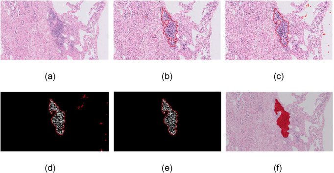
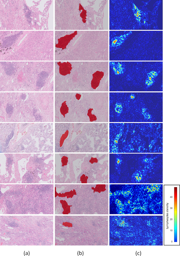
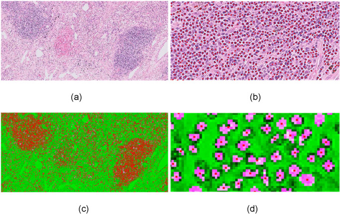
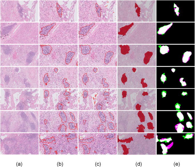
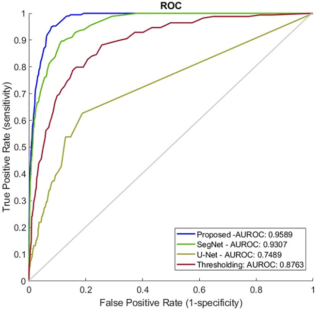
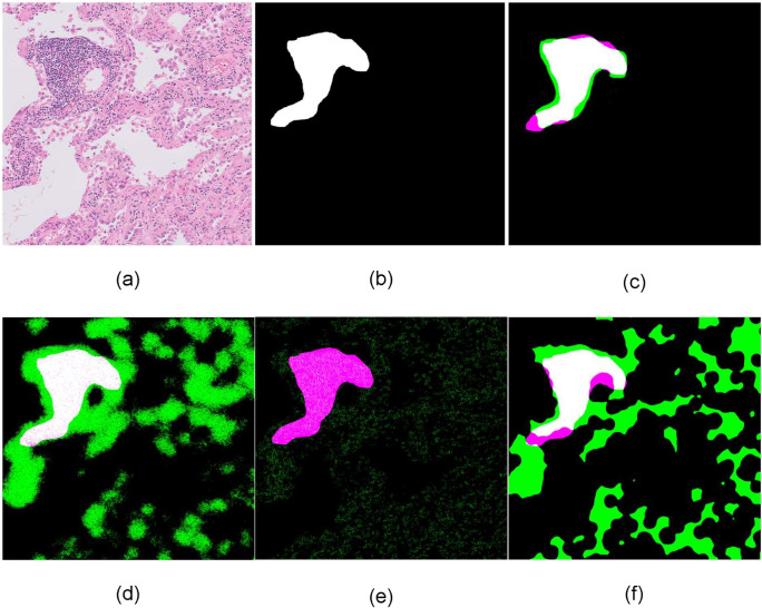
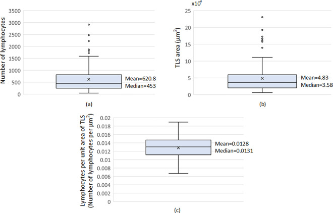
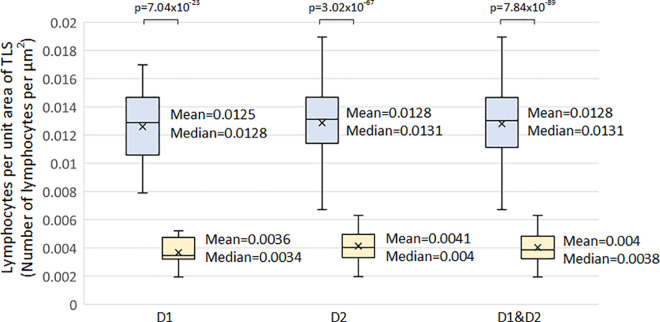
Tertiary lymphoid structures (TLS) are ectopic aggregates of lymphoid cells in inflamed, infected, or tumoral tissues that are easily recognized on an H&E histology slide as discrete entities, distinct from lymphocytes. TLS are associated with improved cancer prognosis but there is no standardised method available to quantify their presence. Previous studies have used immunohistochemistry to determine the presence of specific cells as a marker of the TLS. This has now been proven to be an underestimate of the true number of TLS. Thus, we propose a methodology for the automated identification and quantification of TLS, based on H&E slides. We subsequently determined the mathematical criteria defining a TLS. TLS regions were identified through a deep convolutional neural network and segmentation of lymphocytes was performed through an ellipsoidal model. This methodology had a 92.87% specificity at 95% sensitivity, 88.79% specificity at 98% sensitivity and 84.32% specificity at 99% sensitivity level based on 144 TLS annotated H&E slides implying that the automated approach was able to reproduce the histopathologists' assessment with great accuracy. We showed that the minimum number of lymphocytes within TLS is 45 and the minimum TLS area is 6,245μm2. Furthermore, we have shown that the density of the lymphocytes is more than 3 times those outside of the TLS. The mean density and standard deviation of lymphocytes within a TLS area are 0.0128/μm2 and 0.0026/μm2 respectively compared to 0.004/μm2 and 0.001/μm2 in non-TLS regions. The proposed methodology shows great potential for automated identification and quantification of the TLS density on digital H&E slides.

摘要

三级淋巴结构 (TLS) 是在炎症、感染或肿瘤组织中异位聚集的淋巴样细胞,在 H&E 组织学切片上很容易被识别为离散实体,与淋巴细胞不同。TLS 与癌症预后改善相关,但目前尚无标准化方法来定量其存在。先前的研究使用免疫组织化学来确定特定细胞的存在作为 TLS 的标志物。现在已经证明,这低估了 TLS 的真实数量。因此,我们提出了一种基于 H&E 切片的自动识别和定量 TLS 的方法。随后,我们确定了定义 TLS 的数学标准。通过深度卷积神经网络识别 TLS 区域,并通过椭圆模型进行淋巴细胞分割。这种方法在 144 张具有 TLS 注释的 H&E 切片上具有 92.87%的特异性和 95%的敏感性、88.79%的特异性和 98%的敏感性以及 84.32%的特异性,这意味着自动方法能够非常准确地重现组织病理学家的评估。我们表明,TLS 内淋巴细胞的最小数量为 45,TLS 的最小面积为 6245μm2。此外,我们还表明,TLS 内淋巴细胞的密度是 TLS 外的 3 倍以上。与非 TLS 区域的 0.004/μm2 和 0.001/μm2 相比,TLS 区域内淋巴细胞的平均密度和标准偏差分别为 0.0128/μm2 和 0.0026/μm2。该方法在数字 H&E 切片上自动识别和定量 TLS 密度具有很大的潜力。

https://cdn.ncbi.nlm.nih.gov/pmc/blobs/7a5b/8460026/a0fdcbf3460e/pone.0256907.g009.jpg
https://cdn.ncbi.nlm.nih.gov/pmc/blobs/7a5b/8460026/02d1c845f25f/pone.0256907.g001.jpg
https://cdn.ncbi.nlm.nih.gov/pmc/blobs/7a5b/8460026/f88507d4cf3e/pone.0256907.g002.jpg
https://cdn.ncbi.nlm.nih.gov/pmc/blobs/7a5b/8460026/fdfe5947f47d/pone.0256907.g003.jpg
https://cdn.ncbi.nlm.nih.gov/pmc/blobs/7a5b/8460026/12e11db26420/pone.0256907.g004.jpg
https://cdn.ncbi.nlm.nih.gov/pmc/blobs/7a5b/8460026/470b62f94ae4/pone.0256907.g005.jpg
https://cdn.ncbi.nlm.nih.gov/pmc/blobs/7a5b/8460026/17ee7b8083c7/pone.0256907.g006.jpg
https://cdn.ncbi.nlm.nih.gov/pmc/blobs/7a5b/8460026/38e9724d73a2/pone.0256907.g007.jpg
https://cdn.ncbi.nlm.nih.gov/pmc/blobs/7a5b/8460026/ad5d33317307/pone.0256907.g008.jpg
https://cdn.ncbi.nlm.nih.gov/pmc/blobs/7a5b/8460026/a0fdcbf3460e/pone.0256907.g009.jpg

相似文献

1
Tertiary lymphoid structures (TLS) identification and density assessment on H&E-stained digital slides of lung cancer.肺癌 H&E 染色数字切片中三级淋巴结构(TLS)的鉴定和密度评估。
PLoS One. 2021 Sep 23;16(9):e0256907. doi: 10.1371/journal.pone.0256907. eCollection 2021.
2
Density of tertiary lymphoid structures and their correlation with prognosis in non-small cell lung cancer.三级淋巴结构密度及其与非小细胞肺癌预后的相关性。
Front Immunol. 2024 Aug 13;15:1423775. doi: 10.3389/fimmu.2024.1423775. eCollection 2024.
3
Tumor-infiltrating lymphocyte subsets and tertiary lymphoid structures in pulmonary metastases from colorectal cancer.结直肠癌肺转移灶中的肿瘤浸润淋巴细胞亚群和三级淋巴结构
Clin Exp Metastasis. 2016 Oct;33(7):727-39. doi: 10.1007/s10585-016-9813-y. Epub 2016 Jul 23.
4
Germinal Centers Determine the Prognostic Relevance of Tertiary Lymphoid Structures and Are Impaired by Corticosteroids in Lung Squamous Cell Carcinoma.生发中心决定肺鳞癌中三级淋巴结构的预后相关性,并被皮质类固醇所抑制。
Cancer Res. 2018 Mar 1;78(5):1308-1320. doi: 10.1158/0008-5472.CAN-17-1987. Epub 2017 Dec 26.
5
Tertiary lymphoid structure score: a promising approach to refine the TNM staging in resected non-small cell lung cancer.三级淋巴结构评分:一种有前途的方法,可细化切除的非小细胞肺癌的 TNM 分期。
Br J Cancer. 2021 May;124(10):1680-1689. doi: 10.1038/s41416-021-01307-y. Epub 2021 Mar 15.
6
Exploring the impact of tertiary lymphoid structures maturity in NSCLC: insights from TLS scoring.探讨非小细胞肺癌中三级淋巴样结构成熟的影响:来自 TLS 评分的见解。
Front Immunol. 2024 Sep 20;15:1422206. doi: 10.3389/fimmu.2024.1422206. eCollection 2024.
7
The prognostic significance of tumor-infiltrating lymphocytes assessment with hematoxylin and eosin sections in resected primary lung adenocarcinoma.苏木精和伊红染色切片中肿瘤浸润淋巴细胞评估对切除的原发性肺腺癌的预后意义。
PLoS One. 2019 Nov 19;14(11):e0224430. doi: 10.1371/journal.pone.0224430. eCollection 2019.
8
Peritumoral tertiary lymphoid structure and tumor stroma percentage predict the prognosis of patients with non-metastatic colorectal cancer.肿瘤周围三级淋巴结构和肿瘤间质百分比可预测非转移性结直肠癌患者的预后。
Front Immunol. 2022 Sep 16;13:962056. doi: 10.3389/fimmu.2022.962056. eCollection 2022.
9
Development of Tools for the Selective Visualization and Quantification of TLS-Immune Cells on Tissue Sections.用于在组织切片上选择性可视化和定量TLS免疫细胞的工具的开发。
Methods Mol Biol. 2018;1845:47-69. doi: 10.1007/978-1-4939-8709-2_4.
10
Investigating Tumor-Associated Tertiary Lymphoid Structures in Murine Lung Adenocarcinoma.研究小鼠肺腺癌中肿瘤相关三级淋巴结构
Methods Mol Biol. 2018;1845:259-273. doi: 10.1007/978-1-4939-8709-2_15.

引用本文的文献

1
Three-dimensional assessments are necessary to determine the true, spatially resolved composition of tissues.三维评估对于确定组织的真实、空间分辨组成是必要的。
Cell Rep Methods. 2025 Jun 16;5(6):101075. doi: 10.1016/j.crmeth.2025.101075. Epub 2025 Jun 9.
2
Recent advances in biomarkers for predicting the efficacy of immunotherapy in non-small cell lung cancer.预测非小细胞肺癌免疫治疗疗效的生物标志物的最新进展。
Front Immunol. 2025 May 8;16:1554871. doi: 10.3389/fimmu.2025.1554871. eCollection 2025.
3
Quantitatively defined stromal B cell aggregates are associated with response to checkpoint inhibitors in unresectable melanoma.

本文引用的文献

1
Deep-Learning-Based Characterization of Tumor-Infiltrating Lymphocytes in Breast Cancers From Histopathology Images and Multiomics Data.基于深度学习的乳腺癌组织病理学图像和多组学数据中肿瘤浸润淋巴细胞的特征分析。
JCO Clin Cancer Inform. 2020 May;4:480-490. doi: 10.1200/CCI.19.00126.
2
Tertiary lymphoid structures in cancer - considerations for patient prognosis.癌症中的三级淋巴结构——对患者预后的考量
Cell Mol Immunol. 2020 Jun;17(6):570-575. doi: 10.1038/s41423-020-0457-0. Epub 2020 May 15.
3
Test-time augmentation for deep learning-based cell segmentation on microscopy images.
定量定义的基质B细胞聚集与不可切除黑色素瘤对检查点抑制剂的反应相关。
Cell Rep. 2025 Apr 22;44(4):115554. doi: 10.1016/j.celrep.2025.115554. Epub 2025 Apr 11.
4
Integrative whole slide image and spatial transcriptomics analysis with QuST and QuPath.使用QuST和QuPath进行整合的全玻片图像和空间转录组学分析。
NPJ Precis Oncol. 2025 Mar 12;9(1):70. doi: 10.1038/s41698-025-00841-9.
5
Spatial distribution of tertiary lymphoid structures in the molecular and clinical context of non-small cell lung cancer.非小细胞肺癌分子与临床背景下三级淋巴结构的空间分布
Cell Oncol (Dordr). 2025 Mar 3. doi: 10.1007/s13402-025-01052-x.
6
Spatial characterization of tertiary lymphoid structures as predictive biomarkers for immune checkpoint blockade in head and neck squamous cell carcinoma.三级淋巴结构的空间特征作为头颈部鳞状细胞癌免疫检查点阻断的预测生物标志物
Oncoimmunology. 2025 Dec;14(1):2466308. doi: 10.1080/2162402X.2025.2466308. Epub 2025 Feb 18.
7
A novel approach to digital characterisation of Tertiary Lymphoid Structures in colorectal cancer.一种用于结直肠癌中三级淋巴结构数字表征的新方法。
Front Immunol. 2025 Jan 28;16:1500792. doi: 10.3389/fimmu.2025.1500792. eCollection 2025.
8
Genomic Characteristics Related to Histology-Based Immune Features in Breast Cancer.与乳腺癌基于组织学的免疫特征相关的基因组特征
Mod Pathol. 2025 May;38(5):100736. doi: 10.1016/j.modpat.2025.100736. Epub 2025 Feb 15.
9
Artificial intelligence-based spatial analysis of tertiary lymphoid structures and clinical significance for endometrial cancer.基于人工智能的子宫内膜癌三级淋巴结构空间分析及其临床意义
Cancer Immunol Immunother. 2025 Feb 1;74(3):84. doi: 10.1007/s00262-024-03929-6.
10
Infiltration and subtype analysis of CD3 + CD20 + T cells in lung cancer.肺癌中CD3 + CD20 + T细胞的浸润及亚型分析
BMC Cancer. 2025 Jan 30;25(1):179. doi: 10.1186/s12885-025-13581-7.
基于深度学习的显微镜图像细胞分割的测试时增强。
Sci Rep. 2020 Mar 19;10(1):5068. doi: 10.1038/s41598-020-61808-3.
4
Semi-Supervised Nests of Melanocytes Segmentation Method Using Convolutional Autoencoders.基于卷积自动编码器的半监督黑素细胞巢分割方法。
Sensors (Basel). 2020 Mar 11;20(6):1546. doi: 10.3390/s20061546.
5
Clinical efficacy and safety of anti-PD-1/PD-L1 inhibitors for the treatment of advanced or metastatic cancer: a systematic review and meta-analysis.抗 PD-1/PD-L1 抑制剂治疗晚期或转移性癌症的临床疗效和安全性:系统评价和荟萃分析。
Sci Rep. 2020 Feb 7;10(1):2083. doi: 10.1038/s41598-020-58674-4.
6
Tertiary lymphoid structures improve immunotherapy and survival in melanoma.三级淋巴结构可改善黑色素瘤的免疫治疗和生存率。
Nature. 2020 Jan;577(7791):561-565. doi: 10.1038/s41586-019-1914-8. Epub 2020 Jan 15.
7
Tertiary lymphoid structures in the era of cancer immunotherapy.癌症免疫治疗时代的三级淋巴结构。
Nat Rev Cancer. 2019 Jun;19(6):307-325. doi: 10.1038/s41568-019-0144-6.
8
Recognizing basal cell carcinoma on smartphone-captured digital histopathology images with a deep neural network.利用深度神经网络在智能手机拍摄的数字组织病理学图像上识别基底细胞癌。
Br J Dermatol. 2020 Mar;182(3):754-762. doi: 10.1111/bjd.18026. Epub 2019 Aug 22.
9
U-Net: deep learning for cell counting, detection, and morphometry.U-Net:用于细胞计数、检测和形态测量学的深度学习。
Nat Methods. 2019 Jan;16(1):67-70. doi: 10.1038/s41592-018-0261-2. Epub 2018 Dec 17.
10
A deep learning-based algorithm for 2-D cell segmentation in microscopy images.基于深度学习的显微镜图像二维细胞分割算法。
BMC Bioinformatics. 2018 Oct 3;19(1):365. doi: 10.1186/s12859-018-2375-z.