文献检索文档翻译深度研究
Suppr Zotero 插件Zotero 插件
邀请有礼套餐&价格历史记录

新学期,新优惠

限时优惠:9月1日-9月22日

30天高级会员仅需29元

1天体验卡首发特惠仅需5.99元

了解详情
不再提醒
插件&应用
Suppr Zotero 插件Zotero 插件浏览器插件Mac 客户端Windows 客户端微信小程序
高级版
套餐订阅购买积分包
AI 工具
文献检索文档翻译深度研究
关于我们
关于 Suppr公司介绍联系我们用户协议隐私条款
关注我们

Suppr 超能文献

核心技术专利:CN118964589B侵权必究
粤ICP备2023148730 号-1Suppr @ 2025

基于深度学习的显微镜图像细胞分割的测试时增强。

Test-time augmentation for deep learning-based cell segmentation on microscopy images.

机构信息

Biological Research Centre, Szeged, Hungary.

University of Szeged, Szeged, Hungary.

出版信息

Sci Rep. 2020 Mar 19;10(1):5068. doi: 10.1038/s41598-020-61808-3.


DOI:10.1038/s41598-020-61808-3
PMID:32193485
原文链接:https://pmc.ncbi.nlm.nih.gov/articles/PMC7081314/
Abstract

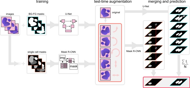
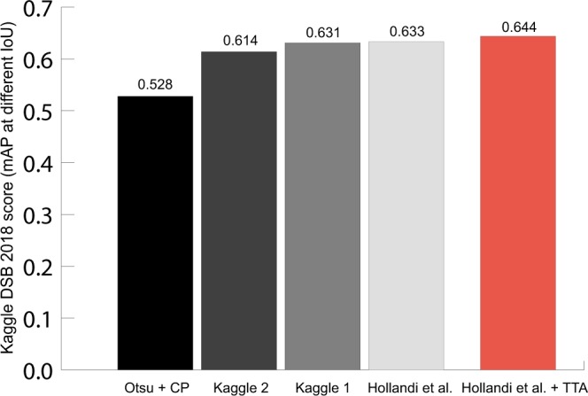
Recent advancements in deep learning have revolutionized the way microscopy images of cells are processed. Deep learning network architectures have a large number of parameters, thus, in order to reach high accuracy, they require a massive amount of annotated data. A common way of improving accuracy builds on the artificial increase of the training set by using different augmentation techniques. A less common way relies on test-time augmentation (TTA) which yields transformed versions of the image for prediction and the results are merged. In this paper we describe how we have incorporated the test-time argumentation prediction method into two major segmentation approaches utilized in the single-cell analysis of microscopy images. These approaches are semantic segmentation based on the U-Net, and instance segmentation based on the Mask R-CNN models. Our findings show that even if only simple test-time augmentations (such as rotation or flipping and proper merging methods) are applied, TTA can significantly improve prediction accuracy. We have utilized images of tissue and cell cultures from the Data Science Bowl (DSB) 2018 nuclei segmentation competition and other sources. Additionally, boosting the highest-scoring method of the DSB with TTA, we could further improve prediction accuracy, and our method has reached an ever-best score at the DSB.

摘要

深度学习的最新进展彻底改变了细胞显微镜图像的处理方式。深度学习网络架构具有大量参数,因此,为了达到高精度,它们需要大量标注数据。一种常见的提高准确性的方法是通过使用不同的增强技术来人工增加训练集。一种不太常见的方法依赖于测试时增强(TTA),它会生成图像的变换版本进行预测,并合并结果。在本文中,我们描述了如何将测试时论证预测方法纳入两种主要的显微镜图像单细胞分析中使用的分割方法。这些方法是基于 U-Net 的语义分割和基于 Mask R-CNN 模型的实例分割。我们的研究结果表明,即使只应用简单的测试时增强(例如旋转或翻转以及适当的合并方法),TTA 也可以显著提高预测准确性。我们利用了来自 Data Science Bowl(DSB)2018 核分割竞赛和其他来源的组织和细胞培养物的图像。此外,通过 TTA 增强 DSB 中得分最高的方法,我们可以进一步提高预测准确性,我们的方法在 DSB 中达到了历史最佳成绩。

https://cdn.ncbi.nlm.nih.gov/pmc/blobs/f148/7081314/8cbbe85a54dc/41598_2020_61808_Fig5_HTML.jpg
https://cdn.ncbi.nlm.nih.gov/pmc/blobs/f148/7081314/76212400b840/41598_2020_61808_Fig1_HTML.jpg
https://cdn.ncbi.nlm.nih.gov/pmc/blobs/f148/7081314/d3b57026acaf/41598_2020_61808_Fig2_HTML.jpg
https://cdn.ncbi.nlm.nih.gov/pmc/blobs/f148/7081314/25bd5a73bf20/41598_2020_61808_Fig3_HTML.jpg
https://cdn.ncbi.nlm.nih.gov/pmc/blobs/f148/7081314/832481949411/41598_2020_61808_Fig4_HTML.jpg
https://cdn.ncbi.nlm.nih.gov/pmc/blobs/f148/7081314/8cbbe85a54dc/41598_2020_61808_Fig5_HTML.jpg

相似文献

[1]
Test-time augmentation for deep learning-based cell segmentation on microscopy images.

Sci Rep. 2020-3-19

[2]
Evaluation of Deep Learning Architectures for Complex Immunofluorescence Nuclear Image Segmentation.

IEEE Trans Med Imaging. 2021-7

[3]
Catheter segmentation in X-ray fluoroscopy using synthetic data and transfer learning with light U-nets.

Comput Methods Programs Biomed. 2020-8

[4]
Combined Transfer Learning and Test-Time Augmentation Improves Convolutional Neural Network-Based Semantic Segmentation of Prostate Cancer from Multi-Parametric MR Images.

Comput Methods Programs Biomed. 2021-10

[5]
Multi-Modality Microscopy Image Style Augmentation for Nuclei Segmentation.

J Imaging. 2022-3-11

[6]
Deep Learning-Assisted Burn Wound Diagnosis: Diagnostic Model Development Study.

JMIR Med Inform. 2021-12-2

[7]
Instance Segmentation of Multiple Myeloma Cells Using Deep-Wise Data Augmentation and Mask R-CNN.

Entropy (Basel). 2022-1-17

[8]
Effect of learning parameters on the performance of the U-Net architecture for cell nuclei segmentation from microscopic cell images.

Microscopy (Oxf). 2023-6-8

[9]
Comparison of Multi-Label U-Net and Mask R-CNN for panoramic radiograph segmentation to detect periodontitis.

Imaging Sci Dent. 2022-12

[10]
Nuclei instance segmentation from histopathology images using Bayesian dropout based deep learning.

BMC Med Imaging. 2023-10-19

引用本文的文献

[1]
Explainable CNN-Radiomics Fusion and Ensemble Learning for Multimodal Lesion Classification in Dental Radiographs.

Diagnostics (Basel). 2025-8-9

[2]
Deep learning-based quantitative analysis of glomerular morphology in IgA nephropathy whole slide images and its prognostic implications.

Sci Rep. 2025-7-2

[3]
Topology-preserving contourwise shape fusion.

Sci Rep. 2025-3-28

[4]
A Novel Pipeline for Adrenal Gland Segmentation: Integration of a Hybrid Post-Processing Technique with Deep Learning.

J Imaging Inform Med. 2025-3-4

[5]
Ensemble Deep Learning Object Detection Fusion for Cell Tracking, Mitosis, and Lineage.

IEEE Open J Eng Med Biol. 2023-6-21

[6]
A Hybrid Transformer-Convolutional Neural Network for Segmentation of Intracerebral Hemorrhage and Perihematomal Edema on Non-Contrast Head Computed Tomography (CT) with Uncertainty Quantification to Improve Confidence.

Bioengineering (Basel). 2024-12-15

[7]
Artificial intelligence correctly classifies developmental stages of monarch caterpillars enabling better conservation through the use of community science photographs.

Sci Rep. 2024-11-7

[8]
LVI-PathNet: Segmentation-classification pipeline for detection of lymphovascular invasion in whole slide images of lung adenocarcinoma.

J Pathol Inform. 2024-8-30

[9]
Enhanced accuracy with Segmentation of Colorectal Polyp using NanoNetB, and Conditional Random Field Test-Time Augmentation.

Front Robot AI. 2024-8-9

[10]
STTA: enhanced text classification via selective test-time augmentation.

PeerJ Comput Sci. 2023-12-19

本文引用的文献

[1]
nucleAIzer: A Parameter-free Deep Learning Framework for Nucleus Segmentation Using Image Style Transfer.

Cell Syst. 2020-5-20

[2]
Expert-validated estimation of diagnostic uncertainty for deep neural networks in diabetic retinopathy detection.

Med Image Anal. 2020-8

[3]
Nucleus segmentation across imaging experiments: the 2018 Data Science Bowl.

Nat Methods. 2019-10-21

[4]
A Deep Learning-Based Approach for High-Throughput Hypocotyl Phenotyping.

Plant Physiol. 2019-10-21

[5]
Aleatoric uncertainty estimation with test-time augmentation for medical image segmentation with convolutional neural networks.

Neurocomputing (Amst). 2019-9-3

[6]
Evaluation of Deep Learning Strategies for Nucleus Segmentation in Fluorescence Images.

Cytometry A. 2019-7-16

[7]
Segmentation of Nuclei in Histopathology Images by Deep Regression of the Distance Map.

IEEE Trans Med Imaging. 2019-2

[8]
Mask R-CNN.

IEEE Trans Pattern Anal Mach Intell. 2018-6-5

[9]
Intelligent image-based in situ single-cell isolation.

Nat Commun. 2018-1-15

[10]
A Dataset and a Technique for Generalized Nuclear Segmentation for Computational Pathology.

IEEE Trans Med Imaging. 2017-3-6

文献AI研究员

20分钟写一篇综述,助力文献阅读效率提升50倍

立即体验

用中文搜PubMed

大模型驱动的PubMed中文搜索引擎

马上搜索

推荐工具

医学文档翻译智能文献检索