• 文献检索
  • 文档翻译
  • 深度研究
  • 学术资讯
  • Suppr Zotero 插件Zotero 插件
  • 邀请有礼
  • 套餐&价格
  • 历史记录
应用&插件
Suppr Zotero 插件Zotero 插件浏览器插件Mac 客户端Windows 客户端微信小程序
定价
高级版会员购买积分包购买API积分包
服务
文献检索文档翻译深度研究API 文档MCP 服务
关于我们
关于 Suppr公司介绍联系我们用户协议隐私条款
关注我们

Suppr 超能文献

核心技术专利:CN118964589B侵权必究
粤ICP备2023148730 号-1Suppr @ 2026

文献检索

告别复杂PubMed语法,用中文像聊天一样搜索,搜遍4000万医学文献。AI智能推荐,让科研检索更轻松。

立即免费搜索

文件翻译

保留排版,准确专业,支持PDF/Word/PPT等文件格式,支持 12+语言互译。

免费翻译文档

深度研究

AI帮你快速写综述,25分钟生成高质量综述,智能提取关键信息,辅助科研写作。

立即免费体验

超声评估膈肌收缩和松弛特性:膈肌位移与食管和膈下压力的相关性。

Ultrasonographic assessment of diaphragmatic contraction and relaxation properties: correlations of diaphragmatic displacement with oesophageal and transdiaphragmatic pressure.

机构信息

Department of Intensive Care Medicine, General Hospital of Thessaloniki Papageorgiou, Thessaloniki, Greece

Department of Intensive Care Medicine, General Hospital of Thessaloniki Papageorgiou, Thessaloniki, Greece.

出版信息

BMJ Open Respir Res. 2021 Sep;8(1). doi: 10.1136/bmjresp-2021-001006.

DOI:10.1136/bmjresp-2021-001006
PMID:34556491
原文链接:https://pmc.ncbi.nlm.nih.gov/articles/PMC8461713/
Abstract

UNLABELLED

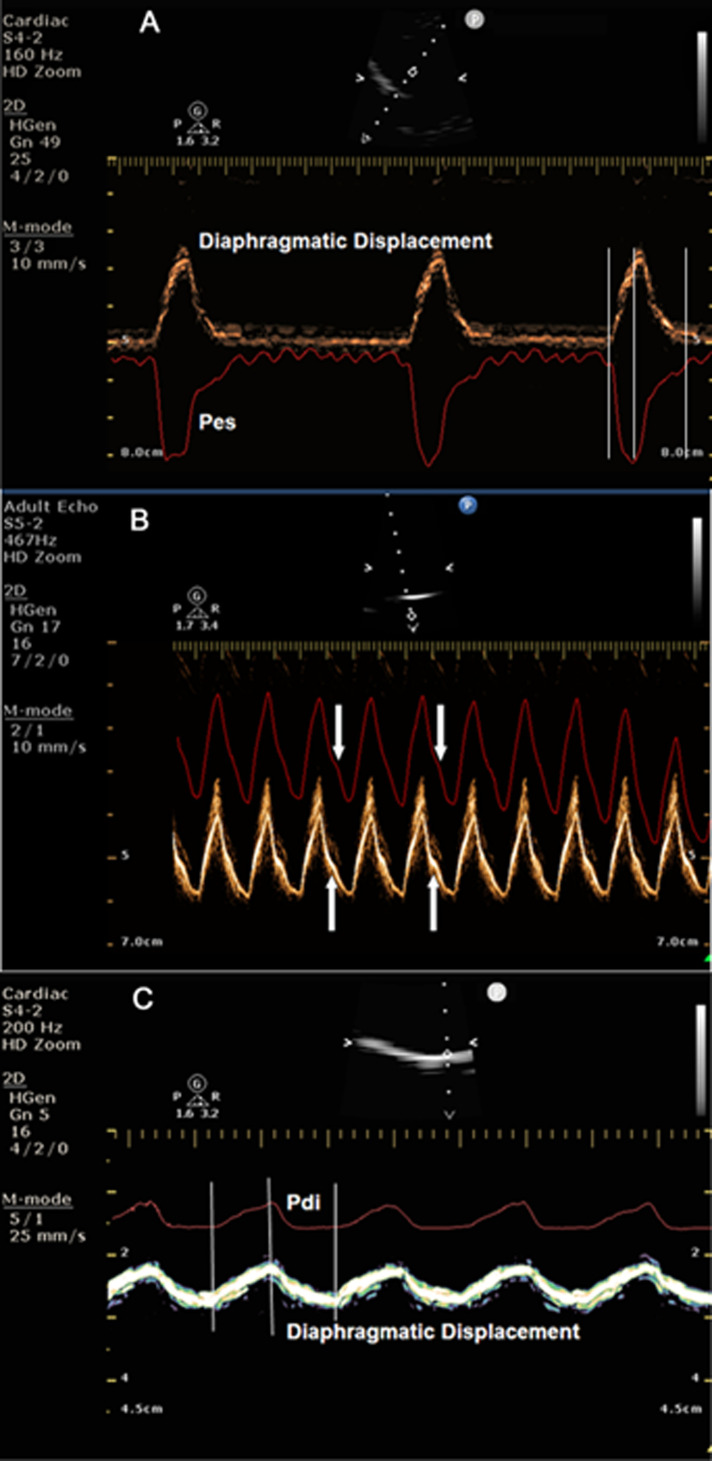
Transdiaphragmatic (Pdi) and oesophageal pressures (Pes) are useful in understanding the pathophysiology of the respiratory system. They provide insight into respiratory drive, intrinsic positive end-expiratory pressure, diaphragmatic fatigue and weaning failure.

BACKGROUND

The use of Pdi and Pes in clinical practice is restricted due to the invasiveness of the technique and the cumbersome equipment needed. On the other hand, diaphragmatic displacement is non-invasively and easily assessed with M-mode ultrasound.

PURPOSE

We observed striking similarities in shape and magnitude between M-mode diaphragmatic displacement, Pes and Pdi pressures. The study aimed to evaluate if the information provided by these two pressures could be obtained non-invasively from the diaphragmatic displacement curve.

MATERIAL AND METHODS

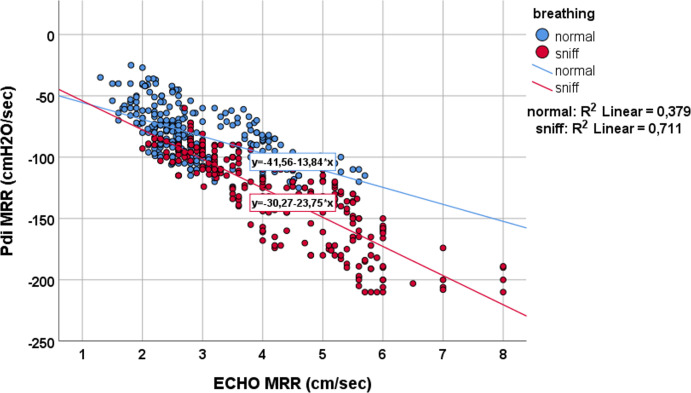
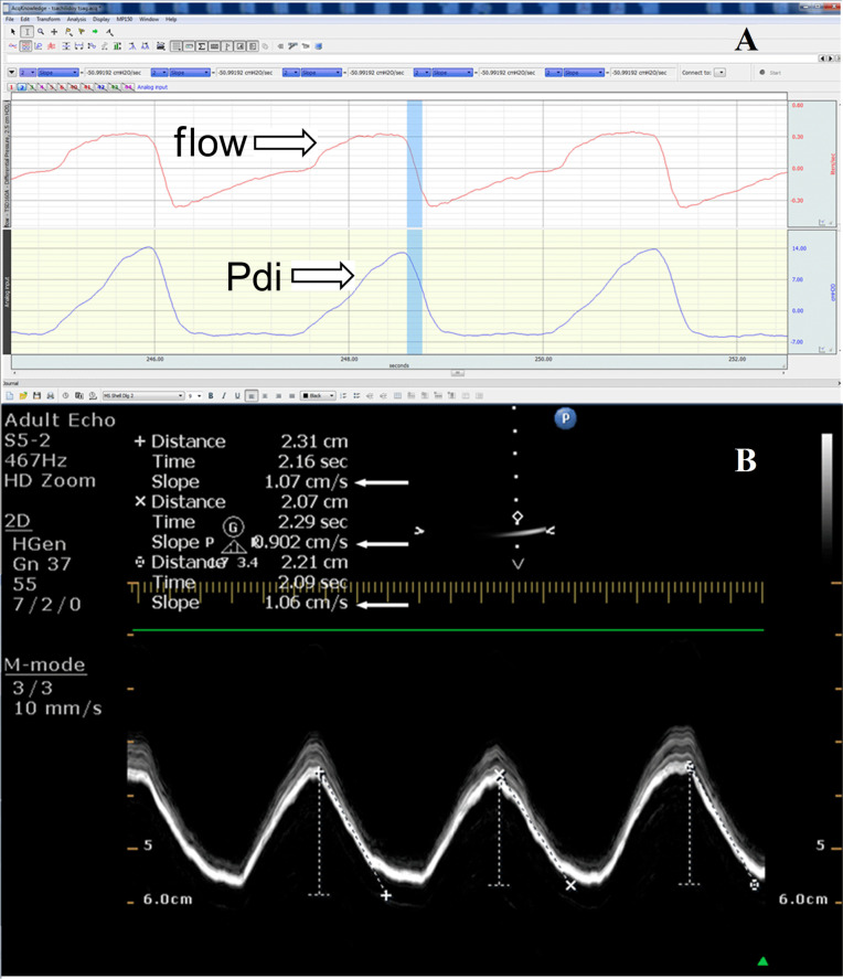
In 14 consecutive intubated patients undergoing a weaning trial, simultaneous recordings of Pes and Pdi pressures and the diaphragmatic displacement were assessed while breathing spontaneously and during a sniff-like manoeuvre. Moreover, the slope of the diaphragmatic displacement curve during relaxation was compared with the maximal relaxation rate (MRR) obtained from the Pdi curve.

RESULTS

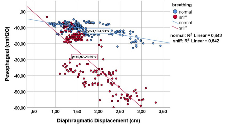
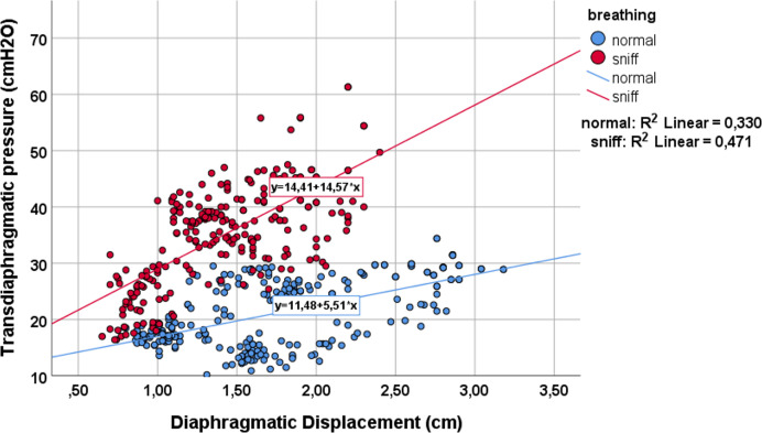
More than 200 breaths were analysed in pairs. Diaphragmatic displacement significantly correlated with Pdi (R=0.33, p<0.001) and Pes (R=0.44, p<0.001), and this correlation further improved during sniff (R=0.47, p<0.001) and (R=0.64, p<0.001), respectively. Additionally, a significant correlation was found between the relaxation slope derived from the diaphragmatic displacement curve and the MRR derived from the Pdi curve, both in normal breathing (R=0.379, p<0.001) and during the sniff manoeuvre (R=0.71, p<0.001).

CONCLUSIONS

M-mode diaphragmatic displacement parameters correlate well with the ones obtained from oesophageal pressure and Pdi, particularly during sniffing. Diaphragmatic displacement assessment possibly offers an alternative non-invasive solution for understanding and clinically monitoring the diaphragmatic contractile properties and weaning failure due to diaphragmatic fatigue.

摘要

目的

我们观察到 M 模式膈肌位移、Pes 和 Pdi 压力之间在形状和幅度上存在显著相似性。本研究旨在评估是否可以从膈肌位移曲线无创地获得这些两种压力提供的信息。

材料和方法

在 14 例连续接受撤机试验的插管患者中,在自主呼吸和嗅探样动作期间评估了 Pes 和 Pdi 压力以及膈肌位移的同步记录。此外,比较了膈肌位移曲线在松弛期间的斜率与从 Pdi 曲线获得的最大松弛率 (MRR)。

结果

对超过 200 对呼吸进行了分析。膈肌位移与 Pdi(R=0.33,p<0.001)和 Pes(R=0.44,p<0.001)显著相关,并且在嗅探时相关性进一步提高(R=0.47,p<0.001)和(R=0.64,p<0.001)。此外,在正常呼吸(R=0.379,p<0.001)和嗅探动作期间(R=0.71,p<0.001),从膈肌位移曲线得出的松弛斜率与从 Pdi 曲线得出的 MRR 之间存在显著相关性。

结论

M 模式膈肌位移参数与从食管压力和 Pdi 获得的参数相关性良好,特别是在嗅探时。膈肌位移评估可能为理解和临床监测膈肌收缩特性以及由于膈肌疲劳导致的撤机失败提供了一种替代的无创解决方案。

https://cdn.ncbi.nlm.nih.gov/pmc/blobs/af72/8461713/fcb633de6899/bmjresp-2021-001006f05.jpg
https://cdn.ncbi.nlm.nih.gov/pmc/blobs/af72/8461713/d0140a0056b1/bmjresp-2021-001006f01.jpg
https://cdn.ncbi.nlm.nih.gov/pmc/blobs/af72/8461713/c9a07ba4c3bd/bmjresp-2021-001006f02.jpg
https://cdn.ncbi.nlm.nih.gov/pmc/blobs/af72/8461713/909506c15f83/bmjresp-2021-001006f03.jpg
https://cdn.ncbi.nlm.nih.gov/pmc/blobs/af72/8461713/b1569e72ff7e/bmjresp-2021-001006f04.jpg
https://cdn.ncbi.nlm.nih.gov/pmc/blobs/af72/8461713/fcb633de6899/bmjresp-2021-001006f05.jpg

相似文献

1
Ultrasonographic assessment of diaphragmatic contraction and relaxation properties: correlations of diaphragmatic displacement with oesophageal and transdiaphragmatic pressure.超声评估膈肌收缩和松弛特性:膈肌位移与食管和膈下压力的相关性。
BMJ Open Respir Res. 2021 Sep;8(1). doi: 10.1136/bmjresp-2021-001006.
2
Effects of muscle group recruitment on sniff transdiaphragmatic pressure and its components.肌肉群募集对嗅吸跨膈压及其组成部分的影响。
Eur J Appl Physiol. 2001 Oct;85(6):593-8. doi: 10.1007/s004210100469.
3
Sniff and Muller manoeuvres to measure diaphragmatic muscle strength.嗅气和米勒动作用于测量膈肌力量。
Respir Med. 2008 Dec;102(12):1737-43. doi: 10.1016/j.rmed.2008.07.004. Epub 2008 Aug 16.
4
Tissue Doppler Imaging of the Diaphragm in Healthy Subjects and Critically Ill Patients.健康受试者和危重症患者膈肌的组织多普勒成像。
Am J Respir Crit Care Med. 2020 Oct 1;202(7):1005-1012. doi: 10.1164/rccm.201912-2341OC.
5
Recruitment of some respiratory muscles during three maximal inspiratory manoeuvres.在三次最大吸气动作过程中某些呼吸肌的募集情况。
Thorax. 1993 Jul;48(7):702-7. doi: 10.1136/thx.48.7.702.
6
The value of sniff esophageal pressures in the assessment of global inspiratory muscle strength.屏气食管压力在评估整体吸气肌力量中的价值。
Am Rev Respir Dis. 1988 Sep;138(3):598-603. doi: 10.1164/ajrccm/138.3.598.
7
The Relationship of Diaphragmatic Ultrasound-Based and Manometric Indices With Difficult Weaning in Tracheostomized Patients.基于膈肌超声和测压指标与气管切开患者脱机困难的关系
Respir Care. 2024 Sep 26;69(10):1323-1331. doi: 10.4187/respcare.11782.
8
Changes in rate of relaxation of sniffs with diaphragmatic fatigue in humans.人类膈肌疲劳时嗅吸松弛速率的变化。
J Appl Physiol Respir Environ Exerc Physiol. 1983 Sep;55(3):731-5. doi: 10.1152/jappl.1983.55.3.731.
9
Maximum relaxation rate of the diaphragm during weaning from mechanical ventilation.机械通气撤机过程中膈肌的最大松弛率。
Thorax. 1994 Jan;49(1):54-60. doi: 10.1136/thx.49.1.54.
10
Evaluation of Respiratory Muscle Strength and Diaphragm Ultrasound: Normative Values, Theoretical Considerations, and Practical Recommendations.评估呼吸肌力量和膈肌超声:正常值、理论考虑和实际建议。
Respiration. 2020;99(5):369-381. doi: 10.1159/000506016. Epub 2020 May 12.

引用本文的文献

1
Association of Diaphragm Involvement Assessed by Ultrasound With Disease Severity in Facioscapulohumeral Muscular Dystrophy.超声评估的膈肌受累与面肩肱型肌营养不良症疾病严重程度的相关性
J Cachexia Sarcopenia Muscle. 2025 Oct;16(5):e70057. doi: 10.1002/jcsm.70057.
2
Diaphragmatic ultrasound and its relationship to breathing effort and load: a prospective observational study.膈肌超声及其与呼吸功和负荷的关系:一项前瞻性观察性研究。
Crit Care. 2025 May 13;29(1):190. doi: 10.1186/s13054-025-05436-1.
3
Influence of an inspiratory muscle fatigue protocol on older adults on respiratory muscle strength, muscle oxygen saturation, and functional capacity: a randomized controlled trial.

本文引用的文献

1
Assessment of diaphragmatic function by ultrasonography: Current approach and perspectives.超声评估膈肌功能:当前方法与展望。
World J Clin Cases. 2020 Jun 26;8(12):2408-2424. doi: 10.12998/wjcc.v8.i12.2408.
2
A New Method for Diaphragmatic Maximum Relaxation Rate Ultrasonographic Measurement in the Assessment of Patients With Diaphragmatic Dysfunction.一种用于评估膈肌功能障碍患者的膈肌最大松弛率超声测量新方法。
IEEE J Transl Eng Health Med. 2018 Oct 11;6:2700710. doi: 10.1109/JTEHM.2018.2868671. eCollection 2018.
3
Interpreting diaphragmatic movement with bedside imaging, review article.
吸气肌疲劳方案对老年人呼吸肌力量、肌肉氧饱和度和功能能力的影响:一项随机对照试验。
BMC Geriatr. 2024 Dec 19;24(1):1015. doi: 10.1186/s12877-024-05610-0.
4
Diaphragm ultrasound and diaphragmatic 2D speckle tracking imaging in acute heart failure: a case series.急性心力衰竭中的膈肌超声与膈肌二维斑点追踪成像:病例系列
Eur Heart J Case Rep. 2024 Nov 28;8(12):ytae632. doi: 10.1093/ehjcr/ytae632. eCollection 2024 Dec.
5
Influence of an inspiratory muscle fatigue protocol on healthy youths on respiratory muscle strength and heart rate variability. A randomized controlled trial.吸气肌疲劳方案对健康青年呼吸肌力量和心率变异性的影响。一项随机对照试验。
Front Physiol. 2024 Aug 23;15:1457019. doi: 10.3389/fphys.2024.1457019. eCollection 2024.
6
Evaluation and modeling of diaphragm displacement using ultrasound imaging for wearable respiratory assistive robot.用于可穿戴呼吸辅助机器人的超声成像膈肌位移评估与建模
Front Bioeng Biotechnol. 2024 Aug 16;12:1436702. doi: 10.3389/fbioe.2024.1436702. eCollection 2024.
7
Influence of an inspiratory muscle fatigue protocol on healthy youths on respiratory muscle strength, vertical jump performance and muscle oxygen saturation: a randomized controlled trial.吸气肌疲劳方案对健康青年呼吸肌力量、垂直跳跃表现和肌肉氧饱和度的影响:一项随机对照试验。
J Transl Med. 2024 Aug 5;22(1):732. doi: 10.1186/s12967-024-05555-3.
8
The Relationship of Diaphragmatic Ultrasound-Based and Manometric Indices With Difficult Weaning in Tracheostomized Patients.基于膈肌超声和测压指标与气管切开患者脱机困难的关系
Respir Care. 2024 Sep 26;69(10):1323-1331. doi: 10.4187/respcare.11782.
9
Ultrasonographic evaluation of the diaphragm in critically ill patients to predict invasive mechanical ventilation.对危重症患者膈肌进行超声评估以预测有创机械通气。
J Intensive Care. 2023 Sep 19;11(1):40. doi: 10.1186/s40560-023-00690-3.
床边成像对膈肌运动的解读,综述文章。
J Crit Care. 2016 Aug;34:56-65. doi: 10.1016/j.jcrc.2016.03.006. Epub 2016 Mar 17.
4
Monitoring patient-ventilator asynchrony.监测患者与呼吸机不同步情况。
Curr Opin Crit Care. 2016 Jun;22(3):246-53. doi: 10.1097/MCC.0000000000000307.
5
The application of esophageal pressure measurement in patients with respiratory failure.食管压力测量在呼吸衰竭患者中的应用。
Am J Respir Crit Care Med. 2014 Mar 1;189(5):520-31. doi: 10.1164/rccm.201312-2193CI.
6
Sonographic evaluation of the diaphragm in critically ill patients. Technique and clinical applications.超声评估危重症患者的膈肌:技术与临床应用。
Intensive Care Med. 2013 May;39(5):801-10. doi: 10.1007/s00134-013-2823-1. Epub 2013 Jan 24.
7
Sonographic assessment of changes in diaphragmatic kinetics induced by inspiratory resistive loading.超声评估吸气阻力负荷引起的膈肌动力学变化。
Respirology. 2013 Apr;18(3):468-73. doi: 10.1111/resp.12011.
8
Diaphragmatic motion studied by m-mode ultrasonography: methods, reproducibility, and normal values.M型超声心动图研究膈肌运动:方法、可重复性及正常值
Chest. 2009 Feb;135(2):391-400. doi: 10.1378/chest.08-1541. Epub 2008 Nov 18.
9
M-mode sonography of diaphragmatic motion: description of technique and experience in 278 pediatric patients.膈肌运动的M型超声检查:技术描述及278例儿科患者的经验
Pediatr Radiol. 2005 Jul;35(7):661-7. doi: 10.1007/s00247-005-1433-7. Epub 2005 Mar 18.
10
Weaning prediction: esophageal pressure monitoring complements readiness testing.撤机预测:食管压力监测辅助撤机准备测试。
Am J Respir Crit Care Med. 2005 Jun 1;171(11):1252-9. doi: 10.1164/rccm.200503-356OC. Epub 2005 Mar 11.